박 영 주
150-950 서울시 영등포구 대림1동948-1번지 한림대학교성심병원 강남 치과/구강악안면외과 Young-Ju Park
Department of Oral and Maxillofacial Surgery, Kangnam Sacred Heart Hospital, College of Medicine, Hallym University
948-1, Daerim-1 Dong, Yeongdeungpo-gu, Seoul 150-950, Korea Tel: +82-2-829-5233 Fax: +82-2-846-9789
E-mail: [email protected]
백서 두개골결손모델에서 실크단백과 골형성단백 이식체가 골재생에 미치는 영향
남정훈1,2∙노경록1,2∙방은오1,2∙유우근1,2∙강응선1,2∙권해용3∙김성곤4∙박영주1,2
1한림대학교 의과대학 강남성심병원 구강악안면외과학교실, 2한림대학교 임상치의학대학원 구강악안면임프란트학과,
3농촌진흥청, 4강릉원주대학교 치과대학 구강악안면외과학교실
The effect of silk fibroin and rhBMP-2 on bone regeneration in rat calvarial defect model
Jeong-Hun Nam
1,2, Kyung-Lok Noh
1,2, Eun-O Pang
1,2, Woo-Geun Yu
1,2, Eung-Sun Kang
1,2, Hae-Yong Kweon
3, Seong-Gon Kim
4, Young-Ju Park
1,21
Department of Oral and Maxillofacial Surgery, Kangnam Sacred Heart Hospital, College of Medicine
2
Department of Oral and Maxillofacial Implantology, Clinical Graduate School of Dentistry, Hallym University
3
National Academy of Agricultural Science, Rural Development Administration
4
Department of Oral and Maxillofacial Surgery, College of Dentistry, Gangneung-Wonju National University
Introduction:This study evaluated the capability of silk fibroin (SF) and recombinant human bone morphogenetic protein-2 loaded SF (SF-BMP) as a bone defect replacement matrix when grafted in a calvarial bone defect of rats in vivo.
Materials and Methods:A total 70 calvarial critical size defects (5.0 mm in diameter) made on 35 adult female Sprague-Dawley rats were used in this study. The defects were transplanted with (1) rhBMP-2 loaded silk fibroin graft (SF-BMP: 0.8+10 μg), (2) Silk fibroin (SF: 10 μg), and (3) no graft material (Raw). The samples were evaluated with soft x-rays, alkaline phosphatase activity, calcium/phosphate quantification, histological and histomorphometric analysis at postoperative 4 and 8 weeks.
Results:The SF-BMP group (48.86±14.92%) had a significantly higher mean percentage bone area than the SF group (24.96±11.01%) at postop- erative 4 weeks.(P<0.05) In addition, the SF-BMP group (40.01±12.43%) had a higher % bone area at postoperative 8 weeks than the SF group (33.26±5.15%). The mean ratio of gray scale levels to the host bone showed that the SF-BMP group (0.67±0.08) had a higher mean ratio level than the SF group (0.61±0.09) at postoperative 8 weeks. These differences were not statistically significant.(P=0.168 and P=0.243, respectively) The ratio of the calcium and phosphate contents of the SF-BMP (0.93±0.22) group was lower than that of the SF (1.90±1.42) group at postoperative 4 weeks. However, the SF-BMP group (0.75±0.31) had a higher Ca/PO4ratio than the SF (0.68±0.04) at postoperative 8 weeks. These differences were not statistically significant.(P=0.126 and P=0.627, respectively) For the bone-specific alkaline phosphatase (ALP) activity, which is recognized as a reliable indicator of the osteoblast function, the SF-BMP (23.71±8.60 U/L) groups had a significantly higher value than the SF group (12.65±
6.47 U/L) at postoperative 4 weeks.(P<0.05) At postoperative 8 weeks, the SF-BMP (21.65±10.02 U/L) group had a lower bone-specific ALP activi- ty than the SF group (16.72±7.35 U/L). This difference was not statistically significant.(P=0.263) For the histological evaluation, the SF-BMP group revealed less inflammation, lower foreign body reactions and higher bone healing than the SF group at postoperative 4 and 8 weeks. The SF group revealed more foreign body reactions at postoperative 4 weeks. However, this immunogenic reaction decreased and the remnant of grafted material was observed at postoperative 8 weeks. For histomorphometric analysis, the SF-BMP group had a significantly longer bone length to total length ratio than those of the SF group at postoperative 4 and 8 weeks.(P<0.05)
Conclusion:The rhBMP-2 loaded silk fibroin graft revealed fewer immunoreactions and inflammation as well as more new bone formation than the pure silk fibroin graft. Therefore, silk fibroin may be a candidate scaffold for tissue engineered bone regeneration.
Key words:Silk, Fibroins, Recombinant human bone morphogenetic protein-2, Bone regeneration, Tissue scaffolds
[paper submitted 2010. 6. 7 / revised 2010. 10.15 / accepted 2010. 10. 19 ] Abstract (J Korean Assoc Oral Maxillofac Surg 2010;36:366-74)
* This study was supported by a grant from the BioGreen21 Program (No. PJ007170201006), Rural Development Administration, Suwon, Republic of Korea.
Ⅰ. 서 론
현대 치의학의 목표는 예견 가능한 방법으로 환자들의 구강건강을 회복시켜주는 것이다. 고령화 사회의 심화와 더불어 치아상실부의 치조골흡수와 위축이 진행된 경우가 많다. 이에 가용골의 높이와 폭경을 늘리기 위하여 골이식 이 널리 사용되고 있다. 그 중 자가골이식술을 골결손부를 회복하기 위한 치료방법으로 많이 채택한다. 자가골이식 은 골세포, 성장인자 등을 함께 이식하여 골형성 측면에서 효과적이나 큰 결손부를 수복하기에는 공급에 한계가 있 고, 골채취를 위한 부가적인 수술, 채취부위의 손상을 가져 온다. 이를 극복하고자 동종골, 이종골 또는 합성골을 사용 할 수 있지만 이는 골전도능만을 제공하는 경우가 많으며, 흡수되지 않고 남아있는 경우도 있어 골유합의 지연을 초 래하기도 한다. 이러한 문제점을 해결하기 위한 방안으로 다양한 지지체와 조직활성인자 조합의 골이식재 연구가 진행 중이다.
골형성 유도 단백질로 알려진 bone morphogenetic protein
(BMP)은 Urist
1가 소개하였고, Mizutani와 Urist2는 인간의 골육종세포로부터 BMP를 분리하였음을 보고하였다. Sato 와 Urist3는 BMP의 아미노산 염기서열을 분석하였고 조직 배 양 에 서 BMP로 부 터 유 도 된 연 골 을 배 양 하 였 으 며 ,Wozney
등4에 의해 recombinant BMP가 알려지게 되었다.기존의 연구에서 보고된 BMP는 포유류의 골조직, 골육종 세포, 치아의 경조직 등에서 분리 추출된 것으로 그 추출량 이 제한되어 있었으며 과정 또한 복잡하였다. 그러나 최근 에 와서는 유전자 조작기술의 진보로 다량의 BMP를 만들 수 있게 되었다. BMP는 다른 비교원성 단백질에 비하여 소 량 존재하며 종간특이성을 가지지 않는다는 특성이 있다5
. BMP는 미분화간엽세포를 연골세포나 조골세포로 분화시
켜 발생하는 것으로 알려져 있고, recombinant human BMP-2 (rhBMP-2)는 적절한 운반 매개체와 함께 조합했을 때 골
결손을 치료하는데 유용하다6-11.
골재생에 있어 그 효과가 이미 잘 알려진 만큼 rhBMP-2의 서방출이 가능한 지지체 의 연구가 많이 시도되고 있다.조직공학에서 지지체는 세포외기질의 구조 및 생물학적 기능과 유사해야 한다. 즉, 기계적으로 공간을 지지하고 수 혜부에 유도분자나 세포를 운반하며 새롭게 형성된 조직 의 구조와 기능을 조절해야 한다. 최근 이러한 전달체로
polymer (hydroxyapatite, hyaluronic acid
등의 천연재료나polylactide-co-glycolide acid, polylactide acid등의 합성 poly- mer)나 hydrogel 등 최근에는 우수한 생체친화성, 적절한
기계적 물성, 생분해성 및 다공성 등의 scaffold가 각광을 받고 있다12-17.
누에고치의 실크에서 추출한 단백질인 silk fibroin (SF) 역시 지지체로서 관심이 고조되고 있고, 이를 이용한 인조 골 연구가 활발히 진행되고 있다. SF은 생체친화성, 느린 분해속도, 적절한 기계적 성질 등 지지체로서 우수한 특징
을 가지고 있다18-20
. 과거에는 silk로 옷을 만들거나 식품, 화
장품 등을 만드는데 사용하였으나, 근래에는 실크막의 섬 유아세포 배양 효과가 확인되었고21,
실크 지지체의 연골 및 골아세포배양 효과도 보고되고 있으며22-25,
국내에서도 연구가 활발하여 실크 나노 지지체를 이용한 골재생에 대 한 보고가 있었다26-28.
본 연구에서는 백서 두개골결손 모델을 통해 SF과
rhBMP-2조합의 골재생능을 측정하여 rhBMP-2의 운반체
로서 SF의 효용성을 평가하고자 하였다.Ⅱ. 연구대상 및 방법 1. SF의 제조
SF은 한국농촌진흥청 잠사과 Biogreen
21 연구부(Suwon,Korea)에서 제공받았다. 제조과정을 요약하면, 누에고치에
서 절각견을 추출하고, 과도한 거품을 방지하기 위해 겉 껍 데기인 세리신을 제거하고 fibroin만 남겨 산가수분해, 중 성염가수분해, 효소가수분해를 시행하고, 전기탈염장치와 겔여과장치로 중화시키고, 동결건조장치와 분무건조장치 를 이용하여 건조분말을 만든 후 스펀지 형태의 이식재로 가공한 것으로 분자량은 약 350 kDa이다.2. rhBMP-2의 준비
실험에 사용할 rhBMP-2 (R&D systems Inc., Minneapolis,
MN, USA)
는 중국 햄스터의 난소세포에서 발현되어human BMP-2로 유전자 재조합된 것으로, 제조사의 설명
에 따라 사용하였다.3. 동물실험군 및 수술
체 중 250-300 gm의 8주 령 된 Sprague-Dawley 백 서
(Oriental Bio Co., Seoul, Korea) 36마리를 사용하였으며, 이
식재와 실험기간에 따라 3개 군으로 분류하여 실험하였다.SF-BMP군, SF군, Raw군은 각 12 마리이며, 군별로 4주, 8
주 기간으로 6마리씩 반분하였다.(Table 1)실험동물은 Xylazine 20 mg/mL (Rompun, Bayer korea
Ltd., Seoul, Korea)과 Tiletamine 25 mg/mL과 Zolazepam 25
mg/mL (Zoletil 50, Virbac Laboratories, Carros, France)의 혼
합액(1:3 부피비, 0.4 cc, 복강 내 주사)을 사용하여 마취하 였다. 전두부를 제모하고 포비돈 솜으로 소독한 후, 비골에 서 후두골 사이의 두개골시상봉합을 따라 약 2 cm가량의 절개를 가한 다음 골막을 포함하는 피판을 거상하여 두정 골까지 노출시켰다. 5 mm trephine bur (Ace Surgical supplyCo., Brockton, USA)와 Max. 32,000 rpm의 저속 드릴 키트
(Sae Yang Machinery Co., Daegu, Korea)를 사용하여 두정부
에 직경 5 mm의 원형결손부를 양쪽에 2개씩 형성하였다.제거한 골창은 정상두정골의 칼슘(Ca)과 인산(PO4
)의 정량
측정에 사용하기 위해 냉동보관하였다. 예상하지 못한 출 혈은 oxidized cellulose (Surgicel, Johnson & Johnson Medical,New Brunswick, NJ, USA)를 이용하여 지혈하였다. 골결손
부에는 10 μg의 SF과 0.8 μg의 rhBMP-2를 이용하여 각 군의 조성에 맞게 이식하였고, 피부는 5-0 nylon (Ethicon, Johnson& Johnson Medical, New Brunswick, NJ, USA)으로 봉합하였
다. 실험 4주, 8주 후 각 실험동물들을 이산화탄소(CO2) 질
식을 통하여 희생시킨 다음 결손부를 포함한 두개골을 채 취하여 방사선학적, 정량적, 조직형태학적 분석을 시행하 였다. 본 실험은 한림대학교 동물실험윤리위원회의 승인하 에 시행하였다.(승인번호: No. Hallym 2009-66)4. 방사선학적 분석
채취된 조직을 캐비닛형 soft x-ray (CMB-2, Softex Co.,
Tokyo, Japan)를 관전압 20 kVp, 관전류 2 mA, 노출시간 2
분의 조건)으로 방사선사진을 채득하였다. 방사선필름(Biomax, Kodak, Rochester, NY, USA)은 디지털스캐너 (Cannon, Tokyo, Japan)를 이용하여 디지털영상으로 변환
하였으며, 골재생부에서 무작위로 추출한 6개 점의 방사선 밀도를 영상분석기(OPTIMAS, version 6.51.199, MediaCybernetics, L.P., Silver Spring, MD, USA)를 이용하여 흑화
도(gray scale)의 평균치를 측정하였다. 또한 획득한 이미지 에 결손부의 원래 면적과 새로 재생한 부위의 외형을 비교 하여 재생면적의 비율을 계산하였다.5. 건조중량, alkaline phosphatase (ALP) 활성도 및 Ca/PO4 정량분석
Soft x-ray촬영 후, 조직학 및 조직형태학적 분석을 위해
군별로 개체의 절반을 할당하고 남은 나머지 개체를 ALP 활성도 및 Ca/PO4정량분석에 이용하였다. 5 mm직경의 원 형결손부를 수술용 가위로 절단하고 골수와 혈액을 제거 하기 위해 phosphate buffered saline (PBS)에 담구어 4℃, 72 시간 동안 단백질을 추출하였다. 불용성 물질을 제거하기 위해 4℃, 13,000 rpm, 10분 동안 원심분리한 다음 동결건조 기(Labconco Co., KC, USA)에 넣어 overnight한 후 정밀저 울을 사용해 건조중량을 측정하였다.1) ALP 활성도 분석
ALP 활성도는 para-nitrophenyl phosphate (pNPP)로 부터 p-nitrophenolate의 시간 의존적인 형성에 의해 측정(405 nm
에서 빛의 최대 흡수)되는 QuantiChrom alkaline phosphataseassay kit (Bioassay Systems, Hayward, CA, USA)를 사용하였
다. 96 microtiter plate well에 뼈 추출물 30 μL를 첨가하였다.반응을 시작하기 위해 샘플 well에 전체 반응 양이 200 μL 가 되도록 working solution (assay buffer, pH 10.5, 5 mM Mg
acetate, 10 mM pNPP)을 신속히 첨가하고 잘 섞어주었다.
405 nm에서 초기 흡광도와 4분 경과 후 흡광도를 microtiter plate reader (Powerwave X340, Bio-Tek instruments, Inc., Winooski, VT, USA) 를 이용하여 측정하였다. ALP 활성도
는 U/mg 뼈 건조중량으로 계산하였으며, 1 U는 실온에서 1 분당 기질 1 μmol의 산물로의 전환으로 정의하였다.2) Ca과 PO
4의 정량분석Ca양과 PO
4의 측정은 QuantiChrom calcium phosphate assaykit (Bioassay Systems, Hayward, CA, USA)를 사용하여 분석
하였다. 건조된 뼈를 회화로에 넣어 700℃, 2시간 동안 희석 재가 될 때까지 태워 Ca과 결합한 유기물 성분을 제거하였 다. 회화가 끝난 시료는 6 N 염산 3 mL을 넣어 4℃ overnight 시킨 다음, Whatman paper #1로 filtering 하였다. Ca양은 10 배로 희석한 시료 5 μL를 96 well plate에 첨가하고 workingreagent 200
μL 첨가 후 실온에서 3분간 반응시켰으며, 612nm에서 흡광도를 microtiter plate를 이용하여 측정하였다.
PO
4양 측정을 위해 100배 희석한 시료 50 μL를 96 well plate 에 첨가하고 reagent 100 μL 첨가 후 실온에서 30분간 반응 시키고 광밀도 620 nm에서 흡광도를 측정한 후 여러 stan-dard의 농도를 비교하여 샘플의 농도를 산출하였다.
6. 조직학 및 조직형태학적 분석
Soft x-ray 촬영 후, 각 시료의 절반을 조직학 및 조직형태
학적 분석을 위해 처리하였다. 각 조직은 10% bufferedformaldehyde에 고정하였으며, 10% formic acid로 탈회하였
다. 시상봉합에 직각으로 골결손부의 적도를 포함하여 양 부위를 trimming 한 후 파라핀에 포매하였다. 블록을 4 μm 두께로 절단한 후 hematoxylin and eosin (H&E) 과 Masson’strichrome (MT)
그리고 osteocalcin 면역염색을 하였다. MT 염 색 을 시 행 한 슬 라 이 드 영 상 은 ArtixScan 4000TF(Microtek, Columbia, MD, USA)으로 영상주사(scan)한 후 SPOT version 4.6 (Diagnostic Instrument, Inc, Sterling. Heights,
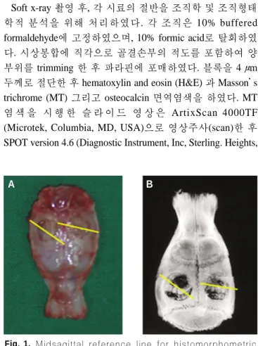
Fig. 1. Midsagittal reference line for histomorphometric analysis. A. Harvested calvaria, B. Soft X-ray image.
A B
MI, USA)으로 화상화(capture)하였다. 조직형태학적 분석은 1.25배의 배율로 이식부위에서 신생골의 길이와 면적을 계
측하고, 전체 길이 및 면적에 대한 신생골의 비율을 Optimas 소프트웨어 프로그램을 통해 분석, 계산하였다.(Fig. 1)7. 통계학적 방법
조직형태학적, 방사선학적, 정량적 분석을 통해 얻은 결 과는 Statview version 5.0.1 (SAS Institute Inc, Cray, NC,
USA)
통계 프로그램을 이용하여 one-way analysis of vari-ance (ANOVA) test 로 분석하였다. 유의성 있는 결과가 관
찰되면 a pairwise multiple test인 Fisher’s protected least sig-nificant difference (PLSD)를 이용하여 각 군을 비교하였다.
P-value가 0.05 이하일 경우 유의성 있는 차이로 간주하였다.
Ⅲ. 결 과 1. 실험동물과 실험술식
모든 동물은 수술 직후 회복되었으나 Raw군 8주차 1마리 가 술후 3일째 사망하였다. 따라서 각 군의 실험동물은 SF-
BMP군 12마리, SF군 12마리, Raw군 11마리 총 35마리의 70개의 백서 두개골결손부를 분석하였다. 수술 4주, 8주 후
창상의 감염이나 예상치 못한 조직반응은 발견되지 않았 다.(Table 1)
2. 방사선학적 분석
수 술 4주 차 에 서 신 생 골 형 성 은 SF-BMP군 (48.86±
14.92%)이 SF군(24.96±11.01%)보다 통계적으로 유의하게
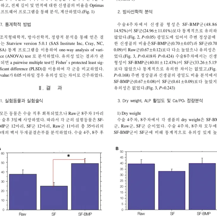
많았다.(Fig. 2, P<0.05) 광밀도에 있어서 주변 정상골에 대 한 신생골의 비율은SF-BMP군(0.70±0.07)과 SF군(0.70±0.09)이 Raw군(0.67±0.12)보다 다소 높았으나 유의성은 없
었다.(Fig. 3, P=0.418와 P=0.424) 수술8주차에서는 신생골 형성이 SF-BMP군(40.01±12.43%)이 SF군(33.26±5.15%) 보다 많았으나 통계적으로 유의한 차이는 없었고,(Fig. 2,P=0.168) 주변 정상골과 신생골의 광밀도 비율 분석에서도 SF-BMP군(0.67±0.08)이 SF군(0.61±0.09)보다 높았지만
유의성은 없었다.(Fig. 3, P=0.243)3. Dry weight, ALP 활성도 및 Ca/PO4 정량분석
1) Dry weight
수술 4주차, 8주차에서 각 샘플의 dry weight은 SF-BMP 군, Raw군, SF군 순이었다. 수술 4주차, 8주차 모두에서
SF-BMP군이 SF군에 비해 통계적으로 유의성 있게 높았
Table 1.Animal grouping
Groups N (sites/animals)
Composition Graft
4 weeks 8 weeks
SF-BMP 12 (6) 10 (6) rhBMP-2 loaded silk fibroin 0.8+10 μg
SF 12 (6) 12 (6) Silk fibroin 10 μg
Raw 12 (6) 12 (5) Raw defect None
(SF: silk fibroin, SF-BMP: recombinant human bone morphogenetic protein-2 loaded SF)
A B
60
50
40
30
20
10
0
50 45 40 35 30 25 20 15 10 5 0
Raw SF SF-BMP Raw SF SF-BMP
Fig. 2.% bone area of densitometric analysis. A. The SF-BMP group (48.86±14.92%) had a significant higher value than that of SF group (24.96±11.01%, *P<0.05) at postoperative 4 weeks. B. The SF-BMP group (40.01±12.43%) had a higher
%bone area than that of SF group (33.26±5.15%) at postoperative 8 weeks. This difference was not, however, statistically significant.(P=0.168)
(SF: silk fibroin, SF-BMP: recombinant human bone morphogenetic protein-2 loaded SF)
다.(Table 2, P<0.05)
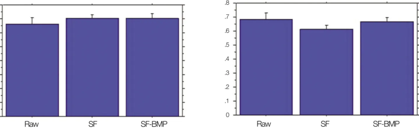
2) ALP 활성도
측정값이 높을수록 PO4함유 substrate의 hydrolysis를 증가 시켜 orthophosphate를 형성하고 Ca흡수를 증진시켜 골재 생의 좋은 지표가 되는 ALP활성도는 4주차에서 SF-BMP 군(23.71±8.60 U/L)이 SF군(12.65±6.47 U/L)보다 유의하 게 높았고(P<0.05), 8주차에서는 통계적으로 유의한 차이 는 없으나 SF-BMP군(21.65±10.02 U/L)이 SF군(16.72±
7.35 U/L)보다 높았다.(Fig. 4, P=0.263)
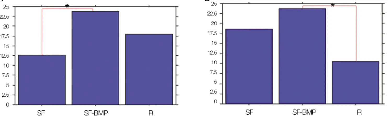
3) Ca과 PO
4의 정량분석Ca의 경우 4주차에서 SF-BMP군(28.67±14.28 mg/dL)이 SF군(12.53±5.01 mg/dL)과 Raw군(12.16±4.63 mg/dL) 보다
유의하게 높았고(P<0.05), 8주차에서는 SF-BMP군 (28.56±20.44 mg/dL)이 SF군(2.83±1.38 mg/dL)보다 유의하게 높았
다.(Fig. 5, P<0.05) PO4의 경우 4주차에서 SF-BMP군(30.77
±12.52 mg/dL)이 SF군(12.28±11.87 mg/dL) 과 Raw군
(12.06±6.99 mg/dL)보다 유의하게 높았고(P<0.05), 8주차에
서는 SF-BMP군(34.09±12.37 mg/dL)이 SF군(4.25±2.34mg/dL)보다 통계적으로 유의성 있게 높았다.(Fig. 6, P<0.05)
수치가 높을수록 maturation level이 높은 골광화도(degree ofmatrix mineralization)를 나타내는 Ca과 PO
4비율(Ca/PO4)의
경우, 4주차에 SF군(1.90±1.42), Raw군(1.15±0.36), SF-BMP군(0.93±0.22) 순이었고, 8주차에서는 SF-BMP군(0.75
±0.31), Raw군(0.71±0.14), SF군(0.68±0.04) 순이었으나 모두 통계적 유의성은 없었다.(P=0.126와 P=0.627)
4) 조직학 및 조직형태학적 분석
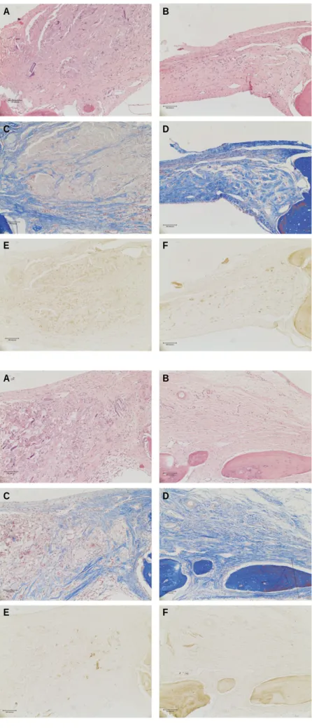
조직학적 소견상 SF-BMP군의 경우, 이물반응과 염증이 적으며 8주로 갈수록 골형성이 빨리 진행되었고, 술후 회복 과 골재생이 잘되는 양상이 관찰되었다.(Fig. 7) SF군의 경 우, 초기에 이물반응과 염증반응이 심하며, 8주로 갈수록 줄 어드는 양상을 보였고, 아직 이식체가 잔존함이 관찰되었 다.(Fig. 8) 조직형태학적 분석에서 신생골 길이는 4주차에
SF-BMP군이 SF군에 비해 유의성 있게 높았고(P<0.05), 8주
차에는 SF-BMP군이 SF군과 Raw군에 비해 유의하게 높았 다.(P<0.05) 신생골 면적에서는 4주차와 8주차 모두에서 SF-BMP군이 다른 군보다 높은 수치를 보였지만 통계적으로 유
의한 차이는 없었다.(Table 3)A B
.8 .7 .6 .5 .4 .3 .2 .1 0
Raw SF SF-BMP Raw SF SF-BMP
Fig. 3.Gray scale ratio to host bone. A. The SF-BMP group (0.70±0.07) and SF group (0.70±0.09) had a higher value than that of the Raw group (0.67±0.12) at postoperative 4 weeks. These differences were not, however, statistically significant.(P=0.418 and P=0.424, respectively) B. At postoperative 8 weeks, the SF-BMP group (0.67±0.08) had a higher value than that of the SF group (0.61±0.09), however, it was not statistically significant.(P=0.243)
(SF: silk fibroin, SF-BMP: recombinant human bone morphogenetic protein-2 loaded SF) .8
.7 .6 .5 .4 .3 .2 .1 0
Table 2.Dry weight
Groups 4 weeks (mg) 8 weeks (mg)
SF-BMP 635.83±21.09 32.17±23.05
SF 611.75±4.03 7.00±2.83
Raw 616.83±13.35 28.17±13.47
(SF: silk fibroin, SF-BMP: recombinant human bone morphogenetic protein-2 loaded SF)
Table 3.Histomorphometric analysis
Groups Bone length (mm) Bone area (mm2)
4 weeks 8 weeks 4 weeks 8 weeks
SF-BMP 0.30±0.26 0.45±0.44 0.013±0.013 0.027±0.041
SF 0.24±0.82 0.13±0.16 0.002±0.007 0.008±0.009
Raw 0.16±0.20 0.97±0.140.011±0.018 0.002±0.002
(SF: silk fibroin, SF-BMP: recombinant human bone morphogenetic protein-2 loaded SF)
A B
25 22.5 20 17.5 15 12.5 10 7.5 5 2.5 0
SF SF-BMP R SF SF-BMP R
Fig. 4.ALP activity. A. The SF-BMP group (23.71±8.60 U/L) had a significant higher value than that of the SF group (12.65±
6.47 U/L) at postoperative 4 weeks.(*P<0.05) B. The SF-BMP group (21.65±10.02 U/L) had a higher value than that of the SF group (16.72±7.35 U/L) at postoperative 8 weeks. However, there was not statistically significant difference.(P=0.263) (ALP: alkaline phosphatase, SF: silk fibroin, SF-BMP: recombinant human bone morphogenetic protein-2 loaded SF)
25 22.5 20 17.5 15 12.5 10 7.5 5 2.5 0
A B
30
25
20
15
10
5
0 SF SF-BMP Raw SF SF-BMP R
Fig. 5. Ca2+ quantification. A. The SF-BMP group (28.67±14.28 mg/dL) had a significant higher value than that of the SF group (12.53±5.01 mg/dL) at postoperative 4 weeks.(*P<0.05) B. The SF-BMP group (28.56±20.44 mg/dL) had a signifi- cant higher value than that of the SF group (2.83±1.38 mg/dL) at postoperative 8 weeks.(*P<0.05)
(SF: silk fibroin, SF-BMP: recombinant human bone morphogenetic protein-2 loaded SF) 40
35 30 25 20 15 10 5 0
A B
35
30
25
20
15
10
5
0 SF SF-BMP Raw SF SF-BMP R
Fig. 6. PO42- quantification. A. The SF-BMP group (30.77±12.52 mg/dL) had a significant higher value than that of the SF group (12.28±11.87 mg/dL) at postoperative 4 weeks.(*P<0.05) B. The SF-BMP group(34.09±12.37 mg/dL) had a signifi- cant higher value than that of the SF group (4.25±2.34 mg/dL)at postoperative 8 weeks.(*P<0.05)
(SF: silk fibroin, SF-BMP: recombinant human bone morphogenetic protein-2 loaded SF) 45
40 35 30 25 20 15 10 5 0
Fig. 7. A. Postoperative 4 weeks.(H&E staining, original magnification x100) B. Postoperative 8 weeks.(H&E staining, original magnification x100) C. Postoperative 4 weeks.(Masson’s trichrome staining, original magnification x100) D. Postoperative 8 weeks.(Masson’s trichrome staining, original magnification x100) E.
Postoperative 4 weeks.(Osteocalcin immunoreac- tive staining, original magnification x100) F.
Postoperative 8 weeks.(Osteocalcin immunoreac- tive staining, original magnification x100)(rhBMP- 2: recombinant human bone morphogenetic pro- tein-2, SF: silk fibroin, SF-BMP: recombinant human bone morphogenetic protein-2 loaded SF)
A B
C D
E F
Fig. 8. A. Postoperative 4 weeks.(H&E staining, original magnification x100) B. Postoperative 8 weeks.(H&E staining, original magnification x100) C. Postoperative 4 weeks.(Masson’s trichrome staining, original magnification x100) D. Postoperative 8 weeks.(Masson’s trichrome staining, original magnification x100) E.
Postoperative 4 weeks.(Osteocalcin immunoreac- tive staining, original magnification x100) F.
Postoperative 8 weeks.(Osteocalcin immunoreac- tive staining, original magnification x100)(rhBMP- 2: recombinant human bone morphogenetic pro- tein-2, SF: silk fibroin)
A B
C D
E F
Ⅳ. 고 찰
본 연구에서는 rhBMP-2의 운반체 및 골재생의 지지체로 서 SF의 효용성을 평가하고자 백서 두개골결손 모델을 이 용하여 rhBMP-2와 SF 이식체의 골재생능을 측정하였다.
누에고치로부터 추출하는 SF은 저렴하고 생체친화성이 좋으며 적절한 기계적 성질 및 느린 분해속도 등의 장점이
있다18-20
.
그러나 SF을 이용한 여러 연구에서 염증 반응과면역 반응이 있음이 보고되었다29,30
.
본 연구에서도 조직학 적으로 순수한 SF을 이식한 경우, 초기에 이물반응과 섬유 화, 비만세포 및 대식세포 등이 관찰되며 염증 반응과 면역 반응을 보인 반면, rhBMP-2를 함께 이식한 경우 초기 염증 이 최소화 되는 양상과 8주에 가서도 SF군보다 염증반응 및 면역반응이 적은 것을 볼 수 있었다. 또한, SF-BMP군은8주차에서 다른 군에 비해 경계부 주변에 골생성 유도가
많이 나타났다. 이로 미루어 rhBMP-2는 SF의 이물반응과 염증반응을 적절히 조절하는데 어느 정도 역할을 했으리 라 여겨지고, SF 또한 rhBMP-2의 지지체 역할을 하였으리 라 판단한다. 방사선학적 분석에서 광밀도와 신생골로 추 정되는 방사선 불투과성 부위의 면적을 측정해 보았을 때,SF-BMP군이 SF군보다 4주, 8주차 모두에서 골재생능이
우수한 것으로 보였고, Ca과 PO4의 정량 분석, ALP 활성도 역시 같은 결과를 보였으며, 이는 조직학적 소견과 일치하 는 것으로 보인다. 실험군이 raw defect의 healing phase에 비해서는 4, 8주 동안 높은 Ca/PO4비율과 ALP활성도를 나 타내는 것으로 보아서 골재생의 새로운 대안재가 될 수 있 다고 여겨진다.본 연구에 사용된 SF은 분자량 350 kDa의 고분자 물질이 고 생체 내에서 서서히 분해되는 절차를 밟는데, 대개 술후
24개월이 걸리는 것으로 알려져 있다
31. 즉, SF이 차지하고
있는 공간이 완전흡수가 일어나면서 재생골로 대체될 때 까지 많은 시간이 소요되리라 예상되며, 이 과정에서 많은 염증세포의 도움이 필요하며, 주변의 재생골이 생성되는 부위와 분리되는 섬유성조직이 차단막처럼 형성되게 되 며, 이는 자가골이식시 재생골의 direct contact이나 느리게 분해되고 공간을 차지하고 있는 합성골 이식재와는 다른 골재생 경로를 가진다고 여겨진다. 또한, 분해되는 동안 면 역반응을 나타내며 임파구 및 다핵성 거대세포 등이 관찰 되는데, 이러한 한계를 극복하고 염증과 이물반응을 줄이 기 위한 방법으로 표면처리, 입자크기의 조절, 간엽줄기세 포 및 각종 성장인자와 조합 등을 꾀할 수 있다. Zhao등32은
in vitro
연구에서 SF이 Rat Genome Database (RGD) 펩타이 드와 화학적 결합 시 골형성이 유의하게 증가됨을 보고하 였다. SF과 hydroxyapatite 복합체의 in vitro연구 중 Kino등33 은 세포부착능과 ALP 활성도의 증가를 보고하였고, Zhao 등34은 SF-hydroxyapatite 복합체가 골모세포의 증식과 생존 에 유리함을 보고하였다. 또한 Kirker-Head 등23은 SF scaf-fold와 간엽줄기세포, 골형성단백을 이용한 골재생능 실험
에서 골이 유의하게 증가하였음을 보고한 바 있다. 최근에 는 SF의 크기를 줄이거나, 분자량을 낮추는 방법으로 생분 해를 6주 이내에 이룰 수 있는 것이 보고되었으며, 저분자 량화 처리한 SF 이식재가 in vitro상에서 ALP활성도나 col-
lagen type 1, fibronectin 값이 dose dependant하게 증가되는
결과를 얻었고, in vivo실험에서도 platelet-rich fibrin과 동시 이식하였을 때 골재생의 좋은 결과가 보고되었다26-28. 또한
최근 발전된 정제기술로 glycosylated protein의 오염을 피 하면 생체친화적이고 비염증성, 면역반응이 없는 형태로 제조할 수 있는 것으로 알려져 있다25.
본 연구와 여러 연구를 종합할 때, SF은 세포나 성장인자 등을 이용한 조직공학적 골재생에서 지지체로서의 능력을 지닌 만큼 그 자체보다는 염증이물반응을 줄이고 세포인 자나 성장인자의 서방출을 위한 전처리나 조절능을 부여 할 수 있도록 향후 부가적인 연구가 필요하다고 생각한다.
Ⅴ. 결 론
본 연구에서는 백서 두개골결손부의 골재생 실험에서
SF, rhBMP-2를 조합하여 다음과 같은 결과를 얻었다.
1. rhBMP-2를 첨가한 SF을 이식한 경우 SF만 이식한 경
우보다 골재생능이 우수하고, 특히 다른 군에 비해 경 계부 주변에서 골생성 유도가 많이 일어났으며, 4주차 에 염증반응이 최소화 되고, 8주차까지 염증 및 이물반 응이 적은 결과를 보였다.2. 본 연구를 통해서 조직공학적 골재생에 있어서 SF 단
독으로 사용하기 보다는 rhBMP-2와 조합하는 것이 더 우수하며, 이는 향후 골재생능의 향상을 위해 SF의 표 면처리나 저분자량화 같은 전처리 및 성장인자와의 조 합에 대한 부가적인 연구를 시사하고 있다고 생각한다.References
1. Urist MR. Bone formation by autoinduction. Science 1965;150:
893-9.
2. Mizutani H, Urist MR. The nature of bone morphogenetic protein (BMP) fractions derived from bovine bone matrix gelatin. Clin Orthop Relat Res 1982:213-23.
3. Sato K, Urist MR. Bone morphogenetic protein-induced cartilage development in tissue culture. Clin Orthop Relat Res 1984;183:180-7.
4. Wozney JM, Rosen V, Celeste AJ, Mitsock LM, Whitters MJ, Kriz RW, et al. Novel regulators of bone formation: Molecular clones and activities. Science 1988;242:1528-34.
5. Sampath TK, Reddi AH. Homolgy of bone-inductive proteins from human, monkey, bovine, and rat extracellular matrix. Proc Natl Acad Sci U S A 1983;80:6591-5.
6. Chai Y, Slavkin HC. Biology of bone induction and its clinical applications. Oral Maxillofac Surg Clin North Am 1994;7:739- 53.
7. Wozney JM. Biology and clinical applications of rhBMP-2. In:
Lynch SE, Genco RJ, Marx RE, eds. Tissue engineering: applica- tions in maxillofacial surgery and periodontics. Chicago:
Quintessence; 1999:103-10.
8. Komaki M, Katagiri T, Suda T. Bone morphogenetic protein-2 does not alter differentiation pathway of committed progenitors of osteoblasts and chondroblasts. Cell Tissue Res 1996;284:9-17.
9. Nam JH, Park JC, Yu SB, Chung YI, Tae GY, Kim JJ, et al.
Bone regeneration with MMP sensitive hyaluronic acid-based hydrogel, rhBMP-2 and nanoparticles in rat calvarial critical size defect (CSD) model. J Korean Assoc Oral Maxillofac Surg 2009;35:137-45.
10. Geiger M, Li RH, Friess W. Collagen sponges for bone regenera- tion with rhBMP-2. Adv Drug Deliv Rev 2003;55:1613-29.
11. Lee JH, Kim SM, Park JC, Sung MA, Yu SB, Nam JH, et al.
Bone regeneration with hyaluronic acid based hydrogel-nanopar- ticle complex and rhBMP-2 in rat critical size defect model.
Tissue Eng Regen Med 2009;6:730-8.
12. Sakiyama-Elbert SE, Hubbell JA. Development of fibrin deriva- tives for controlled release of heparin-binding growth factors. J Control Release 2000;65:389-402.
13. Kim HD, Valentini RF. Retention and activity of BMP-2 in hyaluronic acid-based scaffolds in vitro. J Biomed Mater Res 2002;59:573-84.
14. Chung YI, Ahn KM, Jeon SH, Lee SY, Lee JH, Tae G. Enhanced bone regeneration with BMP-2 loaded functional nanoparticle- hydrogel complex. J Control Release 2007;121:91-9.
15. Saito N, Okada T, Horiuchi H, Ota H, Takahashi J, Murakami N, et al. Local bone formation by injection of recombinant human bone morphogenetic protein-2 contained in polymer carriers.
Bone 2003;32:381-6.
16. Boyan BD, Lohmann CH, Somers A, Niederauer GG, Wozney JM, Dean DD, et al. Potential of porous poly-D,L-lactide-co-gly- colide particles as a carrier for recombinant human bone morpho- genetic protein-2 during osteoinduction in vivo. J Biomed Mater Res 1999;46:51-9.
17. Sung HJ, Meredith C, Johnson C, Galis ZS. The effect of scaf- fold degradation rate on three-dimensional cell growth and angio- genesis. Biomaterials 2004;25:5735-42.
18. Dal Pra I, Freddi G, Minic J, Chiarini A, Armato U. De novo en- gineering of reticular connective tissue in vivo by silk fibroin nonwoven materials. Biomaterials 2005;26:1987-99.
19. Horan RL, Antle K, Collette AL, Wang Y, Huang J, Moreau JE, et al. In vitro degradation of silk fibroin. Biomaterials 2005;26:3385-93.
20. Gosline JM, DeMont ME, Denny MW. The structure and proper- ties of spider silk. Endeavour 1986;10:37-43.
21. Furuzono T, Ueki M, Kitamura H, Oka K, Imai E. Histological reaction of sintered nanohydroxyapatite-coated cuff and its fi- broblast-like cell hybrid for an indwelling catheter. J Biomed
Mater Res Part B Appl Biomater 2009;89:77-85.
22. Wang Y, Blasioli DJ, Kim HJ, Kim HS, Kaplan DL. Cartilage tissue engineering with silk scaffolds and human articular chon- drocytes. Biomaterials 2006;27:4434-42.
23. Kirker-Head C, Karageorgiou V, Hofmann S, Fajardo R, Betz O, Merkle HP, et al. BMP-silk composite matrices heal critically sized femoral defects. Bone 2007;41:247-55.
24. Karageorgiou V, Tomkins M, Fajardo R, Meinel L, Snyder B, Wade K, et al. Porous silk fibroin 3-D scaffolds for delivery of bone morphogenetic protein-2 in vitro and in vivo. J Biomed Mater Res A 2006;78:324-34.
25. Meinel L, Fajardo R, Hofmann S, Langer R, Chen J, Snyder B, et al. Silk implants for the healing of critical size bone defects.
Bone 2005;37:688-98.
26. Jang ES, Park JW, Kweon H, Lee KG, Kang SW, Baek DH, et al. Restoration of peri-implant defects in immediate implant in- stallations by Choukroun platelet-rich-fibroin and silk fibroin powder combination graft. Oral Surg Oral Med Oral Pathol Oral Radiol Endod 2010;109:831-6.
27. Lee EH, Kim JY, Kweon HY, Jo YY, Min SK, Park YW, et al. A combination graft of low-molecular-weight silk fibroin with Choukroun platelet-rich fibrin for rabbit calvarial defect. Oral Surg Oral Med Oral Pathol Oral Radiol Endod 2010;109:e33-8.
28. Kim JY, Choi JY, Jeong JH, Jang ES, Kim AS, Kim SG, et al.
Low molecular weight silk fibroin increases alkaline phosphatase and type I collagen expression in MG63 cells. BMB Rep 2010;43:52-6.
29. Minoura N, Tsukada M, Nagura M. Fine structure and oxygen permeability of silk fibroin membrane treated with methanol.
Polymer 1990;31:265-9.
30. Santin M, Motta A, Freddi G, Cannas M. In vitro evaluation of the inflammatory potential of the silk fibroin. J Biomed Mater Res 1999;46:382-9.
31. Cao Y, Wang B. Biodegradation of silk biomaterials. Int J Mol Sci 2009;10:1514-24.
32. Zhao J, Zhang Z, Wang S, Sun X, Zhang X, Chen J, et al.
Apatite-coated silk fibroin scaffolds to healing mandibular border defects in canines. Bone 2009;45:517-27.
33. Kino R, Ikoma T, Yunoki S, Nagai N, Tanaka J, Asakura T, et al.
Preparation and characterization of multilayered hydroxyapatite/silk fibroin film. J Biosci Bioeng 2007;103:514- 20.
34. Zhao Y, Chen J, Chou AH, Li G, LeGeros RZ. Nonwoven silk fi- broin net/nano-hydroxyapatite scaffold: preparation and charac- terization. J Biomed Mater Res A 2009;91:1140-9.