changes are related to postoperative stability and recurrence as well as the occurrence of obstructive sleep apnea. As inter- est in obstructive sleep apnea has recently increased, studies on changes in stomatognathic tissues after orthognathic sur- gery have become more frequent. In particular, various stud- ies regarding postoperative changes of the tongue and hyoid bone position and the accompanying changes in the airway space have been examined. While several studies have re- ported no significant changes in the pharyngeal airway space after mandibular setback procedures1-4, other studies reported that, after mandibular setback, a significant decrease in pha- ryngeal airway space occurred leading to obstructive sleep apnea5-8. In addition, previous research on the correlation between the amount of mandibular setback and the amount of change in the pharyngeal airway space showed inconsistent results, failing to draw a clear conclusion. Therefore, an accu- rate assessment is needed to determine which changes occur
I. Introduction
Mandibular setback surgery is usually performed for aes- thetic and functional improvement in patients with skeletal class III malocclusion. Recent studies have reported that mandibular setback is accompanied by changes in the hard and soft tissues in the stomatognathic system and that these
Seung Il Song
Division of Oral and Maxillofacial Surgery, Department of Dentistry, Ajou University School of Medicine, 164 WorldCup-ro, Yeongtong-gu, Suwon 16499, Korea
TEL: +82-31-219-5328 FAX: +82-31-219-5329 E-mail: [email protected]
ORCID: http://orcid.org/0000-0001-9504-6471
This is an open-access article distributed under the terms of the Creative Commons Attribution Non-Commercial License (http://creativecommons.org/licenses/by-nc/4.0/), which permits unrestricted non-commercial use, distribution, and reproduction in any medium, provided the original work is properly cited.
CC
Retrospective study on change in pharyngeal airway space and hyoid bone position after mandibular setback surgery
Sung Woon On1, Min Woo Han2, Doo Yeon Hwang1, Seung Il Song1
1Division of Oral and Maxillofacial Surgery, Department of Dentistry, Ajou University School of Medicine, Suwon,
2Department of Dentistry, Gumdan Top General Hospital, Incheon, Korea
Abstract(J Korean Assoc Oral Maxillofac Surg 2015;41:224-231)
Objectives: The purpose of this study was to evaluate changes in the pharyngeal airway space and hyoid bone position after mandibular setback sur- gery with bilateral sagittal split ramus osteotomy (BSSRO) and to analyze the correlation between the amount of mandibular setback and the amount of change in pharyngeal airway space or hyoid bone position.
Materials and Methods: From January 2010 to February 2013, a total of 30 patients who were diagnosed with skeletal class III malocclusion and underwent the same surgery (BSSRO) and fixation method in the Division of Oral and Maxillofacial Surgery, Department of Dentistry at the Ajou University School of Medicine (Suwon, Korea) were included in this study. Lateral cephalograms of the 30 patients were assessed preoperatively (T1), immediately postoperatively (T2), and 6 months postoperatively (T3) to investigate the significance of changes by time and the correlation between the amount of mandibular setback and the amount of change in the airway space and hyoid bone position.
Results: Three regions of the nasopharynx, oropharynx, and hypopharynx were measured and only the oropharynx showed a statistically significant decrease (P<0.01). A significant posterior and inferior displacement of the hyoid bone was found 6 months after surgery (P<0.01). Analysis of the cor- relation between the amount of mandibular setback and the amount of final change in the airway space and hyoid bone position with Pearson’s correla- tion showed no significant correlation.
Conclusion: In this study, the oropharynx significantly decreased after mandibular setback surgery, and changes in the surrounding structures were identified through posteroinferior movement of the hyoid bone during long-term follow-up. Therefore, postoperative obstructive sleep apnea should be considered in patients who plan to undergo mandibular setback surgery, and necessary modifications to the treatment plan should also be considered.
Key words: Mandibular setback surgery, Pharyngeal airway space, Hyoid bone position, Obstructive sleep apnea
[paper submitted 2015. 3. 9 / revised 2015. 4. 13 / accepted 2015. 4. 26]
Copyright Ⓒ 2015 The Korean Association of Oral and Maxillofacial Surgeons. All rights reserved.
geal airway space were excluded. The patients were between 18 and 40 years of age, with an average age of 24 years old, and there were 12 men and 18 women. The study was con- ducted in accordance with the regulations and guidelines of the Ajou University School of Medicine’s Institutional Review Board (AJIRB-MED-MDB-13-236). The amount of mandibular setback was determined by the average of the left and right sides; this average was 8.4 mm.
Patients had preoperative, immediate postoperative, and 6 months postoperative (at least 6 months from the surgery) lateral cephalograms. The head was in the natural head posi- tion (NHP) at the time of cephalogram. Patients were asked to tilt their head forward and backward to induce the NHP.
When they felt comfortable, they were instructed to main- tain that posture and to stare at their eyes in the mirror, after which NHP was finally determined. In order to maintain an unchanged head position and to provide bilateral support, ear rods were not placed next to the external acoustic meatus but just touched the bilateral ears. Additionally, for antero- posterior support, the nasion positioner was in slight contact with the soft tissue nasion. After positioning the head, lateral cephalograms were obtained.
After taking preoperative, immediate postoperative, and 6 months postoperative lateral cephalograms, the airway space in the posterior airway space (PAS), particularly regarding
the hyoid bone after mandibular setback surgery, and further studies regarding the correlations that the parameters have with the amount of mandibular setback are required.
In this study, changes in the hyoid bone position and the amount of airway space in the nasopharynx, oropharynx, and hypopharynx were evaluated after mandibular setback using bilateral sagittal split ramus osteotomy (BSSRO) in patients with skeletal class III malocclusion. Long-term compliance was also evaluated. An analysis of the correlation between the amount of mandibular setback and the amount of change in the airway space and hyoid bone was also performed.
II. Materials and Methods
From January 2010 to February 2013, 30 patients who were diagnosed with skeletal class III malocclusion who un- derwent the same BSSRO and fixation method by the same operator in the Division of Oral and Maxillofacial Surgery, Department of Dentistry at the Ajou University School of Medicine (Suwon, Korea) and received over 6 months of fol- low-ups were selected for this study. Patients who underwent other surgeries, including maxillary surgery, genioplasty, and segmental osteotomy that could affect changes in the pharyn-
C3
P
B
H
A
Go
Me
Ba PNS ANS
S N
Fig. 1. Parameters and landmarks used for cephalometry. Upper horizontal line: Frankfort horizontal plane, lower oblique line: man- dibular plane, vertical line: Sella vertical plane. (S: Sella, N: nasion, Ba: basion, PNS: posterior nasal spine, ANS: anterior nasal spine, A: A point, P: the most inferior point of soft palate, Go: gonion, C3: third cervical vertebra, B: B point, Me: menton, H: hyoid bone) Sung Woon On et al: Retrospective study on change in pharyngeal airway space and hyoid bone position after mandibular setback surgery. J Korean Assoc Oral Maxillofac Surg 2015
S
PNS ANS
A
B
Me H C3
P Go
N
O hp
H hp Nph
Fig. 2. Measurement of the pharyngeal airway space. The airway space measurement was classified into three areas: the nasophar- ynx (Nph), oropharynx (Oph), and hypopharynx (Hph). (S: Sella, N:
nasion, PNS: posterior nasal spine, ANS: anterior nasal spine, A:
A point, P: the most inferior point of soft palate, Go: gonion, C3:
third cervical vertebra, B: B point, Me: menton, H: hyoid bone) Sung Woon On et al: Retrospective study on change in pharyngeal airway space and hyoid bone position after mandibular setback surgery. J Korean Assoc Oral Maxillofac Surg 2015
① Nasopharyngeal airway space (Nph): The distance from PNS to the posterior pharyngeal wall when extending the pal- ate plane that connected the ANS to the PNS.
② Oropharyngeal airway space (Oph): The distance be- tween the anterior pharyngeal wall and posterior pharyngeal wall when drawing a line from the point P parallel to the FH plane.
③ Hypopharyngeal airway space (Hph): The distance be- tween the anterior pharyngeal wall and posterior pharyngeal wall when drawing a line from the most anterior-inferior point of the C3 parallel to the FH plane.
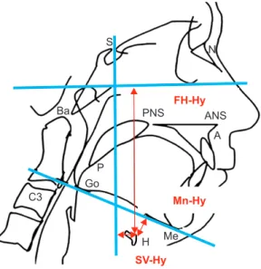
④ SV-Hy: The distance from the SV plane to the most an- terior point of the hyoid bone.
⑤ FH-Hy: The distance from the FH plane to the most an- terior point of the hyoid bone.
⑥ Mn-Hy: The distance from the Mn plane to the most anterior point of the hyoid bone.
⑦ SV-Pog: The distance from the SV plane to the Pog.
The mean and standard deviation of the measurements were determined for each time period, and to test for sig- nificance in the amount changed during the time period, the differences were tested at the 1% and 5% significance levels using a paired t-test. In addition, the correlation between the amount of mandibular setback and the amount of change in and hyoid position of the nasopharynx, oropharynx, and hy-
popharynx were measured by selecting specific measuring points described below; they were compared and analyzed during this time period. The preoperative, immediate post- operative, and 6 months postoperative measurements were named T1, T2, and T3, respectively, and the reference lines used in this study were the Frankfort horizontal (FH) plane and the Sella vertical (SV) plane, which is a vertical line from the Sella (S) to the FH and mandibular (Mn) plane. The measuring points used were S, the most inferior point of the soft palate (P), hyoid bone (H), third cervical vertebra (C3), anterior nasal spine (ANS), posterior nasal spine (PNS), and pogonion (Pog).(Fig. 1)
Seven items were measured in this study. First, the air- way space measurement was classified into three areas: the nasopharynx, oropharynx, and hypopharynx.(Fig. 2) The hyoid bone position was evaluated in three ways: using the horizontal position, vertical position, and the distance to the mandibular inferior border.(Fig. 3) The vertical distance from the SV plane to Pog was measured to evaluate recurrence.
(Fig. 4) The distances were measured up to 0.1 mm using the cephalometric analysis software program (V-ceph 6.0; Os- stem, Seoul, Korea) on lateral cephalograms of the same size.
The seven items measured in this study are as follows:
S N
A
Go P
C3
H Me Ba
SV-Hy FH-Hy
ANS
Mn-Hy PNS
Fig. 3. Measurement of hyoid bone position. The hyoid bone po- sition was evaluated with 3 measuring items using the horizontal position (SV-Hy), vertical position (FH-Hy), and distance to the mandibular inferior border (Mn-Hy). (S: Sella, N: nasion, Ba: ba- sion, PNS: posterior nasal spine, ANS: anterior nasal spine, A:
A point, P: the most inferior point of soft palate, Go: gonion, C3:
third cervical vertebra, Me: menton, H: hyoid bone)
Sung Woon On et al: Retrospective study on change in pharyngeal airway space and hyoid bone position after mandibular setback surgery. J Korean Assoc Oral Maxillofac Surg 2015
Fig. 4. Measurement of SV-Pog. The vertical distance from the Sella vertical (SV) plane to the pogonion (Pog) was measured to evaluate recurrence. (S: Sella, N: nasion, Ba: basion, PNS: poste- rior nasal spine, ANS: anterior nasal spine, A: A point, P: the most inferior point of soft palate, Go: gonion, C3: third cervical vertebra, B: B point)
Sung Woon On et al: Retrospective study on change in pharyngeal airway space and hyoid bone position after mandibular setback surgery. J Korean Assoc Oral Maxillofac Surg 2015
S
Ba PNS ANS
P Go
C3 B
A N
SV-Pog
pharynx showed a significant decrease of 4.3 mm from 18.9 mm preoperatively to 14.6 mm immediately postoperatively (P<0.01). Six months after surgery, it was 16.6 mm, which recovered to 2.0 mm, showing a stable state with a decrease of 2.3 mm (P<0.01) compared to the preoperative state.
The measurement of the hypopharynx, Hph, showed an increase of 0.7 mm immediately postoperatively compared to the preoperative measurement and this state was maintained through the six-month period after surgery, but there was no statistical significance.(Tables 1, 2)
2. Changes in hyoid bone position
The horizontal position of the hyoid bone, SV-Hy, showed posterior movement of 7.1 mm, from 33.0 mm preoperatively to 25.9 mm immediately postoperatively (P<0.01), and the position was 27.2 mm 6 months after surgery, which repre- sented a 1.3 mm recovery. It stabilized at about 5.8 mm pos- teriorly compared to the preoperative position.
The vertical position of the hyoid bone, FH-Hy, showed 12.9 mm of significant inferior movement, from 123.9 mm preoperatively to 136.8 mm immediately after surgery (P<0.01), and it returned to 128.1 mm six months after sur- gery (P<0.01). It ultimately stabilized at about 4.2 mm inferi- orly compared to the preoperative position (P<0.01).
The distance from the hyoid bone, Mn-Hy, showed a 12.0 mm increase, from 13.5 mm preoperatively to 25.5 mm im- mediately after surgery (P<0.01), and showed a slight return, measuring 20.5 mm at six months after surgery (P<0.01). It ultimately stabilized at a position about 7.0 mm away from the preoperative position (P<0.01).
Eventually, the position of the hyoid bone showed move- ment toward the posteroinferior position immediately after the airway space and hyoid bone were analyzed using the
Pearson’s correlation coefficient. All the statistical analyses were done using IBM SPSS Statistics version 22.0 (IBM Co., Armonk, NY, USA).
III. Results
1. Changes in pharyngeal airway space
The measurement of the nasopharynx, Nph, showed a 1.4 mm decrease immediately postoperatively compared to the preoperative measurement and showed a recovery of 0.7 mm at 6 months after surgery. The changes were not statistically significant.
The measurement of the oropharynx, Oph, only showed statistical significance in terms of the airway areas. The oro-
Table 2. Changes of cephalometric variables between T1 and T2, T2 and T3, and T1 and T3
Variable T1-T2 T2-T3 T1-T3
Mean±SD P-value Mean±SD P-value Mean±SD P-value
Nph Oph Hph SV-Hy FH-Hy Mn-Hy SV-Pog
1.4±4.1 4.3±3.4 –0.7±4.7 7.1±6.9 –13.0±8.9 –12.1±10.3
11.1±5.6
0.070
<0.001**
0.405
<0.001**
<0.001**
<0.001**
<0.001**
–0.7±3.8 –2.0±2.8 0.00 ±4.2 –1.29±5.95
8.8±7.6 5.0±7.9 –0.9±2.8
0.329 0.001**
0.961 0.243
<0.001**
0.002**
0.083
0.7±3.5 2.3±2.7 –0.8±4.3 5.83±5.55 –4.2±3.8 –7.1±5.9 10.1±4.8
0.266
<0.001**
0.344
<0.001**
<0.001**
<0.001**
<0.001**
(T1: preoperatively, T2: immediately postoperatively, T3: 6 months postoperatively, SD: standard deviation, Nph: nasopharyngeal airway space, Oph: oropharyngeal airway space, Hph: hypopharyngeal airway space, SV-Hy: the distance from the Sella vertical (SV) plane to the most anterior point of the hyoid bone, FH-Hy: the distance from the Frankfort horizontal plane to the most anterior point of the hyoid bone, Mn-Hy: the distance from the mandibular plane to the most anterior point of the hyoid bone, SV-Pog: the distance from the SV plane to the pogonion)
**Significant at the level of P<0.01.
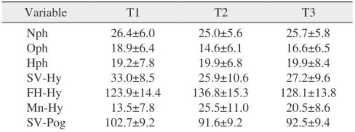
Sung Woon On et al: Retrospective study on change in pharyngeal airway space and hyoid bone position after mandibular setback surgery. J Korean Assoc Oral Maxillofac Surg 2015 Table 1. Data of cephalometric variables obtained at each time
Variable T1 T2 T3
Nph Oph Hph SV-Hy FH-Hy Mn-Hy SV-Pog
26.4±6.0 18.9±6.4 19.2±7.8 33.0±8.5 123.9±14.4
13.5±7.8 102.7±9.2
25.0±5.6 14.6±6.1 19.9±6.8 25.9±10.6 136.8±15.3 25.5±11.0 91.6±9.2
25.7±5.8 16.6±6.5 19.9±8.4 27.2±9.6 128.1±13.8
20.5±8.6 92.5±9.4 (T1: preoperatively, T2: immediately postoperatively, T3: 6 months postoperatively, Nph: nasopharyngeal airway space, Oph:
oropharyngeal airway space, Hph: hypopharyngeal airway space, SV- Hy: the distance from the Sella vertical (SV) plane to the most anterior point of the hyoid bone, FH-Hy: the distance from the Frankfort horizontal plane to the most anterior point of the hyoid bone, Mn- Hy: the distance from the mandibular plane to the most anterior point of the hyoid bone, SV-Pog: the distance from the SV plane to the pogonion)
Sung Woon On et al: Retrospective study on change in pharyngeal airway space and hyoid bone position after mandibular setback surgery. J Korean Assoc Oral Maxillofac Surg 2015
IV. Discussion
Various measuring points and reference lines have been used in previous studies on mandibular setback and airway changes. This study used radiopaque measuring points that were easy to read from lateral cephalograms, including S, P, C3, ANS, PNS, and Pog. This study also used reference lines that could be set clearly, including the FH plane and the SV plane, which is the vertical line from the S to the FH and Mn plane, in order to minimize the measurement error that can occur with lateral cephalograms.
Many studies on mandibular setback and changes in the airway space and hyoid bone have reported inconsistent re- sults. For airway changes, Athanasiou et al.1 reported that the postoperative airway space was decreased, but that, with the elongation of the angle of the cervical vertebra afterwards, the reduced airway space was compensated for; at follow- up, the airway space recovered to equal the preoperative volume. According to several studies2-4, there was no signifi- cant decrease in airway space after mandibular setback. In addition, Tselnik and Pogrel5 reported that the airway space increased immediately after mandibular setback, and they claimed this was because of anterior displacement of the hyoid bone postoperatively and that the airway space eventu- ally stabilized in the decreased state. Greco et al.6 reported that the airway volume of the hypopharynx was significantly decreased immediately after surgery and during long-term follow-up; Enacar et al.7 also reported similar results. Since the hypopharynx that they measured was in the same position as the oropharynx in this study, the present study showed the same results. In studies that compared the two-dimensional (2D) area of the pharynx, Güven and Saraçoğlu8 also reported that the decreased airway space was maintained immediately after surgery and during the long-term follow-up. With re- spect to studies on the hyoid bone, tongue, and airway space change after mandibular setback, Park and Kim9 reported a significant decrease in airway space at the oropharynx and hypopharynx immediately after surgery. However, the air- way space of the hypopharynx at the follow-ups recovered to the preoperative state while the decreased airway space of the oropharynx did not. In the present study, only decreased oropharyngeal airway space was observed with a statistically significant difference and the decrease was maintained during the long-term follow-up. This is thought to be associated with the finding that the oropharyngeal area is directly affected by the mandibular setback and that posteroinferior movement of the hyoid bone is accompanied by movements of the supra- surgery compared to the preoperative position and a slight
return at 6 months after surgery. It ultimately was stabilized in the posteroinferior position, showing that it maintained its position more inferiorly from the mandibular inferior border.
(Tables 1, 2)
3. Evaluation of recurrence
The measurements of SV-Pog were 102.7 mm preopera- tively, 91.6 mm immediately postoperatively, and 92.5 mm 6 months after surgery during the follow-up period. At the final follow-up, the amount of return was about 0.9 mm compared to the measurement immediately after surgery, but there was no statistical significance. Therefore, the results of this study show that the procedure does not result in the recurrence of mandibular setback surgery.(Tables 1, 2)
4. Correlation between the amount of mandibular setback and the amount of change in the airway space and hyoid bone position
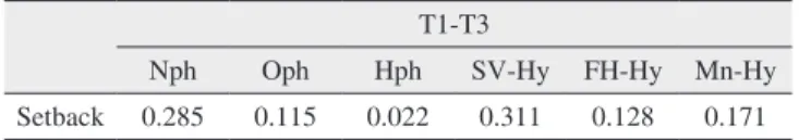
By analyzing the correlation between the amount of man- dibular setback and the amount of change in the airway space and hyoid bone position with Pearson’s correlation, the amount of mandibular setback showed a slightly higher correlation at the horizontal position of the hyoid bone and nasopharyngeal airway space, but overall, there was no sta- tistically significant correlation between them. Therefore, the amount of mandibular setback has no correlation with the amount of change in pharyngeal airway space and the hyoid bone.(Table 3)
Table 3. Pearson’s correlation between mandibular setback and changes of airway space, hyoid bone position
T1-T3
Nph Oph Hph SV-Hy FH-Hy Mn-Hy
Setback 0.285 0.115 0.022 0.311 0.128 0.171 (T1: preoperatively, T2: immediately postoperatively, T3: 6 months postoperatively, Nph: nasopharyngeal airway space, Oph:
oropharyngeal airway space, Hph: hypopharyngeal airway space, SV- Hy: the distance from the Sella vertical plane to the most anterior point of the hyoid bone, FH-Hy: the distance from the Frankfort horizontal plane to the most anterior point of the hyoid bone, Mn-Hy:
the distance from the mandibular plane to the most anterior point of the hyoid bone)
Sung Woon On et al: Retrospective study on change in pharyngeal airway space and hyoid bone position after mandibular setback surgery. J Korean Assoc Oral Maxillofac Surg 2015
collection when the lateral cephalogram is required, cepha- lometric radiographs should be taken at reproducible head positions in each patient. In a study including 30 patients, Malkoc et al.20 demonstrated that airway dimension and tongue and hyoid position measurement were highly repro- ducible on NHP cephalograms. For that reason, in the present study, lateral cephalograms were performed using the method proposed by Solow and Tallgren18. As a result, reproducible airway dimension could be measured.
A limitation in this study was the measurement of lengths using lateral cephalograms only; in addition, polysomnogra- phy required to diagnose true actual obstructive sleep apnea was not performed. Therefore, it was difficult to confirm whether the oropharyngeal decrease occurring postoperative- ly changed the quality of sleep or caused actual obstructive sleep apnea. However, after Partinen et al.21 defined the dis- tance between the posterior pharyngeal wall and tongue base as the line connecting the point B and gonion as the PAS and measured the PAS, they confirmed that the length of the PAS was less than 5 mm. Additionally, they defined the distance from the Mn plane to the hyoid bone as MP-H and reported that if this distance was more than 24 mm, the respiratory failure rate would increase. Riley et al.22 reported that if the PAS was less than 11 mm and MP-H was more than 15.4 mm, obstructive sleep apnea could occur. According to this study, the oropharyngeal decrease necessarily occurred after the mandibular setback surgery, and changes in the surround- ing structures were identified with posteroinferior movement of the hyoid bone through long-term follow-up. Therefore, when performing mandibular setback surgery, it is essential to consider that obstructive sleep apnea can occur postopera- tively through the analysis of lateral cephalograms. In partic- ular, patients who have a tendency to snore and predisposing factors for sleep apnea such as obesity, macroglossia, a short neck, large uvulae, and hypertrophic adenoids should prompt caution; a change to the treatment plan, if necessary, should be considered.
Tselnik and Pogrel5 evaluated airway volume with 2D ar- eas by setting special measurement points and reported that there was a very strong correlation between the amount of mandibular setback and airway volume decrease during long- term follow-up after mandibular setback surgery. Hochban et al.23 also reported that there was a slight correlation between the amount of mandibular setback and airway volume reduc- tion in their study. However, Enacar et al.7 reported that they did not find a correlation between the amount of mandibular setback and the hypopharyngeal airway volume change, a re- hyoid and infrahyoid muscles.
The hyoid bone consists of unstable hard tissues that are wholly supported by soft tissues connecting it to the skull base, mandible, pharynx, and tongue, and it is heavily influ- enced by surrounding tissues. A positional assessment of the hyoid bone is used to evaluate the physiological equilibrium state of the suprahyoid muscles, infrahyoid muscles, and surrounding tissues of the hyoid bone. According to several studies1,7,10,11, the hyoid bone returned to its original preopera- tive position during the follow-up period, and this was due to compensation reactions of the suprahyoid muscles, infrahy- oid muscles, and pharyngeal muscles. Eggensperger et al.12 reported posteroinferior movement of the hyoid bone after mandibular setback with BSSRO, but they assumed that this was due to a skeletal recurrence. Choi et al.13 reported that, after mandibular setback, significant posteroinferior displace- ment of the hyoid bone was shown but the majority recovered vertically over time and there was minimal anterior return.
In the present study, the position of the hyoid bone showed significant posteroinferior movement and slight return during long-term follow-up to a degree that was statistically sig- nificant. The distance to the mandibular inferior border was farther away postoperatively and such posteroinferior dis- placement of the hyoid bone is thought to be associated with the decrease in airway space in the oropharynx found after surgery.
The measurements used in the studies mentioned above were the anterior and posterior length changes from 2D later- al cephalograms, which fail to fully reproduce the actual air- way space compared to three-dimensional analysis methods, including computed tomography and magnetic resonance im- aging. However, Riley and Powell14 demonstrated the effec- tiveness of 2D studies by reporting the correlation between the airway space measured from 2D lateral cephalograms and the airway space volume calculated from three-dimensional computed tomography. In addition, since the lateral cephalo- gram is a preoperative examination typically performed in or- thognathic surgery patients, it was used in this study because it is not financially burdensome to patients, is noninvasive, and it is easy and quickly applied in preoperative analysis.
Airway space can be influenced by head position on lateral cephalogram. Several previous studies demonstrated that craniocervical angulations at different head postures affected pharyngeal diameters in normal subjects15-18. Muto et al.19 also showed a significant correlation between head posture and PAS and reported that a change of 10o in craniocervical inclination altered PAS by about 4 mm. Therefore, for data
Am J Orthod Dentofacial Orthop 1991;100:259-65.
2. Lee DK, Kim SK. Study on the changes in the upper airway fol- lowing osteotomy for the mandibular prognathism. J Korean Dent Assoc 1989;27:1143-53.
3. Chin KS, Shon WS. The relationships between the postoperative stability and the changes in the tongue position, the hyoid bone position and the upper airway size after orthognathic surgery in pa- tients with mandibular prognathism. Korean J Orthod 1993;23:693- 4. Choi JY, Lee SC. A study on changes of hyoid bone position and 705.
upper airway following orthognathic surgery. Kyung Hee Dent J 1993;15:729-42.
5. Tselnik M, Pogrel MA. Assessment of the pharyngeal airway space after mandibular setback surgery. J Oral Maxillofac Surg 2000;58:
282-5.
6. Greco JM, Frohberg U, Van Sickels JE. Long-term airway space changes after mandibular setback using bilateral sagittal split oste- otomy. Int J Oral Maxillofac Surg 1990;19:103-5.
7. Enacar A, Aksoy AU, Sençift Y, Haydar B, Aras K. Changes in hy- popharyngeal airway space and in tongue and hyoid bone positions following the surgical correction of mandibular prognathism. Int J Adult Orthodon Orthognath Surg 1994;9:285-90.
8. Güven O, Saraçoğlu U. Changes in pharyngeal airway space and hyoid bone positions after body ostectomies and sagittal split ra- mus osteotomies. J Craniofac Surg 2005;16:23-30.
9. Park BW, Kim JR. A cephalometric study on changes in pharyn- geal airway space, tongue and hyoid bone positions following the surgical correction of mandibular prognathism. J Korean Assoc Oral Maxillofac Surg 2000;26:164-71.
10. Kawakami M, Yamamoto K, Fujimoto M, Ohgi K, Inoue M, Kirita T. Changes in tongue and hyoid positions, and posterior airway space following mandibular setback surgery. J Craniomaxillofac Surg 2005;33:107-10.
11. Lew KK. Changes in tongue and hyoid bone positions following anterior mandibular subapical osteotomy in patients with Class III malocclusion. Int J Adult Orthodon Orthognath Surg 1993;8:123-8.
12. Eggensperger N, Smolka W, Iizuka T. Long-term changes of hyoid bone position and pharyngeal airway size following mandibular setback by sagittal split ramus osteotomy. J Craniomaxillofac Surg 2005;33:111-7.
13. Choi YH, Kim BK, Choi BJ, Kim YG, Lee BS, Kwon YD, et al.
An investigation of hyoid bone position and airway space in class III malocclusion after orthognathic surgery. J Korean Assoc Maxil- lofac Plast Reconstr Surg 2011;33:401-6.
14. Riley RW, Powell NB. Maxillofacial surgery and obstructive sleep apnea syndrome. Otolaryngol Clin North Am 1990;23:809-26.
15. Harris RS. The effect of extension of the head and neck upon the infrahyoid respiratory passage and the supraclavicular portion of the human trachea. Thorax 1959;14:176-80.
16. Hellsing E. Changes in the pharyngeal airway in relation to exten- sion of the head. Eur J Orthod 1989;11:359-65.
17. Ono T, Otsuka R, Kuroda T, Honda E, Sasaki T. Effects of head and body position on two- and three-dimensional configurations of the upper airway. J Dent Res 2000;79:1879-84.
18. Solow B, Tallgren A. Head posture and craniofacial morphology.
Am J Phys Anthropol 1976;44:417-35.
19. Muto T, Takeda S, Kanazawa M, Yamazaki A, Fujiwara Y, Mizo- guchi I. The effect of head posture on the pharyngeal airway space (PAS). Int J Oral Maxillofac Surg 2002;31:579-83.
20. Malkoc S, Usumez S, Nur M, Donaghy CE. Reproducibility of air- way dimensions and tongue and hyoid positions on lateral cephalo- grams. Am J Orthod Dentofacial Orthop 2005;128:513-6.
21. Partinen M, Guilleminault C, Quera-Salva MA, Jamieson A. Ob- structive sleep apnea and cephalometric roentgenograms. The role of anatomic upper airway abnormalities in the definition of abnor- mal breathing during sleep. Chest 1988;93:1199-205.
22. Riley R, Guilleminault C, Herran J, Powell N. Cephalometric
sult that agreed with that of Chung and Lee’s study24. Güven and Saraçoğlu8 also bolstered these results by reporting that there was no correlation between the amount of mandibular setback and decreased airway volume and position change of the hyoid bone. In the present study, there was no special cor- relation found with Pearson’s correlation, which was used to determine the correlation between the amount of mandibular setback and the amount of change in the airway space and hyoid bone. The reason for this could be that airway space and hyoid bone changes did not occur uniformly according to the amount of mandibular setback due to various environ- mental differences such as the individual structure of hard and soft tissues, the degree of elasticity of the soft tissues, and the degree of obesity.
V. Conclusion
In this study, a significantly reduced oropharynx, postero- inferior movement of the hyoid bone, and changes to the surrounding structures after mandibular setback surgery were identified through long-term follow-up. The results suggest that it is possible to cause obstructive sleep apnea after man- dibular setback surgery, indicating that care is needed when electing to perform mandibular setback surgery, especially for patients who have predisposing factors for sleep apnea such as obesity, macroglossia, a short neck, large uvulae, and hypertrophic adenoids. Therefore, evaluation for obstruc- tive sleep apnea that can occur postoperatively is needed and should be considered when establishing the treatment plan.
Conflict of Interest
No potential conflict of interest relevant to this article was reported.
ORCID
Sung Woon On, http://orcid.org/0000-0001-6192-1530 Min Woo Han, http://orcid.org/0000-0001-7752-6818 Doo Yeon Hwang, http://orcid.org/0000-0002-2500-0897 Seung Il Song, http://orcid.org/0000-0001-9504-6471
References
1. Athanasiou AE, Toutountzakis N, Mavreas D, Ritzau M, Wenzel A.
Alterations of hyoid bone position and pharyngeal depth and their relationship after surgical correction of mandibular prognathism.
Surg 1996;25:333-8.
24. Chung DH, Lee KS. A study on changes of airway, tongue, and hy- oid position following orthognathic surgery. Korean J Orthod 1998;
28:487-98.
analyses and flow-volume loops in obstructive sleep apnea patients.
Sleep 1983;6:303-11.
23. Hochban W, Schürmann R, Brandenburg U, Conradt R. Mandibu- lar setback for surgical correction of mandibular hyperplasia: does it provoke sleep-related breathing disorders? Int J Oral Maxillofac