Effects of Different Knee Flexion Angles According to Three Positions on Abdominal and Pelvic Muscle Activity During Supine Bridging
One-bin Lim1, MSc, PT, Ki-song Kim2, PhD, PT
1Dept. of Physical Therapy, The Graduate School, Yonsei University
2Research Institute for Basic Sciences, Dept. of Physical Therapy, College of Natural Science, Hoseo University
Abstract
1)This study analyzes how different knee flexion angles affect the abdominal and pelvic muscle activity during supine bridging. Twenty healthy subjects participated in the study. We used surface electromyography (EMG) to measure how three different knee flexion angles (100°, 70°, and 40°) affected the activity of the transverse abdominis/internal oblique (TrA/IO), external oblique (EO), biceps femoris (BF), rectus femoris (RF), and gluteus maximus (GM) muscles on the dominant side during supine bridging. The one-way repeated analysis of variance (ANOVA) was used to determine the statistical significance of TrA/IO, EO, BF, RF and GM muscle activity and the GM/BF activity ratio. For the TrA/IO, EO, BF, and GM muscles, supine bridging with different knee flexion angles resulted in significant differences in abdominal and pelvic muscle activity. For the TrA/IO muscles, the post-hoc test demonstrated that muscle activity significantly increased at 40° compared to 70°; however, there were no significant differences between 100° and 70° or 100° and 40°. For the EO muscle, the post-hoc test demonstrated that muscle activity significantly increased at 40° compared to 100° and 70°; no significant difference was observed between angles 100° and 70°. For the BF muscle, the post-hoc test demonstrated that muscle activity significantly increased according to the knee flexion angle (40°>70°>100°). For the GM muscle, the post-hoc test demonstrated that muscle activity significantly increased according to the knee flexion angle (100°>70°>40°). However, for the RF muscle, there was no significant difference.
Additionally, the GM/BF activity ratio significantly increased according to the knee flexion angle (100°>70°>40°). From these results, we can conclude that bridging with a knee flexion of 100° can strengthen the GM muscle, whereas bridging with a knee flexion of 40° is recommended to strengthen the IO, EO, and BF muscles. We can also conclude that knee flexion angles should be modified during supine bridging to increase the muscle activity of different target muscles.
Key Words: Abdominal muscles; Electromyography; Muscle strength.
Introduction
Bridging, also known as the bridge exercise, is commonly used in clinical settings to reinforce lum- bopelvic stability by increasing trunk muscle activity (Marshall and Murphy, 2005; Stevens et al, 2006).
Bridging involves holding the pelvis off the floor in either the supine (back bridges), prone (front or ven- tral bridges), or lateral position (side bridges) (Bjerkefors et al, 2010; Ekstrom et al, 2007; Imai et al, 2010; Kavcic et al, 2004; Lehman et al, 2005;
McGill and Karpowicz, 2009; Stevens et al, 2006).
Patients suffering from low back pain (LBP) (García-Vaquero et al, 2012) or hemiplegia (Kozol et al, 2010) typically employ bridging to strengthen their trunk and hip muscles. Bridging is also used for resistive exercises, including various methods to control body weight. Depending on the client’s con- dition, the complexity of bridging can be modified by a rehabilitation specialist. Previous studies have sug- gested that exercises using unstable devices, such as a therapeutic ball (Marshall and Murphy, 2005;
Corresponding author: Ki-song Kim [email protected]
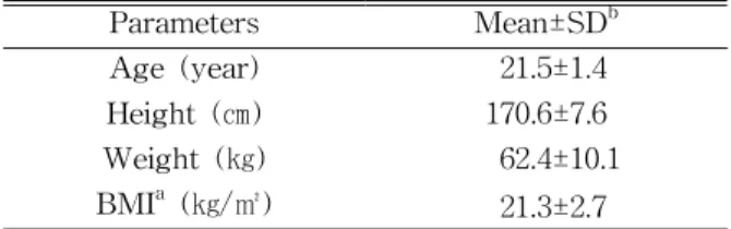
Parameters Mean±SDb
Age (year) 21.5±1.4
Height (㎝) 170.6±7.6
Weight (㎏) 62.4±10.1
BMIa (㎏/㎡) 21.3±2.7
abody mass index, bmean±standard deviation.
Table 1.General characteristics of subjects (N=20) Stevens et al, 2006) or a round foam roll (Kim et al,
2011), increase the trunk’s muscular activity, which helps to stabilize the lumbopelvic spine during bridging.
Previous studies suggested that bridging can strengthen the hip extensor (Andersen et al, 2006;
Ekstrom et al, 2007; Myklebust and Engebretsen, 2004). Clinicians often modify the bridge exercise with different methods to allow for a gradual in- crease in the intensity of the exercise (Lehman et al, 2005). Despite many studies on bridging, no study has investigated how different angles of knee flexion with double leg support affect abdominal and pelvic muscle activity during bridging.
Though the complexity of bridging has been modified for use with various patient populations, the majority of electromyographic (EMG) studies have only analyzed the trunk’s muscular response during conventional bridging. The effects of different knee flexion angles on the abdominal and pelvic muscle activity during bridging has not been examined in previous studies. To our knowledge, the effect of these leg support strategies has only been studied for supine bridging with single or double leg support (Bjerkefors et al, 2010; Ekstrom et al, 2007; Kavcic et al, 2004; Stevens et al, 2006; Stevens et al, 2007).
Previous studies indicated that leg support strategies during bridging exercise could constitute a major challenge to the neuromuscular system and possibly result in higher loads on the spine (Kavcic et al, 2004). In order to help rehabilitation specialists de- termine progressive exercise protocols for the ab- dominal and pelvic musculature, an additional re- search is needed to clarify the effect of leg support using strategies that include knee flexion angle in the supine position. To the best of our knowledge, this is the first study to investigate the effects of different knee flexion angles on abdominal and pelvic muscle activity during supine bridging.
The purpose of this study is to analyze how dif- ferent knee flexion angles (100°, 70°, and 40°) affect the activity of abdominal [transverse abdominis/in- ternal oblique (TrA/IO) and external oblique (EO)]
and pelvic [rectus femoris (RF), biceps femoris (BF), and gluteus maximus (GM)] muscles during supine bridging. The hypothesis of this study is that the abdominal and pelvic muscle activity during supine bridging will differ in various knee flexion angles.
Methods
Subjects
Based on our pilot data and using G*Power 3.1.5 software (Franz Faul, University of Kiel, Kiel, Germany) (Faul et al, 2007), an a priori power anal- ysis was performed to estimate the sample size. The pilot data of 13 asymptomatic subjects were used to achieve an effect size of .43, an alpha level of 5%, and a power of 80%. The estimated desired sample size was 10. Twenty asymptomatic subjects (13 men and 7 women) volunteered to participate in this study (Table 1). Individuals were excluded from par- ticipation in this study if they had a history of ab- dominal or low back pain within six weeks prior to the test or if they were unable to correctly perform a supine bridge.
Instrument
Surface EMG signals were collected using the Noraxon TeleMyo DTS Telemetry system (Noraxon Inc., AZ, USA). EMG data were collected at a sam- pling rate of 1000 ㎐ and analyzed using the Noraxon MyoResearch Master Edition 1.08 XP soft- ware (Noraxon Inc., Scottsdale, AZ, USA). The raw signal was filtered using a digital band-pass filter (Lancosh FIR) between 20 and 450 ㎐ and was also
A B
E
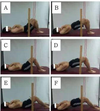
Figure 1. Supine bridge combined with different knee flexion angles (A: knee flexion 100° start, B: knee flexion 100° finish, C: knee flexion 70°
start, D: knee flexion 70° finish, E: knee flexion 40° start, F: knee flexion 40° finish).
notch filtered (60 ㎐, 120 ㎐). The root-mean-square (RMS) values were calculated using a moving win- dow of 50 milliseconds.
Before positioning the electrodes over each muscle, the skin was shaved, abraded, and then cleaned with isopropyl alcohol wipes to reduce the skin resistance.
Disposable Ag/AgCl surface electrodes were posi- tioned parallel to the muscle fibers with a cen- ter-to-center spacing of 2 ㎝. All electrodes were placed on the right side of the following muscles and locations. For the TrA/IO muscles, the electrode was positioned midpoint between the anterior superior iliac spine and the pubic tubercle. For the EO mus- cle, the electrode was positioned 45° obliquely parallel to a line connecting the most inferior point of the costal margin of the ribs and the contralateral pubic tubercle above the anterior superior iliac spine near the level of the umbilicus (Escamilla et al, 2006). For the BF muscle, the electrode was positioned parallel to the muscle fibers on the lateral aspect of the thigh, two thirds of the distance between the tro- chanter and the back of the knee. For the RF mus- cle, the electrode was positioned on the center of the anterior surface of the thigh, approximately half the distance between the knee and the iliac spine. For the GM muscle, the electrode was positioned in the middle of the muscle below the level of the tro- chanter and 1 to 2 inches above the gluteal fold (Criswell, 2011).
Procedures
Each subject was instructed to lie supine with knees flexion. The investigator measured the angle of the knee joint and then placed the participant in the hook-lying position using a 14-inch goniometer (Baseline stainless steel 180° Conzett goniometer, Fabrication Enterprises Inc., NY, USA). The knee joints were bilaterally flexed to 100°, 70°, or 40°
(Figure 1). A target bar was placed so that the sub- ject’s middle aspect of the thigh would slightly touch the bar when the hip joint was fully extended. The subject was asked to touch the target bar with each
supine bridge, which lasted 8 seconds (the initial 3 seconds were the subject moving to the target bar;
and the last 5 seconds were the subject holding the target position). A metronome was used to control the time and speed of the subject’s movement. In order to prevent abduction and adduction of both hip joints, lope guides were aligned with the lower ex- tremities (Kim et al, 2011).
Prior to testing, 30 minutes were spent familiariz- ing the subjects with the standard position and movement. During the familiarization session, each subject received verbal instructions explaining how to correctly perform the supine bridge. Once the subject correctly performed the supine bridge, a test- ing session was scheduled after a 30-minute rest.
During data collection, all subjects performed the bridge under the close supervision of the researcher.
When the subject’s middle aspect of the thigh
C D
F
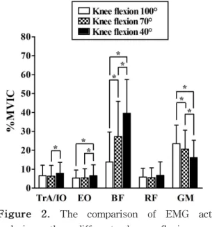
Figure 2. The comparison of EMG activity during the different knee flexion angles (TrA/IO: transverse abdominis/internal oblique, EO: external oblique, BF: biceps femoris, RF:
rectus femoris, GM: gluteus maximus, %MVIC:
%maximal voluntary isometric contraction, error bars: standard deviation, *p<.016).
touched the target bar, the subject isometrically maintained the end position for 5 seconds. This process was performed three times with a 1-minute rest period between each trial. A 3-minute rest was given between each exercise trial to prevent muscle fatigue. Using random numbers generated by the website Randomization.com (available from http://www.randomization.com; accessed 10 June 2013), subjects were randomly assigned to perform the supine bridge at a certain knee flexion angle.
This randomization was done to minimize threats to the study’s internal validity (Youdas et al, 2008).
Data collection
Data on the EMG activity of the TrA/IO, EO, RF, BF, and GM muscles were collected during the su- pine bridge. The mean EMG activity data obtained during the middle 3 seconds of each trial was used for statistical analysis. For normalization, the max- imal voluntary isometric contraction (MVIC), as de-
scribed by Kendall et al (2005), was used to de- termine the reference contraction. For the TrA/IO, each subject was positioned supine and performed a resisted crossed curl-up. The assistant stabilized the subject’s legs. Manual resistance was applied to the left shoulder by the examiner. For the EO, the same procedure was repeated. Manual resistance was ap- plied to the right shoulder. For the RF, the subject seated on the side of the table while they performed knee extension against maximal resistance. For the BF, the subject was positioned prone and performed knee flexion against maximal resistance. For the GM, the subject performed unilateral hip extension from the prone position, while the examiner applied man- ual resistance proximal to the 90° knee flexion. EMG data were recorded during a 5 second reference con- traction, repeated three times, and stored for data analysis. A resting interval of 30~50 seconds was provided between the test trials. The muscle activity was expressed as a percentage of the calculated RMS of MVIC (%MVIC).
Statistical analysis
The Kolmogorov-Smirnov Z-test was performed to investigate whether continuous data approximated a normal distribution. We used a one-way repeated ANOVA to compare the differences in the normalized EMG activity of the TrA/IO, EO, RF, BF, and GM muscles and GM/BF activity ratio (dependent varia- bles) during supine bridges performed at different knee flexion angles (independent variables) with dou- ble leg support. Post-hoc analyses were performed using the Bonferroni test to evaluate the significance of between-exercise pairwise comparisons. The stat- istical significance level was set at α=.05. SPSS ver.
20.0 software was used for all data analysis (SPSS Inc., Chicago, IL, USA).
Results
The comparison of the EMG activity during the
Figure 3. The comparison of GM/BF activity ratio during the different knee flexion angles (GM: gluteus maximus, BF: biceps femoris, KF: knee flexion, error bars: standard deviation, *p<.016).
different knee flexion angles is presented in Figure 2.
A significant difference (F=4.561, p=.025) was noted in TrA/IO muscle activity when the supine bridge was performed at different knee flexion angles. The post-hoc test demonstrated that TrA/IO muscle ac- tivity significantly increased at the 40° knee flexion angle when compared to the 70° knee flexion angle (p=.019). However, there were no significant differ- ences observed between the 100° knee flexion angle and the 70° knee flexion angle (p=1.000) or the 100° knee flexion angle and the 40° knee flexion angle (p=.115).
The difference in the EO muscle activity during the supine bridge performed at different knee flexion angles was significant (F=6.593, p=.007). The post-hoc test demonstrated that the EO muscle ac- tivity significantly increased at the 40° knee flexion angle when compared to the 100° knee flexion angle (p=.033) and the 70° knee flexion angle (p=.004).
However, no significant difference was observed be- tween the 100° knee flexion angle and the 70° knee flexion angle (p=1.000).
The difference in BF muscle activity during the supine bridge performed at different knee flexion an-
gles was significant (F=39.960, p<.001). The post-hoc test demonstrated that BF muscle activity sig- nificantly increased in the order of knee flexion angle (40°>70°>100°; 100° knee flexion angle vs. 70° knee flexion angle, p<.001; 100° knee flexion angle vs. 40°
knee flexion angle, p<.001; 70° knee flexion angle vs.
40° knee flexion angle, p=.002).
The difference in RF muscle activity during the supine bridge performed at different knee flexion an- gles was not significant (F=2.776, p=.089).
The difference in GM muscle activity during the supine bridge performed at different knee flexion an- gles was significant (F=12.559, p<.001). The post-hoc test demonstrated that the GM muscle activity sig- nificantly increased in the order of knee flexion angle (100°>70°>40°; 100° knee flexion angle vs. 70° knee flexion angle, p=.030; 100° knee flexion angle vs. 40°
knee flexion angle, p=.001; 70° knee flexion angle vs.
40° knee flexion angle, p=.042).
The difference in GM/BF activity ratio during the supine bridge performed at different knee flexion an- gles was significant (F=18.649, p<.001). The post-hoc test demonstrated that the GM/BF activity ratio sig- nificantly increased in the order of knee flexion angle (100°>70°>40°; 100° knee flexion angle vs. 70° knee flexion angle, p<.001; 100° knee flexion angle vs. 40°
knee flexion angle, p<.001; 70° knee flexion angle vs.
40° knee flexion angle, p=.001) (Figure 3).
Discussion
The purpose of this study was to analyze how different knee flexion angles (100°, 70°, and 40°) af- fected abdominal (TrA/IO and EO) and pelvic (RF, BF, and GM) muscle activity during supine bridging.
The hypothesis of this study was that using differ- ent knee flexion angles during a supine bridge would generate different activity in the abdominal and pel- vic muscles. The results showed that there were significant differences for the TrA/IO, EO, BF, and GM muscles during the supine bridge according to
different knee flexion angles. However, there was no significant difference in the RF muscle during the supine bridge with different knee flexion angles (Figure 2). Thus, the findings of this study partially validated the research hypothesis.
For the TrA/IO muscle, the post-hoc test showed that muscle activity in the position of knee flexion 40° increased significantly compared to that of 70°
flexion. However, there were no significant differ- ences between 40° and 100° and between 70° and 100°. These results may indicate that the extended lumbar position at 40° causes greater lumbopelvic in- stability, which is caused by decreased action in the GM muscle and plantar flexor of the ankle at more than 100°. Therefore, through the use of lumbar flexion, the activity of the TrA/IO muscle could be increased to improve lumbopelvic stability. However, our conclusions should be confirmed by further re- search that includes using kinematic data to inves- tigate how different angles affect the lumbopelvic stability of flexed knees and ankle joints.
The other mechanism is that the 40° knee flexion position could be lengthened by moving the arm po- sition than 100° knee flexion in the lever system.
The lengthened moving arm would need greater ab- dominal muscle activity for the dynamic equilibrium.
A previous study reported that a more challenging environment and trial can induce greater activity of abdominal muscle during bridging exercises (Santos and Aruin, 2009). The participants expressed physical exertion to perform bridging at 40° knee flexion.
However, this explanation is not adequate because there was no kinematic data and participant’s ex- ertion scale was not measured in this study. Hence, a suitable experimental design for the finding of cor- relation with kinematic and kinetic data is needed in further study.
For the EO muscle, the post-hoc test demon- strated that muscle activity in the position of knee 40° flexion increased significantly compared to that of 70° flexion and of 100° flexion. However, there was no significant difference between 70° flexion and
100° flexion. These results could be explained by the fact that the activity of EO decreased in the position of knee 100° flexion. On the other hand, the activity of the GM muscle increased in the position of knee 100° flexion. This result could be explained by the same methods that were used for TrA/IO muscles.
Additionally, there could be considerable causes for the increase in the EO muscle’s activity. We ob- served that during the start position of the bridge, the participant’s pelvis was tilted more anteriorly in knee 40° flexion than in 70° and 100° flexion. From this start position, participants used their pelvis pos- teriorly to lift the hip during flexed lumbar position.
This trial might increase the activity of the EO muscle in the 40° flexion position than in the 70°
and 100° flexion position.
For the RF muscle, there were no significant dif- ferences among the supine bridge with different knee flexion angles. These results may indicate that bridging with different knee flexions seem to be controlled by hip extensors, such as the GM or BF muscles, instead of the hip flexor, the RF. Because the trunk and the pelvis are lifted and maintained against gravity, hip extensors are likely to play a critical role during bridging. This finding agree with previous studies (Bergmark, 1989; Ekstrom et al, 2007).
For the BF muscle, the post-hoc test revealed significant differences with different knee flexion angles. The muscle activity of the BF muscle in- creased significantly in the order of the knee flexion angle (40°>70°>100°). These results may be the re- sult of the active insufficiency of BF muscles; in other words, the length of the BF muscle is influ- enced by the angle of the knee flexion. In the 100°
of knee flexion, the BF muscle may be under a state of active insufficiency, and, as a result, it cannot generate enough active tension. Conversely, the ac- tive insufficiency of the BF muscle may explain the significantly increased activity of the GM muscle in the 100° of knee flexion. For the GM muscle, the post-hoc test demonstrated that the GM muscle ac- tivity significantly increased according to the order
of the knee flexion angle (100°>70°>40°). Because the GM muscle is a one-joint muscle, it is not af- fected by the degree of the knee flexion range and can produce enough active tension. Compared with 40° of knee flexion, the GM muscle is placed at the lengthened muscle range in the 100° of knee flexion.
Bridging is a commonly used in clinical settings and, in previous studies was recommended to strengthen hip extensors (Andersen et al, 2006; Ekstrom et al, 2007; Myklebust and Engebretsen, 2004).
For the GM/BF activity ratio, the post-hoc test demonstrated that the GM/BF activity ratio sig- nificantly increased according to the order of the knee flexion angle (100°>70°>40°). The reason for this finding can be explained by the reciprocal inhibition. Reciprocal inhibition is when the central nervous system sends a message to the agonist muscle to contract, the tension in the antagonist muscle is automatically inhibited by impulses from alpha motor neurons (Crone, 1993). Increasing RF activation during the supine bridging exercise may decrease the role of the BF activation during hip ex- tension and thus enhancing the GM activation.
This study has several limitations. First, the leg support strategies during bridging exercise may be used in clinical rehabilitation programs of subjects with LBP or hemiplegia. However, the limitation of this study was that the assessed subjects did not suffer from LBP or hemiplegia. Further research is needed to clarify exactly which patients with LBP or hemiplegia should be applied the leg support strat- egies during bridging exercise. Second, kinematic da- ta while performing the supine bridging with differ- ent knee flexion angles was not obtained. Thus, the amount of hip extension and lumbopelvic movements cannot be quantified. Third, although iliofemoral liga- ment tightness and the degree of physiological angle in the hip joint can affect how different knee flexion angles affect the supine bridge, data were not col- lected for each subject. In order to generalize the findings of this study, a kinematic analysis of a pa- tient population is warranted.
Conclusion
The abdominal and pelvic muscle activity was in- vestigated during supine bridging with different knee flexion angles in this study. The findings of the study indicate that bridging with knee flexion 100°
may increase the GM muscle activity and the GM/BF activity ratio, while bridging with a knee flexion of 40° may increase the IO, EO, and BF muscle activity. Therefore, using different knee flex- ion angles when performing the supine bridge may be used to selectively activate target muscles in the abdominal and pelvic area.
References
Andersen LL, Magnusson SP, Nielsen M, et al.
Neuromuscular activation in conventional ther- apeutic exercises and heavy resistance exercises:
Implications for rehabilitation. Phys Ther. 2006;
86(5):683-697.
Bergmark A. Stability of the lumbar spine. A study in mechanical engineering. Acta Orthop Scand Suppl. 1989;230:1-54.
Bjerkefors A, Ekblom MM, Josefsson K, et al. Deep and superficial abdominal muscle activation dur- ing trunk stabilization exercises with and with- out instruction to hollow. Man Ther. 2010;15(5):
502-507.
Criswell E. Cram’s Introduction to Surface Electromyography. 2nd ed. Sudbury, MA, Jones and Bartlett Publishers, 2011:353-369.
Crone C. Reciprocal inhibition in man. Dan Med Bull.
1993;40(5):571-581.
Ekstrom RA, Donatelli RA, Carp KC.
Electromyographic analysis of core trunk, hip, and thigh muscles during 9 rehabilitation exercises. J Orthop Sports Phys Ther. 2007;37 (12):754-762.
Escamilla RF, McTaggart MS, Fricklas EJ, et al. An electromyographic analysis of commercial and
This article was received August 25, 2013, was reviewed August 25, 2013, and was accepted October 29, 2013.
common abdominal exercises: Implications for rehabilitation and training. J Orthop Sports Phys Ther. 2006;36(2):45-57.
Faul F, Erdfelder E, Lang AG, et al. G*Power 3: A flexible statistical power analysis program for the social, behavioral, and biomedical sciences.
Behav Res Methods. 2007;39(2):175-191.
García-Vaquero MP, Moreside JM, Brontons-Gil E, et al. Trunk muscle activation during stabiliza- tion exercises with single and double leg support. J Electromyogr Kinesiol. 2012;22(3):398- 406.
Imai A, Kaneoka K, Okubo Y, et al. Trunk muscle activity during lumbar stabilization exercises on both a stable and unstable surface. J Orthop Sports Phys Ther. 2010;40(6):369-375.
Kavcic N, Grenier S, McGill SM. Quantifying tissue loads and spine stability while performing com- monly prescribed low back stabilization exercises. Spine (Phila Pa 1976). 2004;29(20):
2319-2329.
Kendall FP, McCreary EK, Provance PG et al.
Muscles: Testing and function with posture and pain. 5th ed. Baltimore, MD, Lippincott Williams
& Wilkins, 2005:186-429.
Kim SJ, Kwon OY, Yi CH, et al. Comparison of ab- dominal muscle activity during a single-legged hold in the hook-lying position on the floor and on a round foam roll. J Athl Train. 2011;46(4):
403-408.
Kozol MZ, Filer M, Ring H. Bridging performance of adults with hemiparesis: Sliding of the paretic limb. J Geriatr Phys Ther. 2010;33(1):26-33.
Lehman GJ, Hoda W, Oliver S. Trunk muscle activ- ity during bridging exercises on and off a swiss ball. Chiropr Osteopat. 2005;13:14.
Marshall PW, Murphy BA. Core stability exercises on and off a swiss ball. Arch Phys Med Rehabil. 2005;86(2):242-249.
McGill SM, Karpowicz A. Exercises for spine stabili- zation: Motion/motor patterns, stability pro- gressions, and clinical technique. Arch Phys Med Rehabil. 2009;90(1):118-126.
Myklebust G, Engebretsen L. Rehabilitation of knee injuries. In: Bahr R, Maehlum S, eds. Clinical Guide to Sports Injuries. Champaign, IL, Human Kinetics, 2004:353-359.
Santos MJ, Aruin AS. Effects of lateral perturbations and changing stance conditions on anticipatory postural adjustment. J Electromyogr Kinesiol.
2009;19(3):532-541.
Stevens VK, Bouche KG, Mahieu NN, et al. Trunk muscle activity in healthy subjects during bridging stabilization exercises. BMC Musculoskelet Disord. 2006;7:75.
Stevens VK, Coorevits PL, Bouche KG, et al. The influence of specific training on trunk muscle recruitment patterns in healthy subjects during stabilization exercises. Man Ther. 2007;12(3):
271-279.
Youdas JW, Guck BR, Hebrink RC, et al. An elec- tromyographic analysis of the ab-slide exercise, abdominal crunch, supine double leg thrust, and side bridge in healthy young adults: Implications for rehabilitation professionals. J Strength Cond Res. 2008;22(6):1939-1946.