• 검색 결과가 없습니다.

Eupatorium chinensis var. simplicifolium Root Extract Inhibits the Lipopolysaccharide-Induced Inflammatory Response in Raw 264.7 Macrophages by Inhibiting iNOS and COX-2 Expression

N/A
N/A
Protected

Academic year: 2021

Share "Eupatorium chinensis var. simplicifolium Root Extract Inhibits the Lipopolysaccharide-Induced Inflammatory Response in Raw 264.7 Macrophages by Inhibiting iNOS and COX-2 Expression"

Copied!
8
0
0

로드 중.... (전체 텍스트 보기)

전체 글

(1)

Eupatorium chinensis var. simplicifolium Root Extract Inhibits the Lipopolysaccharide- Induced Inflammatory Response in Raw 264.7 Macrophages by Inhibiting iNOS and COX-2 Expression

Jin-Ho Lee

1†

, Dae-Hyun Kim

1†

, Ji-Won Shin

1

, Sae-Jin Park

1

, Yoon Suk Kim

2

, Yusu Shin

3

, Ji-Yeon Yu

4

and Tack-Joong Kim

1

*

1

Division of Biological Science and Technology, College of Science and Technology, Yonsei University, Wonju 220-710, Korea

2

Department of Biomedical Laboratory Science, College of Health Sciences, Yonsei University, Wonju 220-710, Korea

3

Department of Herbal Crop Research, National Institute of Horticultural and Herbal Science, RDA, Eumseong 369-873, Korea

4

Laboratory of Chemical Genomics, Korea Research Institute of Chemical Technology, Daejeon 305-600, Korea Received July 8, 2012 /Revised August 1, 2012 /Accepted August 6, 2012

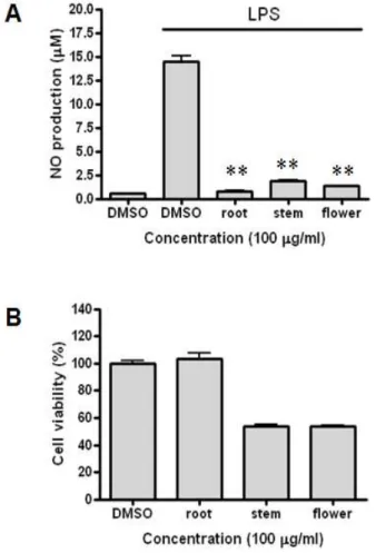
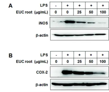
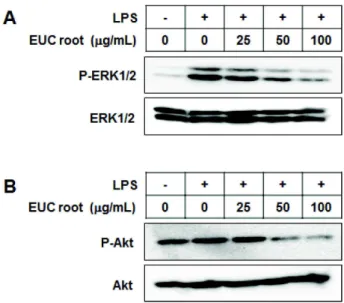
Inflammation is a host defense mechanism that is activated in response to harmful substances or pathogens. However, an excessive inflammatory response is a problem in itself. Macrophages secrete inflammatory mediators such as nitric oxide (NO) or cytokines through various pathways such as the nuclear factor kappa B (NF-κB)-activated pathway after recognizing pathogen-like lipopolysaccharides (LPSs). In this study, anti-inflammatory effects of Eupatorium chinensis var. simplicifolium (EUC) extracts were investigated using LPS-stimulated RAW 264.7 macrophages. The EUC root extract significantly reduced NO production, inducible nitric oxide synthase (iNOS) expression, and cyclooxygenase-2 ex- pression in a concentration-dependent manner. In addition, the EUC root extract reduced phosphor- ylation of mitogen-activated protein kinases and protein kinase B, which is upstream of NF-κB. The EUC root extract also reduced the degradation of inhibitory kappa B. These results indicate that EUC root extract exerts anti-inflammatory effects, which are mediated by inhibition of iNOS expression and the NF-κB pathway.

Key words : Eupatorium chinensis var. simplicifolium, cyclooxygenase-2, inflammation, nitric oxide, NF-κ B pathway

These authors contributed equally to this work.

*Corresponding author

*Tel : +82-33-760-2242; Fax : +82-33-760-2183

*E-mail: [email protected]

Introduction

Inflammation refers to the complex biological response caused by damaged cells and tissues, chemical irritants, or invading organisms such as bacteria [8,17,31,34].

Macrophages, a type of immune cell, are activated by bacte- rial lipopolysaccharide (LPS), interferon-gamma, and gran- ulocyte-macrophage colony-stimulating factor [24].

Activated macrophages play an important role in in- flammatory diseases by releasing various inflammatory cy- tokines such as IL-1β, IL-6, TNF-α as well as other in- flammatory mediators such as nitric oxide (NO) and prosta- glandins [1,18,28]. However, excessive production of in- flammatory mediators can cause severe clinical symptoms, and is associated with various immune-mediated diseases such as rheumatoid arthritis [27].

Toll-like receptors (TLR) are a class of proteins that ini-

tiate the innate immune response by recognizing the patho- gen-associated molecular patterns of pathogens [1,23]. In mammals, 13 kinds of TLR have been identified. Among these, TLR4 forms a complex with CD14 and lymphocyte antigen 96 (MD2), and thus recognizes the complex of LPS, an endotoxin on the outer membrane of gram-negative bac- teria, and LPS-binding protein [7,14,20]. Binding of LPS in- duces a conformational change in TLR4, which allows it to activate protein kinases, inducing an intracellular signaling cascade [24]. Two different types of pathways can be acti- vated upon binding of LPS to the TLR4 receptor: myeloid differentiation primary response gene 88 (MyD88)-dependent pathways, and MyD88-independent pathways [8]. Some ex- amples of these pathways are the mitogen activated protein kinase (MAPK) pathway, nuclear factor kappa B (NF-κB) pathway, and phosphatidylinositol 3-kinase/protein kinase B (PI3K/Akt) pathway [33,35].

MAPKs regulate inflammatory and immune responses by

activating transcription factors such as activator protein-1,

activating transcription factor-2, and NF-κB [20].

(2)

Extracellular signal-regulated kinases (ERKs), Jun N-termi- nal kinases/stress-activated protein kinases, and p38 kinases are all examples of MAPKs associated with LPS-induced ex- pression of inducible nitric-oxide synthase (iNOS) and cyclo- oxygenase-2 (COX-2) in mouse macrophages [32].

NF-κB is a transcription factor that induces expression of inflammatory genes [2]. It preexists in the cytoplasm bound by inhibitory kappa B (IκB), which maintains it in an inactive state [27]. When an appropriate signal is received, phosphor- ylation and degradation of IκB occur, which results in the activation of NF-κB and its translocation into the nucleus [25]. Akt is a component of several pathways in the cell.

This protein phosphorylates Thr23 of IκB kinase, which is also associated with the activation of NF-κB [11,12,30].

Activated and translocated NF-κB subunits in the nucleus bind to NF-κB binding sites, and induce transcription of pro-inflammatory mediators such as iNOS, COX-2, TNF- α, IL-1β, and IL-6 [21].

Nitric oxide is a free radical species produced by activated macrophage that has activity against pathogenic microbes and tumor cells [22,26]. iNOS, a type of NO synthase (NOS), produces high concentrations of NO compared to neuronal or endothelial NOS [15,16]. NO promotes inflammatory re- sponses in several inflammatory diseases such as chronic in- flammation, sepsis, and even death [6]. COX-2, an enzyme necessary for the synthesis of prostaglandins, is also asso- ciated with inflammatory responses [32].

Eupatorium chinensis var. simplicifolium (EUC) is a per- ennial plant in the family Asteraceae that has a wide dis- tribution in Asia. This plant is used in Korean folklore medi- cine to treat various diseases such as influenza, acute bron- chitis, rheumatoid disease, circulatory disease, skin disease, and constipation. However, the pharmacological mecha- nisms of this plant are unclear.

In this paper, it was proved that EUC root extract has anti-inflammatory effects and that it functions by inhibiting the production of iNOS and COX-2 in RAW 264.7 macro- phages by inhibition of the NF-κB pathway.

Materials and Methods Extraction

Eupatorium chinensis var. simplicifolium (EUC) cultivated according to the good agricultural practices method of the Korea Rural Development Administration was harvested in 2009 in Eumseong, Korea. For sample preparation, 200 g of

dried roots, stems, or flowers of EUC plants were extracted three times with 2 L of 99% ethanol at 25°C for three days.

The extracts were filtered through Whatman No. 1 paper and combined followed by concentration using a rotary evaporator (EYELA N-1000, Japan) at 40°C. The extracts of 39 g, 4.53 g, and 3.42 g from the roots, stems, and flowers were obtained, respectively. The extracts were dissolved in dimethylsulfoxide (DMSO) and Dulbecco's Modified Eagle's Medium (DMEM) was then added to achieve a final max- imum DMSO concentration of 0.05%.

Cell culture

Cells from the murine macrophage cell line RAW 264.7 were obtained from the American Type Culture Collection (ATCC, Rockville, MD, USA) and maintained in Dulbecco’s modified Eagle’s medium (DMEM, Sigma-Aldrich, St. Louis, MO, USA) supplemented with 10% heat-inactivated fetal bo- vine serum (FBS, Lonza, Walkersville, MD, USA), 100 U/ml of penicillin (Lonza, Walkersville, MD, USA), and 100 g/ml of streptomycin (Lonza, Walkersville, MD, USA) in a hu- midified atmosphere of 5% CO

2

and 95% air at 37°C.

Nitrite assay

RAW 264.7 macrophages at a concentration of 1×10

5

cells/ml were seeded in 24-well plates and incubated with EUC extracts at various concentrations (0, 25, 50, and 100 g/ml) in the presence or absence of 100 ng/ml of LPS (Sigma-Aldrich, St. Louis, MO, USA) for 18 hr. The cell su- pernatant was mixed with an equal volume of Griess reagent (1% sulfanilamide, 0.1% N-(1-naphthyl) ethylenediamine di- hydrochloride, 2.5% phosphoric acid) for 10 min at room temperature. The absorbance was measured at 550 nm using a microplate reader, and sodium nitrite was used as a standard.

Cytotoxicity assay

RAW 264.7 macrophages (2×10

4

cells/ml) were seeded in 96-well plates and incubated with EUC for 23 hr at 37°C.

After incubation, cells were exposed to 10% of the volume of an EZ-Cytox kit (Daeil Lab Service Co., Ltd., Seoul, Korea) for 1 hr at 37°C. The absorbance at 450 nm was measured using a microplate reader to assess cytotoxicity.

Western blot

For western blot analyses, RAW 264.7 macrophages were

pretreated with a range of concentrations of the EUC extracts

(3)

in the presence or absence of LPS (100 ng/ml) for various times. Then, the cells were washed with 1 X ice-cold phos- phate buffered saline (PBS) and lysed in PRO-PREP lysis buffer (iNtRON Biotechnology, Seongnam, Korea). Protein concentrations were measured using the Bradford assay.

Equal concentrations of cellular protein were separated by 10% SDS polyacrylamide gel electrophoresis and transferred to a polyvinylidene difluoride membrane. After blocking the membranes with 5% non-fat dry milk in TBS containing 0.05% Tween 20 (TBS-T) for 1 hr, each membrane was in- cubated overnight at 4°C in a 1:2,000 dilution of primary antibodies against iNOS, COX-2, phosphorylated forms of ERK1/2, Akt, or full-length IκBα. The membranes were washed three times with TBS-T, and then incubated in a 1:5,000 dilution of horseradish peroxidase-conjugated goat anti-rabbit IgG secondary antibody (Cell Signaling Technology) for 1 hr at room temperature. Bands were de- tected using an enhanced chemiluminescence detection re- agent (GE Healthcare, Buckinghamshire, UK).

Results

Effect of EUC extracts on LPS-induced NO production in RAW 264.7 macrophages

A nitric oxide assay was performed to investigate whether EUC extracts inhibited LPS-induced NO production. After stimulation of RAW 264.7 cells with LPS for 18 hr, NO pro- duction in the culture media increased markedly, to as high as 14.23±0.53 μM. The pretreatment of the cells with EUC root extract, stem extract, and flower extract, however, de- creased the concentration of LPS-induced NO production to 0.81±0.16, 1.86±0.14, and 1.33±0.04 μM, respectively, com- pared to that produced in response to treatment with LPS alone (Fig. 1A). In addition, if the EUC extracts themselves have a toxic effect on cells, this effect must be distinguished from that caused by NO production, which also reduces the viability of LPS-treated cells. To confirm the cell cytotoxicity of EUC, it was investigated by incubating RAW 264.7 cells with the various EUC extracts for 24 hr and then assessing cell death using the EZ-Cytox kit. The cell viability of cells pretreated with the root, stem, and flower extracts was 103.07±8.31, 53.92±1.72, and 53.92±0.97%, respectively. Both EUC stem and flower extracts were cytotoxic, but not the EUC root extract (Fig. 1B).

In the LPS-induced RAW 264.7 cells pretreated with 25, 50, and 100 μg/ml of EUC root extract, the NO levels were

Fig. 1. Effects of EUC root, stem and flower extracts on nitric oxide production by RAW 264.7 macrophages and eval- uation of the cytotoxicity of these extracts. (A) Effect of EUC root, stem and flower extracts on nitric oxide pro- duction in RAW 264.7 cells stimulated with LPS. Cells were pretreated with various concentrations of EUC or DMSO for 30 min, followed by LPS (100 ng/ml) stim- ulation for 18 hr. The amount of nitrite in the culture supernatant was quantified using Griess reagent. (B) Cytotoxicity of EUC root, stem and flower extracts in RAW 264.7 cells. Cells were treated with 100 μg/ml of root, stem and flower extracts of EUC. After a 23-h in- cubation, cytotoxicity was evaluated using the EZ-Cytox kit. The results represent the averages of four similar ex- periments, and are expressed as means±SD. (** p <0.01) 5.68±2.23, 2.02±1.28, and 1.06±0.04 μM, respectively (Fig. 2A).

As shown in Fig. 2B, the cell viability of RAW 264.7 cells

pretreated with 25, 50, and 100 μg/ml of EUC root extract

was 94.71±16.72, 94.03±11.75, and 92.18±7.66%, respectively,

indicating that EUC root extract concentrations of 25 to 100

μ g/ml did not adversely affect the viability of RAW 264.7

cells. From these results, it was concluded that EUC root ex-

tract markedly inhibited the production of NO in LPS-in-

duced RAW 264.7 cells in a concentration-dependent manner.

(4)

Fig. 2. Root extract of EUC dose-dependently decreases nitric oxide production in RAW 264.7 macrophages. (A) EUC root extract dose-dependently decreased nitric oxide production in RAW 264.7 cells stimulated with LPS.

Cells were pretreated with various concentrations of EUC root extract or DMSO for 30 min, and then stimu- lated with LPS (100 ng/ml) for 18 hr. The amount of nitrite in the culture supernatant was quantified using Griess reagent. (B) EUC root extract was not cytotoxic to RAW 264.7 cells. Cells were treated with various doses of EUC or DMSO. After a 23 hr incubation, cytotoxicity was evaluated using the EZ-Cytox kit. The results repre- sent the averages of four similar experiments, and are expressed as means±SD. (** p <0.01)

Effect of EUC root extract on the LPS-induced expression of iNOS and COX-2 in RAW 264.7 macrophages

To investigate whether the suppression of NO production was due to the down-regulation of iNOS expression, the ex- pression of the iNOS protein was investigated. In addition, to determine the effect of the EUC root extract on COX-2 expression, the expression levels of COX-2 protein was analyzed. iNOS expression at EUC root extract concen-

Fig. 3. EUC root extract decreases protein expression of iNOS and COX-2 in LPS-stimulated RAW 264.7 macrophages.

Cells were pretreated with EUC root extract or DMSO for 30 min, and then stimulated with LPS. After 6 or 12 hr stimulation to measure iNOS (A) or COX-2 (B) ex- pression, respectively, the cells were lysed and the ly- sates were examined by western blotting. Western blot bands were analyzed by densitometry and the values given are the percent of control. The results represent the average of four similar experiments, and are ex- pressed as means±SD.

trations of 25 and 50 μg/ml was mildly reduced, but it was markedly suppressed by an EUC root extract concentration of 100 μg/ml (Fig. 3A). As a result, the EUC root extract inhibited iNOS protein expression in LPS-induced RAW 264.7 cells in a concentration-dependent manner. COX-2 ex- pression, similar to iNOS expression, was also inhibited by EUC root extract in a concentration-dependent manner (Fig.

3B).

Effect of the EUC root extract on the LPS-induced phosphorylation of ERK1/2 and Akt in RAW 264.7 macrophages

Nextly, it was investigated the effect of the EUC root ex-

tract on the downstream signaling molecules ERK1/2 and

Akt. The phosphorylation of ERK1/2 and Akt by western

blot analysis was confirmed. As shown in Fig. 4, EUC root

extract suppressed the LPS-induced phosphorylation of

ERK1/2 and Akt in a concentration-dependent manner in

LPS-treated RAW 264.7 macrophages.

(5)

Fig. 4. EUC root extract inhibits the phosphorylation of ERK1/2 and Akt in LPS induced RAW 264.7 cells. Cells were pre-incubated with EUC root extract or DMSO for 30 min prior to stimulation with LPS for 30 min (for ERK1/2) or 45 min (for Akt). Total cell lysates were then obtained and examined by western blotting analysis us- ing the antibodies indicated in the figure. Western blot bands were analyzed by densitometry and the values provided are percentages of the control. The results rep- resent the average of four similar experiments, and are expressed as means±SD.

Fig. 5. EUC root extract prevents IκBα degradation in LPS-in- duced RAW 264.7 cells. Cells were treated with EUC root extract or DMSO for 30 min followed by stimulation with LPS for 30 min. Total cell lysates were then ob- tained and IκBα levels were evaluated by western blot- ting analysis. Western blot bands were analyzed by den- sitometry and the values provided are percentages of the control. The results represent the average of four similar experiments.

Effect of EUC root extract on the LPS-induced degradation of IκB in RAW 264.7 macrophages

The IκB degradation was also investigated because this is a marker of activation of the NF-κB pathway. IκB protein levels were reduced by treatment with LPS alone. The EUC

root extract restored IκB protein levels, however, in a con- centration-dependent manner in LPS-induced RAW 264.7 macrophages (Fig. 5).

Discussion

The plant EUC belongs to the family Asteraceae. A num- ber of plants of this family are traditionally used in folk med- icines because of their pharmacologic effects [4,13].

However, few studies have investigated the underlying pharmacological mechanisms [13]. Chakaravarty et al. used ethanolic leaf extracts of Eupatorium adenophorum spreng (EA), family Asteracaeae, and reported that the extracts had anti-inflammatory potential [4]. This plant grows at an alti- tude of 800~2050 m, and is used as an ingredient in folk remedies for treating oral and skin sores [4]. These authors showed that EA extracts increased the expression of TNF-α and inhibited that of IL-1β, COX-2, as well as decreased hy- droxyl radical generation, thus the extracts had a general anti-inflammatory effect [4]. Eupatorium perfoliatum L., a North America native species widely used by native Indians, has also been shown to have anti-inflammatory effects [13].

Extracts of this plant inhibited NO release in LPS-induced RAW 264.7 cells and downregulated the expression of che- mokines (CCL2, CCL22, CXCL10) and cytokines (IL-1α, IL-1 β , and CSF-3) [13].

NO, a secretory product of mammalian cells, carries out important functions in homeostasis and host defense [26].

Excessive NO production, however, is associated with vari- ous diseases that involve inflammation, such as athero- sclerosis, rheumatoid arthritis, and septic shock [9,29]. Thus, inhibition of NO production in inflammatory diseases is a potentially useful treatment strategy. In this study, EUC root extract was confirmed to significantly reduce LPS-induced NO production by RAW 264.7 macrophages. This suggests that EUC root extract could potentially be used to treat inflammation. In more detail, EUC root extract reduced iNOS protein levels in a concentration-dependent manner and concentration-dependently reduced COX-2 expression, which is associated with prostaglandin production that trig- gers pain or swelling when overproduced. Thus, the EUC root extract interfered directly with upstream mediators in the LPS-induced iNOS and COX-2 synthesis pathways.

MAPKs have been shown to be activated by LPS-induced

up-regulation of iNOS expression [5,31]. In addition, the

PI3K/Akt signal pathway is involved in NF-κB activation

(6)

via IκB degradation, and LPS-activated NF-κB in macro- phages induces transcript expression of iNOS and COX-2 [12,19,21]. NF-κB is a heterodimeric complex consisting of two subunits, p65 and p50, and can bind to a specific cyto- kine promoter sequence [3,10]. NF-κB, which is usually in- hibited by IκB, is activated by LPS-induced phosphorylation, and the p65 subunit then functions as a transcription factor and induces expression of specific genes such as that encod- ing iNOS [27]. In our study, EUC root extract concen- tration-dependently inhibited the phosphorylation of ERK1/2 and Akt and the degradation of IκB. EUC root ex- tract therefore appears to reduce the expression of iNOS and COX-2 genes by inhibition of the MAPK pathway and NF-κB activation.

Numerous studies have investigated the anti-in- flammatory effects and underlying pharmacological mecha- nisms of various botanical extracts using LPS-induced mac- rophages [4,6,35]. Chao et al. reported ethyl acetate extracts of Angelica sinensis inhibited NF-κB luciferase activity in LPS-induced macrophages [6]. Moreover, extracts of the plant Sanguisorba officinalis were reported to exert anti-in- flammatory effects by suppressing NF-κB [35]. Although ex- tracts of plants in the genus Eupatorium show anti-in- flammatory effects that appear to be mediated by inhibition of NO or pro-inflammatory cytokine production [13], the af- fected pathways and inflammatory mediators involved are not yet clear. In this study, it was proved that EUC root extract not only reduces NO production, but also directly affects the MAPK and NF-κB pathways. However, because multiple pathways controlling various cytokines and asso- ciated molecules may be involved, further extensive studies are required to investigate the anti-inflammatory mecha- nisms of EUC root extract in detail. In addition, because plants in the genus Eupatorium have also been reported to have anti-bacterial and oxidative effects, these should be ex- plored in future studies.

In conclusion, EUC root extract inhibits the expression of COX-2 and iNOS and the production of NO by inhibiting the NF-κB pathway; specifically, the root extract inhibits ERK1/2 and Akt phosphorylation and IκB degradation.

These findings suggest that EUC root extract has potential applications as an anti-inflammatory agent.

Acknowledgements

This research was supported by the Industrialization

Support Program for Agricultural and Forestry Biotechnology (810007-03-2-SB220), Ministry for Food, Agriculture, Forestry and Fisheries, Republic of Korea.

References

1. Akira, S., Uematsu, S. and Takeuchi, O. 2006. Pathogen rec- ognition and innate immunity. Cell 124, 783-801.

2. Bonizzi, G. and Karin, M. 2004. The two NF-kappaB activa- tion pathways and their role in innate and adaptive immunity. Trends Immunol . 25, 280-288.

3. Carter, A. B., Knudtson, K. L., Monick, M. M. and Hunninghake, G. W. 1999. The p38 mitogen-activated pro- tein kinase is required for NF-kappaB-dependent gene expression. The role of TATA-binding protein (TBP). J. Biol.

Chem . 274, 30858-30863.

4. Chakravarty, A. K., Mazumder, T. and Chatterjee, S. N. 2011.

Anti-inflammatory potential of ethanolic leaf extract of eu- patorium adenophorum spreng. Through alteration in Production of TNF-α, ROS and expression of certain genes.

Evid. Based Complement Alternat. Med . 471074, 1-10 5. Chan, E. D. and Riches, D. W. 2001. IFN-gamma + LPS in-

duction of iNOS is modulated by ERK, JNK/SAPK, and p38(mapk) in a mouse macrophage cell line. Am. J. Physiol.

Cell Physiol . 280, C441-C450.

6. Chao, W. W., Hong, Y. H., Chen, M. L. and Lin, B. F. 2010.

Inhibitory effects of Angelica sinensis ethyl acetate extract and major compounds on NF-kappaB trans-activation activ- ity and LPS-induced inflammation. J. Ethnopharmacol . 129, 244-249.

7. Chen, T. L., Chang, C. C., Lin, Y. L., Ueng, Y. F. and Chen, R. M. 2009. Signal-transducing mechanisms of ket- amine-caused inhibition of interleukin-1 beta gene ex- pression in lipopolysaccharide-stimulated murine macro- phage-like Raw 264.7 cells. Toxicol. Appl. Pharmacol . 240, 15-25.

8. Datla, P., Kalluri, M. D., Basha, K., Bellary, A., Kshirsagar, R., Kanekar, Y., Upadhyay, S., Singh, S. and Rajagopal, V.

2010. 9,10-dihydro-2,5-dimethoxyphenanthrene-1,7-diol, from Eulophia ochreata, inhibits inflammatory signalling medi- ated by Toll-like receptors. Br. J. Pharmacol . 160, 1158-1170.

9. Detmers, P. A., Hernandez, M., Mudgett, J., Hassing, H., Burton, C., Mundt S., Chun, S., Fletcher, D., Card, D. J., Lisnock, J., Weikel, R., Bergstrom, J. D., Shevell, D. E., Hermanowski-Vosatka, A., Sparrow, C. P., Chao, Y. S., Rader, D. J., Wright, S. D. and Puré, E. 2000. Deficiency in inducible nitric oxide synthase results in reduced athero- sclerosis in apolipoprotein E-deficient mice. J. Immunol . 165, 3430-3435.

10. Ghosh, S., May, M. J. and Kopp, E. B. 1998. NF-kappa B and Rel proteins: evolutionarily conserved mediators of im- mune responses. Annu. Rev. Immunol . 16, 225-260.

11. Guha, M. and Mackman, N. 2002. The phosphatidylinositol

3-kinase-Akt pathway limits lipopolysaccharide activation

of signaling pathways and expression of inflammatory me-

(7)

diators in human monocytic cells. J. Biol. Chem . 277, 32124-32132.

12. Hattori, Y., Hattori, S. and Kasai, K. 2003. Lipopolysaccharide activates Akt in vascular smooth muscle cells resulting in induction of inducible nitric oxide synthase through nuclear factor-kappa B activation. Eur. J. Pharmacol . 481, 153-158.

13. Hensel, A., Maas, M., Sendker, J., Lechtenberg, M., Petereit, F., Deters, A., Schmidt, T. and Stark, T. 2011. Eupatorium perfoliatum L.: phytochemistry, traditional use and current applications. J. Ethnopharmacol . 138, 641-651.

14. Hiransai, P., Ratanachaiyavong, S., Itharat, A., Graidist, P., Ruengrairatanaroj, P. and Purintrapiban, J. 2010.

Dioscorealide B suppresses LPS-induced nitric oxide pro- duction and inflammatory cytokine expression in RAW 264.7 macrophages: The inhibition of NF-kappaB and ERK1/2 activation. J. Cell. Biochem . 109, 1057-1063.

15. Hwang, Y. H., Kim, M. S., Song, I. B., Lim, J. H., Park, B.

K. and Yun, H. I. 2009. Anti-inflammatory effects of talosin A via inhibition of NF-kappaB activation in lipo- polysaccharide-stimulated RAW 264.7 cells. Biotechnol. Lett . 31, 789-795.

16. Jacobs, A. T. and Ignarro, L. J. 2001. Lipopolysaccharide-in- duced expression of interferon-beta mediates the timing of inducible nitric-oxide synthase induction in RAW 264.7 macrophages. J. Biol. Chem . 276, 47950-47957.

17. Jung, H. W., Yoon, C. H., Park, K. M., Han, H. S. and Park, Y. K. 2009. Hexane fraction of Zingiberis Rhizoma Crudus extract inhibits the production of nitric oxide and pro- inflammatory cytokines in LPS-stimulated BV2 microglial cells via the NF-kappaB pathway. Food Chem. Toxicol . 47, 1190-1197.

18. Kim, B. H., Lee, K. H., Chung, E. Y., Chang, Y. S., Lee, H., Lee, C. K., Min, K. R. and Kim, Y. 2006. Inhibitory effect of chroman carboxamide on interleukin-6 expression in re- sponse to lipopolysaccharide by preventing nuclear fac- tor-kappaB activation in macrophages. Eur. J. Pharmacol . 543, 158-165.

19. Lee, H. J., Joo, M., Abdolrasulnia, R., Young, D. G., Choi, I., Ware, L. B., Blackwell, T. S. and Christman, B. W. 2010.

Peptidylarginine deiminase 2 suppresses inhibitory {kappa}B kinase activity in lipopolysaccharide-stimulated RAW 264.7 macrophages. J. Biol. Chem . 285, 39655-39662.

20. Lee, S. J. and Lim, K. T. 2009. Inhibitory effect of ZPDC glycoprotein on the expression of inflammation-related cytokines through p38 MAP kinase and JNK in lipo- polysaccharide-stimulated RAW 264.7 cells. Inflamm. Res . 58, 184-191.

21. Lee, T. Y., Lee, K. C., Chen, S. Y. and Chang, H. H. 2009.

6-Gingerol inhibits ROS and iNOS through the suppression of PKC-alpha and NF-kappaB pathways in lip- opolysaccharide-stimulated mouse macrophages. Biochem.

Biophys. Res. Commun . 382, 134-139.

22. Ma, J. S., Kim, W. J., Kim, J. J., Kim, T. J., Ye, S. K., Song, M. D., Kang, H., Kim, D. W., Moon, W. K. and Lee, K. H.

2010. Gold nanoparticles attenuate LPS-induced NO pro- duction through the inhibition of NF-kappaB and IFN-be-

ta/STAT1 pathways in RAW 264.7 cells. Nitric Oxide . 23, 214-219.

23. Martin, M., Rehani, K., Jope, R. S. and Michalek, S. M. 2005.

Toll-like receptor-mediated cytokine production is differ- entially regulated by glycogen synthase kinase 3. Nat.

Immunol . 6, 777-784.

24. Martinez, F. O. 2011. Regulators of macrophage activation.

Eur. J. Immunol . 41, 1531-1534.

25. Moon, E. Y. and Pyo, S. 2007. Lipopolysaccharide stimulates Epac1-mediated Rap1/NF-kappaB pathway in Raw 264.7 murine macrophages. Immunol. Lett. 110, 121-125.

26. Nathan, C. 1992. Nitric oxide as a secretory product of mam- malian cells. FASEB J . 6, 3051-3064.

27. Oh, J. H., Kang, L. L., Ban, J. O., Kim, Y. H., Kim, K. H., Han, S. B. and Hong, J. T. 2009. Anti-inflammatory effect of 4-O-methylhonokiol, compound isolated from Magnolia officinalis through inhibition of NF-kappaB. Chem. Biol.

Interact . 180, 506-514.

28. Park, P. H., Kim, H. S., Jin, X. Y., Jin, F., Hur, J., Ko, G.

and Sohn, D. H. 2009. KB-34, a newly synthesized chalcone derivative, inhibits lipopolysaccharide-stimulated nitric ox- ide production in RAW 264.7 macrophages via heme oxy- genase-1 induction and blockade of activator protein-1. Eur.

J. Pharmacol . 606, 215-224

29. Petros, A., Bennett, D. and Vallance, P. 1991. Effect of nitric oxide synthase inhibitors on hypotension in patients with septic shock. Lancet . 338, 1557-1558.

30. Rajaram, M. V., Ganesan, L. P., Parsa, K. V., Butchar, J. P., Gunn, J. S. and Tridandapani, S. 2006. Akt/Protein kinase B modulates macrophage inflammatory response to Francisella infection and confers a survival advantage in mice. J. Immunol . 177, 6317-6324.

31. Reddy, D. B. and Reddanna, P. 2009. Chebulagic acid (CA) attenuates LPS-induced inflammation by suppressing NF-kappaB and MAPK activation in RAW 264.7 macrophages. Biochem. Biophys. Res. Commun . 381, 112-117.

32. Sareila, O., Korhonen, R., Kärpänniemi, O., Nieminen, R., Kankaanranta, H. and Moilanen, E. 2008. Janus kinase 3 in- hibitor WHI-P154 in macrophages activated by bacterial en- dotoxin: differential effects on the expression of iNOS, COX-2 and TNF-alpha. Int. Immunopharmacol . 8, 100-108.

33. Takeda, K. and Akira, S. 2004. TLR signaling pathways.

Semin. Immunol . 16, 3-9.

34. Wang, Y. H., Shen, Y. C., Liao, J. F., Lee, C. H., Chou, C.

Y., Liou, K. T. and Chou, Y. C. 2008. Anti-inflammatory ef- fects of dimemorfan on inflammatory cells and LPS-induced endotoxin shock in mice. Br. J. Pharmacol . 154, 1327-1338.

35. Yu, T., Lee, Y. J., Yang, H. M., Han, S., Kim, J. H., Lee, Y., Kim, C., Han, M. H., Kim, M. Y., Lee, J. and Cho, J.

Y. 2011. Inhibitory effect of Sanguisorba officinalis ethanol extract on NO and PGE₂ production is mediated by sup- pression of NF-κB and AP-1 activation signaling cascade.

J. Ethnopharmacol . 134, 11-17.

(8)

초록:Raw 264.7 대식세포에서 등골나물 뿌리 추출물의 염증반응 조절 분자 iNOS와 COX-2 발현 억제 효과

이진호

1

․김대현

1

․신지원

1

․박세진

1

․김윤석

2

․신유수

3

․유지연

4

․김택중

1

*

(

1

연세대학교 과학기술대학 생명과학기술학부,

2

연세대학교 보건과학대학 임상병리학과,

3

농촌진흥청 국립

원예특작과학원,

4

한국화학연구원)

염증반응은 유해한 물질이나 병원체에 대항하여 활성화되는 생체 방어 기전이다. 그러나 과도한 염증반응은

그 자체가 생체에 좋지 않은 영향을 미칠 수 있다. 대식세포는 지질다당류와 같은 병원체를 인식한 후, NF-κB

경로의 활성화를 포함한 다양한 경로를 통하여 산화질소와 같은 염증매개인자들을 분비하는 면역세포이다. 본

연구에서는 지질다당류로 활성화시킨 RAW 264.7 대식세포를 이용하여 등골나물(Eupatorium chinensis var. sim-

plicifolium) 뿌리, 줄기 그리고 꽃 추출물들의 항염증 효과를 알아보았다. 그 중 등골나물 뿌리의 추출물은 농도

의존적으로 산화질소의 생성을 감소시켰으며, 산화질소 합성유도효소(inducible nitric oxide synthase)와 고리형

산소화효소-2(cyclooxygenase-2)의 발현을 통계적으로 유의하게 감소시켰다. 또한 등골나물 뿌리의 추출물은 NF-

κ B 경로에 있는 MAP (mitogen activated protein) 인산화효소와 단백질 인산화효소 B (protein kinase B)의 활성

화를 감소시켰으며, 억제적 kappa B (inhibitory kappa B)의 분해 또한 감소시키는 것을 관찰하였다. 이러한 결과

는 등골나물 뿌리의 추출물이 NF-κB 경로와 산화질소 합성유도효소 발현의 억제를 통하여 항염증작용을 나타낼

수 있음을 제시한다.

수치

Fig. 1. Effects of EUC root, stem and flower extracts on nitric oxide production by RAW 264.7 macrophages and  eval-uation of the cytotoxicity of these extracts
Fig. 3. EUC root extract decreases protein expression of iNOS and COX-2 in LPS-stimulated RAW 264.7 macrophages.
Fig. 4. EUC root extract inhibits the phosphorylation of ERK1/2 and Akt in LPS induced RAW 264.7 cells

참조

관련 문서

The expression of reporter genes driven by the ZAT1 promoter was observed in the tissues undergoing maturation such as border cell of root cap, endodermis,

Inhibitory effects of classified methanol extracts of Smilax china L on the COX-2 and iNOS expression of human colorectal cancer cell lines... Inhibitory

In other words, pomegranate extract has an effect of inhibiting the progression of alveolar bone loss due to periodontal inflammation by reducing the expression of COX-1 and

Null hypotheses of this study was there is no difference in root canal length determination between conventional and heat-treated Ni-Ti files by 2

Fig 4 : Inhibitory effects of classified methanol extracts of Acalypha australis the COX-2 protein expression of the human oral cavity carcinoma KB cells....

4 &gt; Effects of Taro on COX-2 expression and iNOS expression(hot water) in human thyroid cancer cells. The cells were pretreated for 48hours with either

And Western Blotting was used to see the effects of Cnidium officinale MAKINO extracts on inducible Nitric Oxide Synthase (iNOS) and cyclooxygenase-2 (COX-2) expression..

Taken together, these results suggested that latex containing the ficin inhibited the cell growth and induced apoptosis by caspase and Bcl-2 family signaling pathway in