Available at http://www.formulastudy.comHFS
Original Article / 원저
3T3L-1 지방전구세포와 고지방식이로 유도된 비만 마우스 모델에서 모과 추출물의 항비만 효과와 억제 기전
김다혜
1, 권보라
1, 김상준
1, 김홍준
2, 정승일
1, 유강열
1, 김선영
1*1
(재)전주농생명소재연구원,
2우석대학교 한의과대학
Anti-obese Effects and Signaling Mechanisms of Chaenomeles sinensis extracts in 3T3-L1 Preadipocytes and Obese Mice Fed a High-fat Diet
Da-Hye Kim
1, Bora Kwon
1, Sang Jun Kim
1, HongJun Kim
2, Seung-Il Jeong
1, Kang-Yeol Yu
1, Seon-Young Kim
1*1
Jeonju AgroBio-Materials Institute,
2Department of Korean Medical Prescription, College of Korean Medicine, Woosuk University
ABSTRACT
Obesity is one of the most serious health problem because it induced numerous metabolic syndrome and increases the incidence of various disease, including diabetes, hypertension, dyslipidemia, atherosclerosis, and cancer. In 3T3-L1 adipocytes, increases in reactive oxygens species (ROS) occur with lipid accumulation.
NADPH oxidase, producing superoxide anion, may contribute to the development of obesity-associated insulin resistance and type 2 diabetes. In this study, we elucidated the effect of Chaenomeles sinensis koehne extract (CSE) against the development of obesity and the inhibition mechanisms in 3T3-L1 preadiocytes. CSE decreased triglyceride content and inhibited the expression of adipogenic transcription factors including peroxisome proliferator-activated receptor (PPAR)γ, CCAT/enhancer binding protein (C/EBP)α and sterol regulatory element-binding protein (SREBP-1). In addition, CSE highly increased antioxidant activity in a dose-dependent manner. CSE remarkably reduced intracellular ROS increase and NAD(P)H oxidase activity, NOX1, NOX4, Rac1 protein expression, and phosphorylation of p47phox and p67phox
We also studied the effect of CSE on weight gain induced by high-fat diet. The oral treatment of CSE (500 mg/kg, body weight) in diet-induced obese (DIO) mice showed decrease in triglyceride and adipocyte size.
ⓒ 2017 The Korean Medicine Society For The Herbal Formula Study
This paper is available at http://www.formulastudy.com which permits unrestricted non-commercial use, distribution, and reproduction in any medium, provided the original work is properly cited.
Therefore, these results indicate that the effect of CSE on anti-obese effects, adipocyte differentiation and reducing triglyceride contents as well as adipocyte size in obese mice, may be associated with inhibition of NAD(P)H oxidase-induced ROS production and adipose transcription factors. These results showed the potential to inhibit the obesity by CSE treatment through controlling the activation of NAD(P)H oxidase in vitro and in vivo obese model
Key words : Chaenomeles sinensis, 3T3-L1, Obesity, NADPH oxidase, reactive oxygen species
Ⅰ. 緖論53)
식생활의 서구화로 인해 비만인구는 전 세계적으로 폭발적인 증가를 보이고 있다. 일반적으로 체질량지수 (BMI) 25 이상을 과체중, 30 이상이면 비만으로 정의 하지만 일본이나 우리나라의 경우에는 25 이상을 비 만으로 정의하고 있다1). 이 기준을 적용할 경우 2015년 국민 건강 영양 조사 결과에 의하면 비만의 유병률은 남자 39.6%, 여자 28.8%이었다2).
비만의 발생은 지방전구 세포의 분화 및 adipogenesis 과정에 의하여 지방세포내의 triglyceride 축적으로 발생되며, 지방세포의 형성과정에 관여하는 주요 전 사인자로는 CCAAT-enhancer-binding protein α (C/EBPα)3)와 peroxisome proliferator activated receptor γ (PPARγ)가 있다4). C/EBPα는 preadipocytes 의 mitosis를 억제하는 것으로 알려져 있고, PPARγ는 preadipocytes는 물론 myoblasts, C3H10T1/2 stem cells의 adipocyte differentiation을 유발하는 것으로 알려져 있다3-5). PPARγ와 직접 혹은 간접적으로 상호작 용하는 또 다른 전사인자로서 Adipocyte determination and differentiation-dependent factor 1/sterol response element binding protein 1 (ADD1/SREBP1)은 지방 세포내의 지방합성을 촉진시키는 유전자이다6). SREBP1c 는 지방과 간 조직의 fatty acid와 cholesterol 대사 에 매우 중요한 역할을 가지는 전사인자이며 지방세 포에서 PPARγ의 발현을 유도하고 PPARγ의 ligand-binding domain의 활성화와 ligand 생산을 촉진시킨다7). 이 들 전사인자들은 지방세포의 분화 시 발현되어 상호 작용을 통해 adiponectin, fatty acid synthetase (FAS), perilipin, fatty acid binding protein 4 (FABP4)
등의 지방분화 마커 유전자들의 발현을 증가시킨다 3). 다양한 요인들로 인해 생성된 과도한 reactive oxygen species (ROS)는 인체의 항산화 항상성을 파 괴시킴으로 산화적 스트레스를 유발하고 이에 기인한 노화, 당뇨, 비만, 암 등을 비롯한 각종 질병의 주요 한 원인으로 작용한다8). 최근 연구에 따르면, ROS의 발생과 지방 축적이 밀접한 관계가 있는 것으로 보고 되었다. 지방세포 분화과정에서 지방 합성과 관련된 대사경로에서 nicotinamide adenin dinucleoide phosphate (NADPH)가 생성되며, 생성된 NADPH oxidase (NOX)에 의해 NADP-로 산화되면서 superoxide를 생성하며 이때, ROS 생성을 유도하는 것으로 보고되었다9). NOX1, NOX2 (gp91phox), NOX3, NOX4, NOX5, DUOX1, DUOX2가 NOX의 소단위로 이루어져 있고, 대식세포 에서 gp91phox가 발견된 이래로 homolog가 발견되 어 family를 구성하였다9).
모과 (Chaenomeles sinensis (Thouin) Koehne, CS) 는 장미목과 (Rosaceae) 식물인 모과나무 및 동 속식물의 성숙과실을 건조한 것으로 한의학적으로 모 과가 비위를 조화시키며 습을 제거하는 약물로 인식 되어 급성 위장병, 각기병, 근육통, 관절염, 신경통, 감기, 기관지염, 폐렴, 기침, 인후염 등에 대한 처방 에 많이 사용하였다. 또한 최근에 항산화, 항바이러 스, 항응혈, 혈당저하 등에 대한 효능이 보고되었다
10,11). 본 연구에서는 모과 추출물 (CS extract, CSE) 의 마우스 유래 지방 전구세포인 3T3-L1 세포의 분 화 억제 효능과 지방세포 형성에 관여하는 주요한 전 사인자인 C/EBPα, PPARγ 및 SREBP-1c의 발현과 항산화 효능 및 활성산소 생성에 관여하는 효소인 NOX 활성화 억제 효능을 평가하였다. 또한, 고지방
* Corresponding author : Seon-Young Kim. Jeonju ArgoBio-Materials Institute, Pharmacetical Materials R&DB Part. 111-27, Wonjangdong-gil, Deokjin-gu Jeonju-si, Jeollabuk-do, 54810, Republic of Korea
Tel. 82-63-711-1053, Fax. 82-63-711-1051, E. Mail. [email protected], [email protected]
∙Received : November 10, 2017 / Revised : November 19, 2017 / Accepted : November 25, 2017
식이로 유도된 비만 마우스 모델에서 모과 추출물의 체중 증가 억제 효능을 분석하였다.
Ⅱ. 재료 및 방법
1. 시약 및 재료
Dulbecco’s-modified Eagle’s medium (DMEM), fetal bovine serum (FBS), and newborn calf serum (NCS)는 Invitrogen (Carlsbad, CA, USA)사에서 구 입하였다. Insulin, dexamethasone (DEX), 3-isobutyl-1- methylxanthine (IBMX), Oil red O, paraformaldehyde solution, and 2', 7'-dichlorodihydrofluorescein diacetate (H2DCF-DA)는 Sigma-Aldrich (St. Louis, MO, USA)에서 구입하였다. 1차 항제 중 β-actin은 Sigma- Aldrich에서 구입하였고, NOX1, NOX4, Rac1, and p47phox는 Santa Cruz Biotechnology (SantaCruz, CA, USA), SREBP-1, C/EBPα, PPARγ, FABP4 는 Cell Signaling (Beverly, MA, USA)에서 구입하 였다. 이외 시약은 Sigma-Aldrich에서 구입하였다.
2. 모과 추출물(CSE)의 제조
본 연구에 사용된 모과는 경남 울산 광명당 제약에 서 구입하여 우석대학교 한의학대학 본초학교실에서 기원의 진위와 품질 상태를 검정한 후 정선하여 사용 하였다. 모과를 분쇄기로 균질화시킨 후, 추출물 (CSE)을 얻기 위하여 모과 분말 4 kg을 위하여 증류 수 20 L와 혼합하여 90℃에서 3 시간 동안 추출하였 다. 추출 용액은 여과하고 동결건조기(FDU-2100, EYELA, Japan)로 건조하여 분말형태로 회수하였다.
건조한 추출물은 -80℃에서 보관하면서 사용하였다
3. 3T3-1 지방전구세포 분화 및 세포 생존율 분석 3T3-L1 preadipocytes는 ATCC (American Type Culture Collection, USA)에서 구입하여 사용하였 다. 세포는 10% NCS, antibiotic-antimycotic를 함 유한 DMEM (Dulbecco's Modified Eagle Medium, Thermo Scientific)에서 37℃, 5% CO2조건으로 배 양하였다. 분화를 위해 세포주를 100% confluence 시킨 후 MDI (Isobutylmethylxanthine 0.5 mM, dexametason 1 mM, insulin 10 μg/ml)와 10% FBS (fetal bovine serum, Thermo Scientific)를 함유한 DMEM 배지를 사용하여 2일간 배양하였다. 2일 후
(D-2) insulin 10 μg/ml 만을 첨가한 DMEM (containing 10% FBS)으로 교체하여 2일 추가 배양한 뒤, D-5일 째부터 10% FBS만을 포함한 DMEM 배지로 교체하 여 D-8이 되는 시점까지 배양하였다. 시료는 분화 배지 교환 시 매번 동일 농도로 처리하였다. 모과 추 출물이 지방세포의 세포생존에 미치는 영향을 확인하 기 위하여 96 well microplate에 1 x 10⁴ cells이 되 도록 분주하여 24시간 동안 배양하였다. 3T3-L1 cell에 추출물을 처리하고 48시간 후 5 mg/ml의 3-[4,5-dimethylthiazol-2-yl]-2,5-diphenyl tetrazolium bromide (MTT) 용액 10 ml를 첨가하 고 4 시간 추가 배양 후 침전물을 DMSO (dimethyl sulfoxide)를 가하여 용해시킨 뒤 ELISA를 이용하여 540 nm에서 흡광도를 측정하였다. 대조구의 흡광도 를 기준으로 생존율을 산출하였다. 또한, 지방생성 유도 시킨 후 세포생존율을 비교하였다.
4. Oil Red O staining 및 triglyceride (TG) 생성량 분석
3T3-L1 preadipocyte의 세포분화 및 지방축적에 미치는 영향을 비교하기 위하여 분화가 종료된 후 10% formalin 수용액으로 고정하고 Oil Red O working solution (Oil Red O : DW = 3 : 2)으로 지방을 염색하였다. 염색된 Oil Red O는 100%
isopropanol을 이용하여 추출 후 ELISA (Perkin Elmer, Victor2 1420, Multilabel Counter)를 이용 하여 490 nm에서 흡광도를 측정하였다. TG 생성 저해 능은 triglyceride assay kit (Asanpharm, Korea)를 사용하여 측정하였다.
5. 항산화 효능 분석
모과 추출물의 항산화능 분석은 DPPH (1,1-diphenyl- 2-picryl hydrazyl) radical 소거능과 ABTS (2,2’- Azinobis[3-ehylbenzothiazoline-6-sulfonic acid]-diammonium salt) radical 소거능12), SOD (superoxide dismutase) 활성능으로 분석하였고13), 3T3-L1 지방세포 분화 후 DCF-DA (5 μM) 를 사용 하여 세포내 ROS (reactive oxygen species)14)생성 정도를 분석하였다.
6. Immunoblotting
분화된 3T3-L1 cells을 PBS 1회 세척하고
ice-cold RIPA buffer (10 mM Tris-HCl, pH 7.5, 0.1% NP-40, 0.5% sodium deoxycholate, 0.1% SDS, 1 mM sodium orthovandate, 120 mM sodium chloride, 1 mM phenylmethylsulfonyl floride, 10 μg/ml leupeptin, 1 μg/ml aprotonin)을 넣어 lysis 시킨 후 10,000 X g에서 10 분간 원심분리하여 상층액을 얻었다. 동일한 양의 단백질(20 μg/ml)을 SDS- polyacrylamide gel을 사용하여 전기영동 후 PVDF membrane (Bio-Rad Lab., Hercules, CA, USA)에 transfer 하였다. 5% skim milk (0.1% Tween 20 containing PBS, PBST) 용액에서 1시간 동안 non- specific binding site를 blocking한 뒤 1차 항체 [anti-C/EBPɑ, anti-SREBP-1, anti-FABP4, anti-PPARɤ, anti-NOX1, anti-NOX4, anti-Rac1, anti-p47-phox, anti-p67-phox, anti-β-actin (1:2,500)]로 상온 에서 1시간 30분간 incubation하고 2차 항체(anti-rabbit IgG or anti-mouse IgG linked with horseradish peroxidase, SantaCruz Biotechnology, Inc. USA) 로 상온에서 1시간 incubation 하였다. ECL solution을 이용하여 antibody-bound protein을 detection하고 relative protein expression은 Image J software (NIH Image J 1.47, Bethesda, MD)로 정량하였다15).
7. 고지방 식이에 의한 비만 마우스 모델
본 연구에 사용한 C57BL/6J 수컷 마우스(20 ~ 22 g)는 Damul Science (Dejeon, Korea)에서 구입하였 다. 1주간 사육실에서(12 h/12 h light/dark cycle condition)에서 순화시킨 다음 실험에 사용하였다.
실험군은 총 5개 군으로 군당 6마리씩 분리한 후, 각 군은 정상 대조군(ND), 고지방식이군(60% kcal fat, D12942, Research Diets, New Brunswick, NJ), 저 용량 투여군 (50 mg/kg body weight, CSE I), 중용 량 투여군 (250 mg/kg body weight, CSE II), 고용 량 투여군(500 mg/kg body weight, CSE III)으로 구분하였으며, 추출물은 멸균수에 희석하여 매일 일 정한 시간에1회씩 12주간 투여하였으며, 대조군은 멸 균수만 투여하였다. 주 1회 체중과 식이섭취량을 측 정하였다. 실험 종료는 하루전에 절식 후 혈액과 부 고환지방(white adipose tissue, WAT), 간 조직을 적출하고 혈액 분석과 hematoxylin-eoxin (H&E) 염색 후 조직 변화를 비교하였다.
8. 통계 분석
각 실험 결과는 평균(mean) ± 표준편차(standard deviation, SD) 로 나타내었고, 각 데이터의 통계 분 석은 unpaired Student’s t-test를 통해 P값이 0.05 미만(P <0.05) 인 경우 유의성이 있는 것으로 판단하 였다.
Ⅲ. 결 과
1. 모과 추출물이 3T3-L1 preadipocyte의 aipogenesis 에 미치는 영향
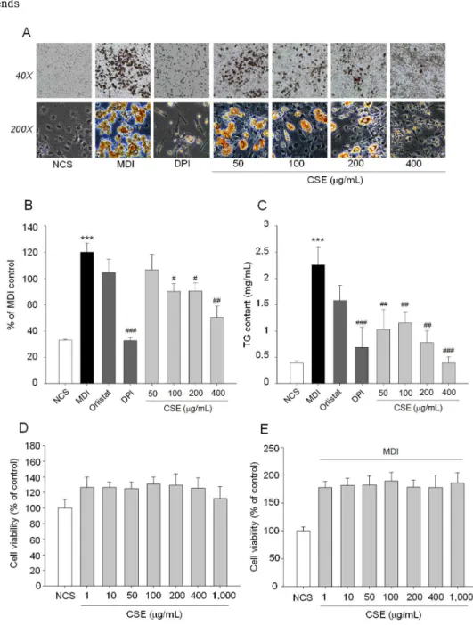
모과 추출물의 항비만 효능을 조사하기 위해 MDI로 분화 유도한 3T3-L1 preadipocyte의 adipogenesis 에 대한 영향을 조사한 결과 Fig. 1A와 1B에 나타낸 바와 같이 농도의존적인 지방세포 분화 억제능이 관 찰되었고 100 μg/ml이상의 농도에서부터 유의한 억 제능을 보였다. Lipid droplet은 preadipocytes의 분 화과정에서 생성되며 phospholipid layer에 둘러싸인 중성지방(triglyceride, TG)로 구성되어 있으며, 중성 지방은 중요한 에너지원이나 과도하게 생성되면 지방 세포에 축적되어 비만의 원인으로 작용한다16,17). 모 과 추출물을 처리하여 생성된 TG 함량을 측정한 결 과 농도 의존적으로 TG 생성이 억제되는 것을 확인 하였으며, 가장 낮은 처리 농도인 50 μg/ml 에서부 터 유의한 억제 효과를 보였다(Fig. 1C). 모과 추출 물의 안전성과 지방생성억제 효능이 세포 독성에 의 한 것인지를 확인하기 위하여 3T3-L1 세포에서 1 ~ 1,000 μg/ml 범위에서 7개 농도를 선정하여 3T3-L1 분 화 전후 모두 독성을 유발하지 않는 것으로 확인하였 다(Fig. 1D, 1E). 결과적으로 모과 추출물은 지방 세 포 분화 억제를 통해 항비만 효능을 보이는 것으로 판단되었다.
2. 모과 추출물의 항산화능 분석
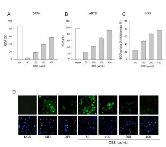
모과 추출물의 항산화능을 분석하기 위해 DPPH, ABTS radical소거능과 SOD 효소 활성 유도효과를 분석하였다. DPPH radical 소거 활성은 농도 의존적 으로 증가하였으며, 90% 수준의 소거활성을 보이는 양성대조군인 ascorbic acid (AA)와 비교하였을 때 고농도인 400 μg/ml의 처리농도에서 60% 이상의 소거활성을 나타내었다(Fig. 2A). ABTS radical 소 거활성 또한 DPPH radical 소거능과 유사한 경향을
보였으며 고농도에서는 양성대조군인 trolox와 유사 한 소거활성을 보였다(Fig. 2B). SOD 효소 활성 증 가 또한 ABTS, DPPH radical 소거 활성에서 보인 바와 같이 농도 의존적으로 활성을 증가시키는 것으 로 나타났다(Fig. 2C). 3T3-L1 preadipocytes를 분 화 시킨 후 세포내 ROS 생성량을 DCF-DA assay를 이용하여 측정하였다. 세포내 ROS 발생시 DCF-DA가 ROS에 의해 산화되어 DCF (2’,7’-dichlorofluorescein) 으로 전환되면서 형광을 나타낸다. 이러한 원리를 이 용해 분석한 결과는 Fig. 2D와 같다. 모과 추출물을 처리하지 않은 대조군과 비교한 결과 100 μg/ml 이 상의 농도에서부터 ROS 생성이 감소하는 것을 확인 할 수 있었다.
3. 모과 추출물이 3T3-L1 adipocytes에서 adipogenesis 조절인자와 NOXs 활성에 미치는 영향
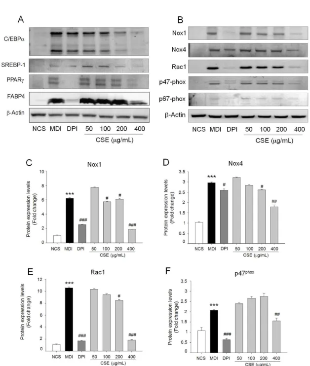
모과 추출물이 3T3-L1 세포의 지방축적 억제효과가 adipogenesis를 조절하는 주요한 전사인자로 알려진 C/EBPα, SREBP-1, PPARγ의 발현에 미치는 영향을 단백질 발현으로 비교하였다(Fig. 3A). C/EBPα, SREBP-1, PPARγ의 발현은 상호작용을 통해 세포내 lipid droplet 생성 및 세포 비대 등과 같은 형태학적 변형과 더불 어 FABP4 등의 adipogenic protein의 발현을 유도 하는 것으로 알려져 있다5). 3T3-L1 분화 시 모과 추 출물을 처리한 군은 처리하지 않은 군에 비해 C/EBPα, SREBP-1, PPARγ의 발현이 농도가 증가함에 따라 유의하게 감소하는 것을 확인할 수 있었고, 고 농도 인 400 μg/ml에서는 분화를 유도하지 않은 수준으로 발현이 억제되었으며 대표적인 adipogenic protein인 FABP4의 발현 또한 400 μg/ml에서 현저히 억제되었 다(Fig. 3A). 양성대조군인 DPI (diphenylenediodonium)은 NOX inhibitor로 알려져 있다18). DPI 처리에 의해 3T3-L1의 adipogenesis (Fig. 1) 와 TG 생성 억제 및 ROS 생성을 억제한 결과(Fig. 2D)로 미루어 모과 추출물 또한 NOX의 활성을 억제하여 adipogenesis 를 억제할 것으로 예측할 수 있었다. 이에 모과 추출 물이 3T3-L1 세포의 adipogenesis 유도시 NOXs 활 성과 관련된 단백질 발현과 인산화에 미치는 영향을 분석한 결과 200 μg/ml 이상의 농도에서 NOX1, NOX4, Rac1, p47-phox, p67-phox의 과발현을 억 제하였다(Fig. 2). 따라서, 모과 추출물은 NOX 활성 저해를 통해 3T3-L1의 adipogenesis를 억제함으로
항 비만 효과를 나타낼 수 있을 것으로 판단된다.
4. 모과 추출물이 고지방식이로 유도된 비만 마우스 모델에서 항비만 효과
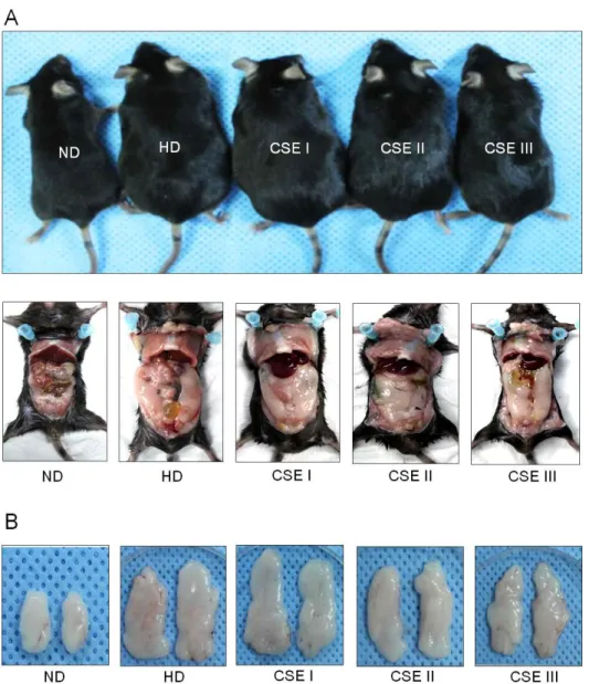
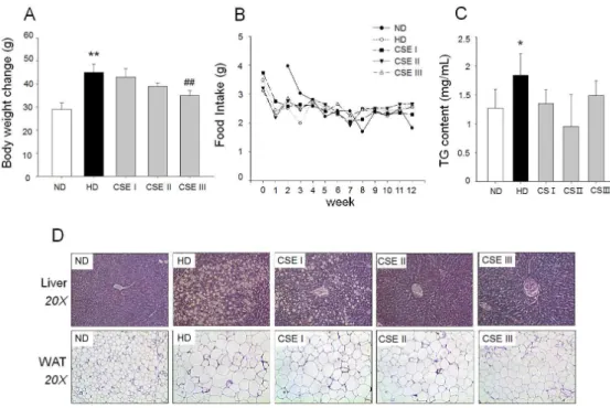
모과 추출물의 고지방식이로 유도한 비만 마우스에서 체중 증가에 미치는 영향을 조사하기 위해서 정상 식 이군(ND), 고지방 식이군(HD), 저용량 투여군(50 mg/kg body weight, CSE I), 중용량 투여군(250 mg/kg body weight), 고용량 투여군(500 mg/kg body weight)로 나누어 12주간 투여한 결과 고지방 식이군 은 정상군에 비해 체중이 증가한 것을 육안으로 확인 할 수 있었으며 복부 지방과 부고환 지방도 증가하였 다. 모과 추출물은 투여한 군은 고지방 식이군에 비 해 체중 증가 및 지방 축적이 감소하는 것이 관찰되 었다(Fig. 4). 12주후 각 군의 체중을 비교한 결과 정상군 평균 체중, 26.2 ~ 31.8 g 대비, 고지방 식 이군은 41.4 ~ 48.6 g으로 체중 증가가 유의하게 확 인되었고, 모과 추출물 투여군의 경우 저용량 투여군 과 중용량 투여군에서는 유의한 차이가 없었으나 고 용량 투여군은 32.8 ~ 37.2 g으로 유의한 체중 증가 억제 효과를 보였다(Fig. 5A). 12주간 식이 섭취량은 각 군별로 차이가 없었다(Fig. 5B). 혈중 TG 분석결 과, 고지방 식이군에서는 대조군 대비 유의한 증가를 보였고 모과 추출물 투여군은 유의성은 없었으나 고 지방 식이군에 비해 감소하는 경향을 보였다(Fig. 5C).
간 조직과 부고환 지방(epididymal white adipose tissue, WAT)의 조직학적 분석 결과 고지방 식이군은 정상군 에 비해 지방 축적의 증가가 확인되었고, 부고환 지 방조직의 지방세포 크기가 증가하였다. 반면 모과 추 출물을 처리한 군에서는 고농도 투여군에서 간 조직 내 지방 축적과 지방세포 비대의 억제 효과를 보였다 (Fig. 5D).
Ⅳ. 고찰
현재까지 비만의 예방과 치료를 위한 약물이 사용되고 있으나 효능 대비 부작용으로 장기간 복용 및 직접적 인 치료효과를 기대하기 어렵다. 이러한 이유로 다양 한 천연 소재 발굴에 대한 연구가 진행되고 있다.
모과는 항산화, 항바이러스, 항응혈, 혈당저하 등에 대한 효능이 보고되었으며, 사포닌, 구연산, 비타민 C, 플라보노이드, 리그난, 페놀성 화합물19)등을 다량
함유하며 면역력 증가 및 감기 예방, 피로 개선 효능 이 알려져 있다. 본 연구에서는 모과 추출물의 지방 세포 분화에 억제능과 관련 기전, 고지방 식이로 유 도된 마우스 비만 모델에서 체중 증가 억제 효능을 분석하였다.
지방세포는 에너지 항상성 조절, 식욕조절, 비만, 인슐린 작용과 관련된 생리활성 물질의 분비 기능을 가지고 있다20,21). 지방분화(adipogenesis)는 전지방세포 (preadipocytes)가 지방세포(adipocytes)로 분화되는 과정으로 지방세포 형성 및 지질 축적에 주요한 역할 을 한다22). 최근 adipogenesis를 조절하는 전사인자 및 유전자 조절을 통한 비만 예방을 위한 연구에 주 요한 타겟은 adipogenesis를 유도하는 전사인자는 대 표적으로 CCAAT/enhancer binding proteins (C/EBPs) 가 있다. C/EBP는 α, β, γ, δ, ε및 CHOP-10의 6종류가 현재까지 보고되었으며, 특히 C/EBP α, β, δ가 adipogenesis와 밀접한 관계가 있는 것으로 알려져 있다. C/EBPβ와 δ는 분화 초기에 발현되어 조절하는 것으로 알려져 있으며, C/EBPd는 분화과정 에 진행되면서 발현이 감소하고 C/EBPb는 C/EBPα 와 PPARγ가 의 발현을 증가시켜 adipogenesis를 유발하는 것으로 알려져 있다22,23). C/EBPα와 PPAR γ는 상호작용 함으로 발현이 증가하며 adipogenic protein인 FABP4 의 과발현을 유도하므로 지방세포 분화에 매우 주요한 전사이다. 이외 SREBP1c는 지방 과 간 조직의 fatty acid와 cholesterol 대사에 매우 중요한 역할을 가지는 전사인자이다7). 모과 추출물이 3T3-L1 세포의 adipogenic 전사인자인 PPARγ, C/EBPα, SREBP-1c의 단백질 발현과 adipogenic protein인 FABP4의 과발현을 억제한 것으로 보아 모과 추출물이 지방세포 생성 억제를 통해 비만을 조 절하는 것으로 평가할 수 있다(Fig.1, Fig. 3A).
산화스트레스는 인슐린 저항성, 비만, 심혈관 질환 등을 포함하여 다양한 질병에 주요한 요인으로 알려 져 있다24). 특히, adipocyte 분화에 다양한 전사인자 가 관여하지만 최근 다양한 연구결과에 따르면 산화 스트레스가 대사성 질환에 매우 중요한 인자로서 인 체 및 마우스에서 산화스트레스가 비만과 지방 축적 에 관여하는 것으로 보고되었다24). 또한, 비만 마우스 모 델에서 지방세포 내 ROS (reactive oxygen species) 가 특이적으로 증가하였고 NOX의 발현이 증가한다 고 밝혀졌다25). 본 연구 결과에서도 모과 추출물이
DPPH, ABTS, SOD 분석법으로 평가한 결과에서 항 산화 효능이 농도 의존적으로 증가하는 것을 확인 할 수 있었고, adipogenesis 과정에서 증가하는 세포내 ROS 생성을 억제하는 결과를 보여 주었다(Fig. 2).
NOXs family 중에서 NOX4가 adipocytes에서 ROS 생성에 주요한 역할을 하는 것으로 밝혀졌다26). NOX 는 현재까지NOX1, NOX2, NOX4, NOX5, DOUX1, DOUX2의 7가지 family가 보고되었다9). NOX family 중 NOX4는 신장 조직에서 처음 밝혀졌으며, 신장 조직 이외에 근육, 간, 백색 및 갈색지방에 발현되어 있으 며, 이외 NOX family 중 NOX1이 지방 분화 시 발 현이 증가하는 것으로 보고되었다26). NOXs 의 활성 화는 p22-phox, p47-phox, p67-phox등의 subunit과 결합을 통하여 증가하고 Rac1은 이들 subunit의 인 산화를 유도한다. NOX4의 경우 특이적으로 p47-phox subunit만 결합하여 활성을 증가시키는 것으로 알려 졌다9,27). 3T3-L1 adipocytes에서 NOX1, NOX4, Rac1, p47-phox, p67-phox 단백질이 과발현되는 것을 모과 추출물이 억제하는 것으로 보아 모과 추출 물이 NOX 활성 억제를 통해 ROS 생성과 adipogenesis 를 억제하는 것으로 추측할 수 있다(Fig. 3).
모과 추출물의 비만 억제 효능에 대한 추가적인 근 거는 고지방 식이로 유도된 동물 모델에서 모과 추출 물을 12주간 경구 투여한 결과로 확인할 수 있었다.
모과 추출물을 고지방 식이 비만 마우스 모델에 저농 도, 중농도, 고농도군으로 나누어 경구 투여하였을 때 고농도 투여군에서 체중, 혈중 중성지방 함량이 고지방 식이군 대비 감소하는 것을 확인하였고, 간 조직에 지방 축적과 지방 조직 비대를 억제하는 것으 로 보아 모과 추출물이 비만 억제에 효과가 있음을 보여준다.
Ⅴ. 결론
본 연구의 결과는 모과 추출물이 NOX 활성 저해를 통 해 세포내 ROS 생성 억제, 지방 세포 분화 및 축적 억제, 지방 분화에 관여하는 전사 인자의 과발현을 억제하여 비만을 제어하는 효과가 있음을 보여주었 다. 또한 고지방 식이로 유도된 비만 마우스 모델에 서도 고용량 처리군에서 체중 증가 및 혈중 중성지 방, 간 지방 축적, 지방 세포 비대를 조절하는 것을 확인하였다. 본 연구 결과는 모과 추출물의 기능성분
에 대한 연구가 수행되어야 하지만 모과 추출물이 비 만 조절에 효과적인 소재로서 가능성을 보여주었다.
감사의 글
본 결과물은 중소벤처기업부의 지역주력산업육성 (R&D) 기술개발사업(No. R0004380)과 산업통상자원 부 풀뿌리기업육성사업(No. R0005297)의 지원으로 이루어졌습니다.
References
1. Rao S, Parab-Waingankar P. Performance of waist circumference relative to BMI in predicting risk of obesity and hypertension among affluent Indian adults. Health 2013;Vol.05No.08:7.
2. Korea Health Statistics 2015. Korea National Health and Nutrition Examination Survey (KNHANES VI-3) 2015.
3. MacDougald OA, Lane MD. Transcriptional regulation of gene expression during adipocyte differentiation. Annu Rev Biochem 1995;64:345-73.
4. Brandes R, Arad R, Bar-Tana J. Inducers of adipose conversion activate transcription promoted by a peroxisome proliferators response element in 3T3-L1 cells. Biochemical Pharmacology 1995;50:1949-51.
5. Villacorta L, Schopfer Francisco J, Zhang J, Freeman Bruce A, Chen YE. PPARγ and its ligands: therapeutic implications in cardiovascular disease. Clinical Science 2009;116:205-18.
6. Spiegelman BM, Flier JS. Adipogenesis and Obesity: Rounding Out the Big Picture. Cell;87:
377-89.
7. Horton JD, Goldstein JL, Brown MS. SREBPs:
activators of the complete program of cholesterol and fatty acid synthesis in the liver. The Journal of Clinical Investigation 2002;109:1125-31.
8. Matsuda M, Shimomura I. Increased oxidative stress in obesity: Implications for metabolic syndrome, diabetes, hypertension, dyslipidemia, atherosclerosis, and cancer. Obesity Research
&Clinical Practice 2013;7:e330-e41.
9. Bedard K, Krause K-H. The NOX Family of ROS-Generating NADPH Oxidases: Physiology and Pathophysiology. Physiological Reviews 2007;87:245-313.
10. Han Y-K, Kim Y-S, Natarajan S, Kim W-S, Hwang J-W, Jeon N-J, Jeong J-H, Moon S-H, Jeon B-T, Park P-J. Antioxidant and Anti- Inflammatory Effects of Chaenomeles sinensis Leaf Extracts on LPS-Stimulated RAW 264.7 Cells. Molecules 2016;21:422.
11. Sancheti S, Sancheti S, Seo S-Y. Antidiabetic and antiacetylcholinesterase effects of ethyl acetate fraction of Chaenomeles sinensis (Thouin) Koehne fruits in streptozotocin- induced diabetic rats. Experimental and Toxicologic Pathology 2013;65:55-60.
12. Floegel A, Kim D-O, Chung S-J, Koo SI, Chun OK. Comparison of ABTS/DPPH assays to measure antioxidant capacity in popular antioxidant-rich US foods. Journal of Food Composition and Analysis 2011;24:1043-8.
13. Weydert CJ, Cullen JJ. MEASUREMENT OF SUPEROXIDE DISMUTASE, CATALASE, AND GLUTATHIONE PEROXIDASE IN CULTURED CELLS AND TISSUE. Nature protocols 2010;5:51-66.
14. Baret P, Septembre-Malaterre A, Rigoulet M, Lefebvre d’Hellencourt C, Priault M, Gonthier M-P, Devin A. Dietary polyphenols preconditioning protects 3T3-L1 preadipocytes from mitochondrial alterations induced by oxidative stress. The International Journal of Biochemistry &Cell Biology 2013;45:167-74.
15. Choi YJ, Kim DH, Kim SJ, Kim J, Jeong S-I, Chung CH, Yu K-Y, Kim S-Y. Decursin attenuates hepatic fibrogenesis through interrupting TGF-beta-mediated NAD(P)H oxidase activation and Smad signaling in vivo and in vitro. Life Sciences 2014;108:94-103.
16. Chitraju C, Mejhert N, Haas JT, Diaz-Ramirez LG, Grueter CA, Imbriglio JE, Pinto S, Koliwad SK, Walther TC, Farese RV, Jr. Triglyceride
Synthesis by DGAT1 Protects Adipocytes from Lipid-Induced ER Stress during Lipolysis. Cell Metabolism;26:407-18.e3.
17. Cao Y. Adipose tissue angiogenesis as a therapeutic target for obesity and metabolic diseases. Nat Rev Drug Discov 2010;9:107-15.
18. Massart C, Giusti N, Beauwens R, Dumont JE, Miot F, Sande JV. Diphenyleneiodonium, an inhibitor of NOXes and DUOXes, is also an iodide-specific transporter. FEBS Open Bio 2014;4:55-9.
19. Du H, Wu J, Li H, Zhong P-X, Xu Y-J, Li C-H, Ji K-X, Wang L-S. Polyphenols and triterpenes from Chaenomeles fruits: Chemical analysis and antioxidant activities assessment. Food Chemistry 2013;141:4260-8.
20. Rajala MW, Scherer PE. Minireview: The Adipocyte
—At the Crossroads of Energy Homeostasis, Inflammation, and Atherosclerosis. Endocrinology 2003;144:3765-73.
21. Chen L, Chen R, Wang H, Liang F. Mechanisms Linking Inflammation to Insulin Resistance.
International Journal of Endocrinology 2015;2015:9.
22. Lowe CE, O'Rahilly S, Rochford JJ. Adipogenesis at a glance. Journal of Cell Science 2011;124:
2681-6.
23. Ntambi JM, Young-Cheul K. Adipocyte differentiation and gene expression. J Nutr 2000;130:3122s-6s.
24. Furukawa S, Fujita T, Shimabukuro M, Iwaki M, Yamada Y, Nakajima Y, Nakayama O, Makishima M, Matsuda M, Shimomura I.
Increased oxidative stress in obesity and its impact on metabolic syndrome. J Clin Invest 2004;114:1752-61.
25. Houstis N, Rosen ED, Lander ES. Reactive oxygen species have a causal role in multiple forms of insulin resistance. Nature 2006;440:944-8.
26. Mouche S, Mkaddem SB, Wang W, Katic M, Tseng Y-H, Carnesecchi S, Steger K, Foti M, Meier CA, Muzzin P, Kahn CR, Ogier-Denis E, Szanto I. Reduced expression of the NADPH oxidase NOX4 is a hallmark of adipocyte differentiation. Biochimica et Biophysica Acta (BBA) - Molecular Cell Research 2007;1773:1015-27.
27. Babior BM. NADPH oxidase. Current Opinion in Immunology 2004;16:42-7.
Figure legends
Fig. 1. Effects of Chaenomeles sinensis extract (CSE) adipogenesis and lipid accumulation in 3T3-L1 cells. (A) Post-confluent 3T3-L1 preadipocytes (day 0) were cultured in the differentiation medium in the presence or absence of CSE (50 ~ 400 μg/ml) and DPI (1 nM) from day 0 to 6. Cell differentiation was examined at day 8 by Oil Red O staining. (B) Stained intracellular oil droplets were eluted with isopropanol and quantified by spectrophotometrical analysis at 500 nm. (C) Intracellular triglyceride (TG) contents in 3T3-L1 adipocytes. Data represent the mean ± SD of at least three independent experiments. (D, E) Cytotoxicity of CSE in preadipocytes and adipocytes. Cell viability was evaluated using a colorimetric assay based on MTT assay after CSE treatment with the indicated concentration for 24 h. *P <0.05 versus NCS; ***P <0.001 versus NCS. #P <0.05, ##P <0.01, ###P <0.001 versus MDI (differentiation medium).
Fig. 2. Antioxidant effects of CSE. (A) DPPH radical scavenging activity and (B) ABTS radical scavenging activity. (C) Superoxide dismutase (SOD) activity. (D) Preadipocytes were plated on coverslips, and induced to differentiation in the presence or absence of CSE (50, 100, 200 and 400 μg/ml) and NOX inhibitor (Diphenyleneiodonium, DPI, 1 nM) for 24 h. Cells were stained with ROS-sensitive dye 2’,7’-dichlorofluorescein diacetate (DCFDA) and observed under a fluorescence microscope. AA, ascorbic acid. Values are means ± SD of at least three independent experiments.
Fig. 3. Effect of CSE on adipogenic protein overexpression NOX activation in 3T3-L1 cells. (A) Modulation of adipogenic transcription factors; SREBP-1, C/EBPα, PPARγ and adipogenic protein;
FABP4 by CSE were evaluated by immunoblot analysis. (B) The protein level of NOX1, NOX4, Rac1, p47phox, and p67phox was determined by immunoblotting. 3T3-L1 cells were differentiated in the absence or presence of CSE for differentiation period. CSE (50, 100, 200 and 400 μ g/mL) treatment blocked NOX protein overexpression in adipocytes. (C, D, E, and F) Relative expression of each proteins corresponding to β-actin was represented. Values are means ± SD of at least three independent experiments. ); ***P <0.001 versus NCS. #P <0.05, ##P <0.01,
###P <0.001 versus MDI.
Fig. 4. Effects of CSE on high fat diet-induced obese mice model. Mice were treated with CSE at dose of 0 (vehicle), 50, 250, and 500 mg/kg body weight daily injection. Mice on normal diet (ND) used as controls were intragastrical injected with vehicle. (A) Representative mice and (B) Epididymal fat pads. HD (high-fat diet), CSE I (50 mg/kg body weight), CSE II (250 mg/kg body weight), CSE III (500 mg/kg body weight).
Fig. 5. Comparison of final body weight and lipid accumulation after CSE treatment. (A) Body weight after CSE treatment. (B) Food intake in ND, HD, and CSE treatment group for 12 weeks. (C) Effect of CSE on serum TG content level. (D) Representative histology of white adipose tissue (WAT) and liver section from each experimental group. Hematoxylin and eosin staining of WAT and liver sections were viewed under a microscope. Values are means ± SD of at least three independent experiments.; *P <0.05, **P <0.01 versus ND. ##P <0.01 versus HD.