Copyright ⓒ 2013, The Microbiological Society of Korea
Lipopolysaccharide 유도된 Raw264.7 세포주에서 전사조절인자 NF-κB와 IRF-1의 공동작용에 의해 조절되는 PD-1 발현연구
최은경1․이수운2․이수웅1,*
1인제대학교 의과대학 미생물학교실, 다발성골수종전문연구센터, 2인제대학교 해운대백병원 구강악안면외과
PD-1 Expression in LPS-Induced Raw264.7 Cells Is Regulated via Co-activation of Transcription Factor NF-κB and IRF-1
Eun-Kyoung Choi1, Soo-Woon Lee2, and Soo-Woong Lee1*
1Department of Microbiology, Advanced Research Center for Multiple Myeloma, College of Medicine, Inje University, Busan 614-735, Republic of Korea
2Department of Oral and Maxillofacial Surgery, Haeundae Paik Hospital, Inje University, Busan 612-896, Republic of Korea
(Received November 28, 2013 / Accepted December 23, 2013)
Programmed Death-1 (PD-1) is one of the important immune-inhibitory molecules which was expressed in T cells, B cells, NKT cells, and macrophages activated by various immune activating factors. Lipopolysaccharide (LPS), the major component of the outer membrane of Gram-negative bacteria, is one of the crucial immunogens for PD-1 expression. However, there are only a few reports on the expression mechanisms of PD-1 in innate immune cells. In this study, we investigate the expression mechanisms of PD-1 in LPS-stimulated Raw264.7 cell lines by RT-PCR, Western Blot, flow cytometry as well as ChIP assay and co-immunoprecipitation. When Raw264.7 cells were stimulated with LPS, PD-1 expression was greatly up-regulated via PI3K and p38 signaling. Primary macrophages isolated from LPS-injected mice were also shown the increased expression of PD-1. In promoter assay, NF-κB and IRF-1 binding regions in mouse PD-1 promoter are important for PD-1 expression. We also found that the co-activation of NF-κB and IRF-1 is indispensable for the maximum PD-1 expression. These results indicate that the modulation of PD-1 expressed in innate immune cells could be a crucial for the disease therapy such as LPS-induced mouse sepsis model.
Keywords: IRF-1, lipopolysaccharide, NF-κB, programmed Death-1, Raw264.7
*For correspondence. E-mail: [email protected]; Tel.: +82-51-890- 6450; Fax: +82-51-891-6004
이상적인 면역반응은 숙주의 조직 또는 기관에 최소한의 손 상을 입히고, 침입한 병원체를 효과적으로 제거하지만 과도한 면역반응은 숙주세포에 치명적인 손상을 주게 된다(Mueller et al., 1989). 따라서 면역세포들은 적절한 면역반응을 유지하는 조절기전을 가지고 있다. 면역반응의 조절에 관여하는 세포막 단백질들을 공동자극분자(costimulatory molecules)라 하며, 주 로 항원제시세포(antigen presenting cells)에 발현되어 면역반응 의 이차 신호(secondary signal) 또는 공동자극 신호(costimulatory signal)를 전달한다(Mueller et al., 1989). 공동자극분자 중 가장 많이 연구된 것은 B7-1 (CD80)과 B7-2 (CD86)이며, T 세포에 발현되는 CD28 또는 CTLA-4의 라이간드로서 면역반응을 조절 한다(Freeman et al., 1989; Azuma et al., 1993).
Programmed Death-1 (PD-1)은 세포사멸과정에서 발현되는 세포막분자로 발견되었으며, anti-CD3 항체를 주사한 생쥐의 가 슴샘, 비장과 림프절에 상주하는 T 세포에서 발현된다고 알려졌 다(Ishida et al., 1992; Agata et al., 1996). 또한, PD-1은 다양한 면역활성인자에 의해 활성화된 T 세포, B 세포, NKT 세포 및 대 식세포(macrophages)에서도 발현된다고 알려졌다(Agata et al., 1996; Keir et al., 2007; Cho et al., 2008). PD-1의 구조는 IgV-like 세포외 부위, 막통과 부위 그리고 immunoreceptor tyrosine-based inhibitory motif (ITIM) 및 immunoreceptor tyrosine-based switch motif (ITSM)를 포함하는 세포내 부위로 구성된 type I 막단백질이며(Zhang et al., 2004; Parry et al., 2005), 구조적 상 동성에 기초해서 CD28계 수용체 구성원으로 분류된다(Latchman et al., 2001). 라이간드인 PD-L1 (B7-H1) 및 PD-L2 (B7-DC)와 의 결합은 세포내 부위의 ITSM tyrosine의 인산화를 유도하고, 신호매개체인 SHP-2를 소집하며, 이로 인해 anti-CD3 항체로
자극된 T 세포의 증식과 사이토카인 분비를 저해하고(Freeman et al., 2000; Carter et al., 2002), B 세포 수용체에 의해 유도되 는 신호전달과정도 방해하는 것으로 알려졌다(Okazaki et al., 2001). PD-L1 및 PD-L2은 호흡기 과민반응에서 T 세포기능을 분극화하고 조절하는데 중요하다고 알려져 왔다(Singh et al., 2011). 또한, PD-L1의 결핍은 심한 천식(asthma)을 유도하는 반 면, PD-L2의 결핍은 호흡기 과민반응을 완화하고 염증반응도 최소화 한다고 보고하였다(Singh et al., 2011). Nishimura 등 (1998)에 의해 수행된 PD-1 결손 생쥐의 분석에서 PD-1은 B 세 포의 성장과 분화의 음성적 조절에 관여하는 인자로 알려졌으 며, PD-1과 PD-1 라이간드의 결합이 T 세포의 활성에 영향을 주 기 때문에 염증반응의 범주를 결정하는 중요인자로 평가되었다 (Freeman et al., 2000). 최근 연구에서 PD-1은 비특이 면역세포 에서 매우 낮은 수준으로 발현되다가 IFN-α 및 다양한 미생물 산물(Lipopolysaccharide, LPS; Lipoteichoic Acid, LTA)에 의 해 발현이 크게 증가되어 미생물 감염에 따른 염증반응 및 패혈 증(sepsis)과 관계된 면역세포들에서 새로운 면역조절기능의 존 재 가능성이 제시되어 왔다(Huang et al., 2009; Yao et al., 2009).
패혈증은 감염에 대한 전신성 염증반응 증후군(systemic inflammatory response syndrome; SIRS)으로 정의되며, 부적절 한 염증 반응의 증폭에 의하여 발생한다고 알려져 있다(Angus et al., 2001). 이를 기초로 한 다양한 면역치료법이 염증반응 억 제에 초점을 맞추어 개발이 되었으나, 그러한 치료법들의 효과 가 미비함으로 인해 패혈증에 대한 새로운 접근 방법이 필요하 게 되었다(Cohen, 2002; Martin et al., 2003). 패혈증의 병인으 로 가장 많이 알려진 것은 그람음성 세균의 세포벽 외부에 존재 하는 지질다당질(lipopolysaccharide, LPS)이며, 실험동물이나 인체에 주사하면 중증 패혈증과 패혈성 쇼크를 유발할 수 있다 (Parant et al., 1977). 혈류로 유리된 LPS는 혈장 내 LPS 결합단 백질(LPS binding protein, LBP)에 의하여 대식세포와 CD14 수 용체에 전달되어 CD14/TLR4 복합체를 형성 한다(Jiang et al., 2005). 이 복합체는 다양한 내부 신호를 통해 염증성 사이토카 인, prostaglandin과 같은 지질 대사물, 보체, 응고인자 등 주요 면역 체계를 활성화 시킨다(Annane et al., 2005). 또한, TNF-α 와 interleukin 1β와 같은 사이토카인과 케모카인의 분비를 통해 선천면역반응과 획득면역반응의 활성을 유도할 뿐만 아니라 과 도한 생물학적 반응을 유도하여 조직손상을 유발한다고 알려져 있다(Hsu and Wen, 2002). Yao 등(2009)이 수행한 최근 연구에 서 PD-1 결손 생쥐가 Listeria 감염에 대해 우수한 선천면역기전 및 생존성 향상을 나타낸다고 보고 하였다. 이는 미생물감염 과 정에 PD-1이 중요한 역할을 하며, PD-1의 발현제어를 통해 과 도한 염증반응, 또는 패혈증의 억제 또는 완화를 유도할 수 있음 을 추론해 볼 수 있다. 하지만, LPS와 같은 미생물 산물에 의한 PD-1의 발현 기전, 동물모델에서 침입 병원체의 제어과정에서 PD-1의 발현기전 및 기능에 관한 연구는 미비한 실정이다. 따라 서, 본 연구에서는 대식세포주 Raw264.7 및 LPS 유도 동물모델 을 이용하여 LPS에 의한 PD-1의 발현기전을 밝히고 패혈증 연 구의 기초자료로 활용하고자 한다.
재료 및 방법
시약 및 항체
Escherichia coli Serotype Lipopolysaccharide(026:B6)는 Sigma-Aldrich (USA)에서 구입하였다. LY294002, PD98059, SP600125 및 SB203580은 Calbiochem (USA)에서 구입하였다.
PE-conjugated anti-mouse PD-1 (clone J43), TLR4, CD11b (clone M1/70), CD11c과 FcR blocker (anti-mouse CD16/CD32 2.4G2) 항체들은 eBioscience (USA)에서 구입하였다. ERK, p38, IRF-1 및 Actin 항체들은 Santa Cruz (USA)에서 구입하였 으며, JNK 및 AKT 항체들은 Cell Signaling Technology (USA) 로부터 구입하였다.
세포주 및 조직유래 면역세포의 배양
Raw264.7 세포주는 American Type Culture collection (ATCC, USA)으로부터 분양 받았으며, 표준화된 조건에서 배양 하였다. 이 연구에 사용된 8주령 수컷 C57BL/6 생쥐의 실험계 획은 인제대학교 의과대학 동물 실험실의 연구 승인을 받아(승 인번호 2010-018) 오리엔트바이오(Korea)에서 구입하여 사용 하였다. 생쥐 복강 유래 대식세포 및 비장 유래 대식세포는 표준 화된 방법으로 분리 후 사용하였다.
역전사효소중합연쇄반응(RT-PCR) 및 실시간 중합효소연쇄반응 (Real-time PCR)
RT-PCR을 위한 세포 RNA(2 μg)는 TRIzol reagent (Invitrogen, USA)를 사용하여 추출하였으며, 역전사효소(M-MLV reverse transcriptase)를 이용하여 cDNA를 합성하였다. PD-1 (F: 5′
-TTC GAA TTC ACC ATG CAA-3′, R: 5′-GTC ATC GTC TTT GTA GTC CAT-3′), IRF-1 (F: 5′-ATG GAT TGG GGG GTC TGA GGT-3′, R: 5′-TTC ACA TTT CAC TGG CTT GGG A-3′), GAPDH (F: 5′-TTC ACC ACC ATG GAG AAG GC-3′, R: 5′-GGC ATG GAC TGT GGT CAT GA-3′)의 발현 은 특이적인 primer (Bioneer, Korea)를 합성하여 이용하였다.
Real-time PCR에 사용된 RNA는 RNase-free DNase (Promega, USA)로 genomic DNA를 제거한 cNDA를 합성하였고, Real-time PCR은 SYBR Supermix kit (Bio-Rad, USA)과 Cycler system (Bio-Rad)을 이용 하였다.
유세포 분석(Flow cytometric analysis)
세포는 BD Biosciences (USA)에서 구입한 항체를 이용하여 염색하였다. 세포를 FACS buffer (PBS containing 1% FCS and 0.1% NaN3)로 씻고 비특이적인 결합을 막기 위해 FcR blocker 로 4℃에서 10분 동안 반응시켰다. PE-conjugated anti-mouse PD-1 항체를 첨가하여 4℃에서 30분간 반응시킨 후 FACSort와 CellQuestPro software (BD Science)로 분석하였다.
Western blot
배양된 Raw264.7 세포주 (1 × 10⁶/ml)에 lysis buffer를 처리 하여 표준화된 방법으로 세포단백질을 얻었다. 반응은 10%
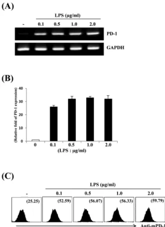
Fig. 1. Dose-dependency of PD-1 expression in LPS-stimulated Raw264.7 cells. Raw264.7 cells were treated with PBS or the indicated concentrations of LPS for 6 h. Total RNAs were isolated from the cells and analyzed for mouse PD-1 mRNA expression by (A) RT-PCR and (B) real-time PCR. (C) Raw264.7 cells were stimulated with the indicated concentrations of LPS for 24 h. Cells were stained with PE-conjugated anti-mouse PD-1 antibody and analyzed by flow cytometry. Numbers indicate the mean fluorescence intensity (MIF) for the gated PD-1 positive cell population.
SDS-PAGE (sodium dodecyl sulfate-polyacrylamide gel)에 30 μg의 단백질을 전기영동한 후 5% skim milk가 포함된 1 × TBST (25 mM Tris–HCl; pH 7.4, 137 mM NaCl, 2.68 mM KCl, 0.05% Tween 20) 용액에서 각각의 특이적인 항체를 사용 하여 2시간동안 반응 시켰으며, ECL 용액으로 반응시킨 후 LAS-3000 SYSTEM (Fuji Photo Film, Japan)으로 측정하였다.
Transient transfection과 luciferase assay
Plasmid의 transient transfection은 Lipofectamine 2000 (Invitrogen)을 사용하여 시약회사에서 제시된 표준화된 방법으 로 수행 하였으며, Luciferase assay은 Dual Luciferase Reporter Assay System (Promega)을 사용하여 반응시킨 후 Victor III (Perkin Elmer, USA)를 사용하여 측정하였다.
Chromatin Immunoprecipitation (ChIP) assay
ChIP assay는 ChIP assay kit (Millipore Corporation, USA) 를 사용하여 시약회사에서 제시된 표준화된 방법으로 수행하였
다. 최종 침전된 protein A/antibody/histone/DNA 복합체를 phenol/chrorofrom 용액을 처리하여 순수 DNA를 얻은 후 PD-1 프로모터(promoter) 부위의 PCR 증폭에 주형으로 사용하였다.
면역침강법(co-immunoprecipitaion)
면역침강법에 사용된 세포단백질은 Raw264.7 세포에 1 μg/ml 의 LPS를 처리 후 protease inhibitors (PMSF 1 mM, 1 × PIC)가 첨가된 lysis buffer (50 mM Tris, 150 mM NaCl, 1% NP-40;
pH 8.0)로 용해시킨 후 표준화된 방법으로 준비하였다. 준비된 세포단백질액에 IRF-1 항체 또는 p65 항체와 반응시킨 후 agarose bead를 이용하여 단백질 침전물을 얻었으며, 30 μl의 1
× dye를 넣고 10분간 끓여 SDS-PAGE를 수행하였다.
데이터 분석
각각의 데이터 값은 세 개가 하나의 실험으로 얻어졌으며, mean ± standard error mean (SEM)으로 표현되었다.
결 과
LPS에 의한 PD-1의 발현분석
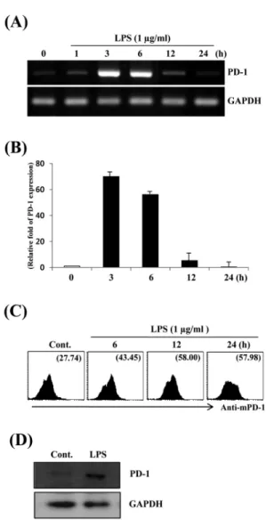
대식세포주 Raw264.7에서 LPS 농도에 따른 programmed death-1 (PD-1) 발현 양상을 알아보기 위하여 다양한 농도의 LPS로 6시간 동안 처리한 후 RT-PCR, real-time PCR 및 NF-κB 를 이용하여 분석하여 보았다. LPS 처리된 Raw264.7 세포들은 PD-1 발현이 매우 높게 유도 되었으나 대조군에서는 매우 낮았다 (Fig. 1A). 이러한 발현양상은 real-time PCR 및 유세포 분석에 서도 유사하게 나타났다. Real-time PCR 결과에 의하면 0.1 μ g/ml의 LPS 처리시 PD-1의 발현은 대조군에 비하여 약 25배 증 가하였으며(Fig. 1B), LPS에 의한 PD-1 유전자의 발현 양상은 NF-κB를 이용한 세포표면 PD-1 단백질발현 결과와도 일치 하 였다(Fig. 1C). LPS 처리에 따른 PD-1 발현의 시간 의존성을 알 아보기 위하여 Raw264.7 세포주를 1 μg/ml 농도의 LPS로 처리 한 후 다양한 시간에서 PD-1 발현을 분석하여 보았다. PD-1의 발 현은 3시간부터 급격히 증가하여 6시간까지 유지되었으며, 12 시간 이후로 현저히 감소되는 발현양상을 보였다(Fig. 2A and 2B).
또한 Raw264.7 세포주의 세포표면에 발현된 PD-1 단백질은 6시 간부터 증가하여 12시간에 최대치를 나타내었으며, 24시간 이 후까지 발현이 유지되었다(Fig. 2C). LPS에 의한 PD-1 발현의 정도는 Western blot 분석에서 또한 확인할 수 있었다(Fig. 2D).
LPS에 의한 세포 신호전달 경로분석
PD-1 발현을 유도하는 신호전달경로를 분석하기 위하여 Raw264.7 세포주를 LPS로 자극 하였을 때 ERK, JNK, Akt 및 p38의 인산화를 확인 할 수 있었다(Fig. 3A). LPS에 의한 PD-1 발현이 ERK, JNK, Akt 또는 p38의 인산화와 연관이 있는지를 특정 신호전달경로를 차단하는 다양한 저해제(inhibitor)를 사용 하여 확인하여 보았다. Fig. 3B의 결과에서 LPS에 의한 PD-1 발 현은 LY294002 (PI3K inhibitor) 및 SB203580 (p38 inhibitor) 에 의해서 기저수준까지 저해되었다. 또한, SP600125 (JNK
Fig. 2. Time-dependency of PD-1 expression in LPS-stimulated Raw264.7 cells. Raw264.7 cells were treated with PBS or LPS (1 μg/ml) for the indicated times. Total RNAs were isolated from the cells and analyzed for mouse PD-1 mRNA expression by (A) RT-PCR and (B) real-time PCR. (C) Raw264.7 cells were stimulated with PBS (Cont.) or LPS (1 μg/ml). At the indicated times, cells were stained with PE-conjugated anti-mouse PD-1 antibody and analyzed by flow cytometry. Numbers indicate the mean fluorescence intensity (MIF) for the gated PD-1 positive cell population. (D) Raw264.7 cells were treated with PBS (Cont.) or 1 μg/ml LPS for 24 h. Whole cell lysates were isolated and were subjected to Western Blotting with anti-mouse PD-1 antibody.
Fig. 3. LPS signaling pathway and effects of various inhibitors on LPS-induced PD-1 expression. (A) Raw264.7 cells were stimulated with LPS (1 μg/ml) for the indicated times. Cell lysates were subjected to SDS-PAGE, blotted onto polyvinylidene difluoride membrane, and then protein phosphorylation was detected with biotinylated anti-p-ERK, anti-p-JNK, anti-p-AKT, and anti-p-p38 antibody. The reaction was developed by the avidin-biotinylated peroxidase complex and ECL. (B) Raw264.7 cells were pretreated for 1 h with the indicated inhibitors and then stimulated with LPS (1 μg/ml) in the presence or absence of the same inhibitor for 6 h.
Total RNAs were prepared and analyzed by RT-PCR for PD-1 or GAPDH. NT (no treatment); LY294002 (PI3K inhibitor); SB203580 (p38 inhibitor); SP600125 (JNK inhibitor); U0126 (ERK inhibitor);
AG490 (JAK inhibitor); PDTC (NF-κB inhibitor).
inhibitor), U0126 (ERK inhibitor) 및 PDTC (NF-κB inhibitor) 에 의해서도 부분적으로 감소 되었다. 이에 반하여 JAK/STAT 저 해제인 AG490은 PD-1 발현에 거의 영향을 주지 않았다(Fig. 3B).
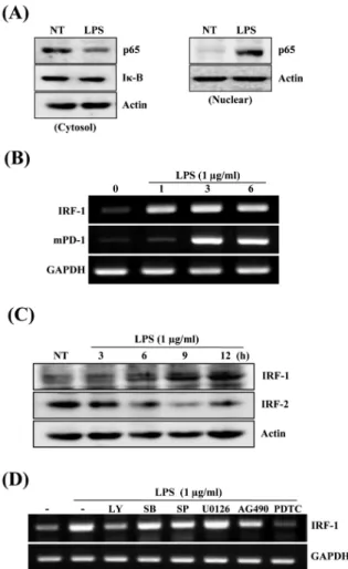
LPS에 의한 PD-1 발현에 관련된 전사조절인자의 활성분석 LPS에 의한 NF-κB의 활성을 확인하기 위하여 Raw264.7 세 포주를 1 μg/ml 농도의 LPS로 30분 동안 처리한 후 세포질단백 질과 핵단백질내에 존재하는 NF-κB의 농도변화를 확인하여 보
았다. Fig. 4A 결과에서 알 수 있듯이 세포질에 존재했던 많은 양의 NF-κB p65가 LPS 처리 후 핵내로 이동되었으며 세포질에 존재하던 Iκ-B의 양도 크게 감소하였다. Salkowski 등(1999)은 IRF-1과 IRF-2 유전자 결핍 동물모델을 이용하여 LPS에 의한 IL-12의 최적발현에 전사조절인자 IRF-1 및 IRF-2가 필수적인 요소임을 증명하였다. 또한, Blanco 등(2000)도 LPS에 의한 Cox-2 발현에 IRF-1 및 IRF-2가 중요하다고 보고하였다. 이와 유사하게 PD-1 프로모터의 분석에서도 interferon-stimulated response element (ISRE) 부위가 interferon regulatory factor 1
Fig. 4. Effects of transcription factors NF-κB and IRF-1 in LPS-stimualted Raw264.7 cells. (A) Raw264.7 cells were treated with PBS or LPS (1 μg/ml) for 30 min, and used to isolate the nuclear and cytosolic protein. Cytosolic (left panel) and nuclear extracts (right panel) were subjected to SDS-PAGE, and detected with anti-p65 antibody. After stripping, cytosolic Iκ-B (left panel) was detected with anti-Iκ-B antibody. (B) Raw264.7 cells were treated with PBS or LPS (1 μg/ml). At the indicated time, PD-1 and IRF-1 expression were analyzed by RT-PCR. (C) Raw264.7 cells were stimulated with LPS (1 μg/ml) for the indicated times. Cell lysates were subjected to SDS-PAGE, blotted onto polyvinylidene difluoride membrane, and then detected with biotinylated anti-IRF-1 or IRF-2 antibody. (D) Raw264.7 cells were pretreated with the indicated inhibitors for 1 h, and stimulated with LPS (1 μg/ml) in the presence or absence of the inhibitors for 6 h. Total RNAs were prepared and analyzed by RT-PCR for PD-1 or GAPDH.
(IRF-1) 결합부위와 100% 일치됨을 알 수 있었다. 따라서 LPS 에 의한 PD-1의 발현과 IRF-1의 활성이 연관이 있을 것으로 추 정되며, 이를 규명하기 위하여 PD-1과 IRF-1 발현 관계를 알아 보았다. LPS에 의한 IRF-1의 발현은 PD-1의 발현보다 앞서 1시 간 만에 발현이 매우 높게 유도 되었다. 또한, IRF-1 단백질도 3 시간부터 증가하기 시작하여 12시간까지 지속되었으며(Fig.
4B), 단백질 수준에서도 유사한 발현 형태를 나타내었다(Fig.
4C). 이와는 반대로 IRF-2의 발현은 LPS 처리 후 현저히 감소하 였다. PD-1 발현과 유사하게 신호전달경로를 차단하는 다양한
저해제(inhibitors)를 이용하여 IRF-1 발현 억제를 확인하여 보 았다. Raw264.7 세포주를 LY294002 (PI3K inhibitor), SB203580 (p38 inhibitor), SP600125 (JNK inhibitor), U0126 (ERK inhibitor), AG490 (JAK/STAT inhibitor), PDTC (NF-κB inhibitor)로 각각 1시간 전처리 후 LPS를 6시간 동안 처리하여 IRF-1의 발현을 확인하여 보았다. IRF-1의 발현은 PDTC (NF-κB inhibitor)에 의해서 완전히 억제되었으며, LY (PI3K inhibitor)에 의해서 40% 정도 발현이 감소되었다. 그러나 나머지 저해제에 대해서는 거의 영향 받지 않았다(Fig. 4D).
LPS에 의한 PD-1의 발현에 전사조절인자 NF-κB와 IRF-1의 상호작용 규명
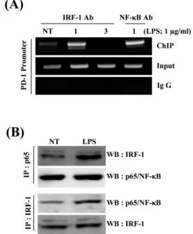
상기 연구결과를 종합해보면 LPS에 의한 PD-1 발현에 전사 조절인자 NF-κB와 IRF-1이 공동작용 할 가능성을 예측 할 수가 있다. 이를 증명하기 위하여 PD-1 유전자의 전사조절부위에 전 사조절인자인 NF-κB와 IRF-1의 결합을 ChIP assay (Chromatin Immunoprecipitation Assay)를 이용하여 확인하여 보았다. 이를 위하여 LPS로 처리된 Raw264.7 세포주 및 대조군으로부터 NF- κB 및 IRF-1에 대한 항체를 이용하여 회수된 DNA를 주형으로 PD-1 프로모터를 PCR 증폭한 결과 LPS로 처리한 후 IRF-1과 NF-κB에 대한 항체로 회수된 각각의 검체에서 PD-1 프로모터 부위가 대조군에 비해 크게 증가되었다(Fig. 5A). IRF-1과 NF-κB 의 상호작용을 확인하기 위하여 두 단백질 간의 결합을 면역 침 강법(co-immunoprecipitation)을 통해 확인하여 보았다. Raw264.7 세포주에 LPS를 처리한 후 얻은 세포질용액을 NF-κB p65 항체 로 면역침전 시키고, 침전된 단백질에 포함된 IRF-1 단백질의 양 을 IRF-1 항체를 이용하여 western blot으로 분석하였다. 결과 5B에서 NF-κB p65 항체로 침전한 단백질에는 IRF-1 단백질이 포함되어 있었으며, LPS 처리된 검체에서 IRF-1 단백질의 양이 유의하게 증가되었다. 또한 IRF-1 항체를 이용한 단백질 침전물 에는 NF-κB 단백질이 포함되어 있었으며, LPS 처리된 검체에 서 NF-κB 단백질의 양이 유의하게 증가되었다. 따라서 이들 결 과는 LPS에 의한 PD-1의 전사조절에 NF-κB 및 IRF-1이 공동 으로 작용하고 있음을 직접 보여주는 증거이다.
LPS 유도 동물 패혈증 모델에서 PD-1 발현분석
LPS 처리된 생쥐 대식세포주 Raw264.7에서 PD-1 발현이 확 인되었기 때문에 패혈증과 유사한 조건인 LPS 유도 동물모델에 서도 PD-1 발현이 유도되는지 확인하여 보았다. 이를 위하여 10 mg/kg의 LPS를 C57BL/6 생쥐의 꼬리정맥에 주사 후 24시간에 비장과 복강 삼출액을 채취하여 대식세포에 발현된 PD-1을 유 세포분석기를 이용하여 분석하여 보았다. PBS를 주사한 대조군 에서 분리한 비장유래 대식세포는 PD-1이 낮게 발현되고 있는 데 반하여, LPS 주사된 생쥐의 비장유래 대식세포는 PD-1 발현 이 현저하게 증가되었다 (Fig. 6A). 또한 복강유래 대식세포에서 도 LPS 처리된 군에서 PD-1 발현이 현저하게 증가 되었다 (Fig.
6B). 이상의 결과를 종합해 보면 LPS에 자극된 대식세포에서 PD-1의 발현은 전사조절인자 NF-κB 와 IRF-1의 공동작용에 의 해 발현이 극대화 되며, LPS에 의한 PD-1의 발현은 대식세포주
Fig. 5. Binding of IRF-1 and NF-κB to PD-1 promoter and analysis of their interaction. (A) Raw264.7 cells were treated with 1 μg/ml LPS for the indicted times. Cell lysates were isolated and analyzed in the chromatin immunoprecipitation (ChIP) assay by using an anti-IRF-1 or anti-NF-κB and primer pairs specific for promoter of the PD-1 gene. Nonimmunoprecipitated lysates (input) served as positive controls. (B) Immunoprecipitation was performed with anti-p65 antibody and the lysates of Raw264.7 cells stimulated with LPS as described in the materials and methods, followed by immunoblot analysis of IRF-1 or, p65 as control after stripping (upper panel). In lower panel, immune blot was analysed with anti-p65 antibody after the immunoprecipitation using anti-IRF-1 antibody or with anti-IRF antibody after stripping as described above.
Fig. 6. PD-1 expression by LPS in mouse splenic and peritoneal macrophages. C57BL/6 (B6) mice were injected with PBS or 10 mg/kg LPS for 24 h. (A) Splenocytes were purified from spleen and then double-stained with FITC-conjugated anti-mouse CD11b and PE-conjugated anti-mouse PD-1 antibody. Cells were analyzed by flow cytometry. (B) Peritoneal macrophages were isolated from peritoneal exudative cells. Cells were stained with PE-conjugated anti-mouse PD-1 antibody, and analyzed by flow cytometry.
Numbers indicate the mean fluorescence intensity (MIF) for the gated CD11b and PD-1 double positive (A) or PD-1 single positive (B) cell population.
Raw264.7 및 비장 또는 복강유래 대식세포에서 유사한 발현기 전에 의해 유도되고 있음을 알 수 있다.
고 찰
본 연구에서는 LPS 처리된 대식세포주인 Raw264.7 세포에 서 programmed death-1 (PD-1)의 발현기전을 분석하였다.
PD-1 발현을 유도하는 신호전달분석에서 LPS 처리된 대식세포 주인 Raw264.7에서 PD-1의 발현은 PI3K 및 p38 신호전달 경로 를 통하여 주로 이루어지며(Fig. 3B), 전사조절인자 NF-κB 및 IRF-1이 중요한 역할을 한다(Fig. 4A, 4C, 5A, and 5B). Cho 등 (2008)에 의하면 컴퓨터프로그램을 이용한 PD-1 프로모터의 분 석에서 LPS에 의해 유도될 수 있는 전사조절인자들의 결합부위 는 P1 (-1400 bp) construct 내에 대부분 포함되어 있었으며, 이 보다 큰 -3000 bp를 포함하는 construct는 LPS에 대한 반응에서 P1 construct와 유사한 결과를 나타내었다. 보다 특이적인 점은 LPS에 의한 PD-1의 발현에 두 개의 전사조절인자인 IRF-1 및 NF-κB가 공동으로 작용하고 있다는 점이다. 이러한 결과는 Hsu 등 (2002)이 수행한 LPS-mediated ROS의 발현연구 및 Kim 등
(2000)이 수행한 LPS-induced c-fos의 발현에도 두 개의 전사조 절인자에 의해 유도되는 것과 일치하며, PD-1의 전사조절에 NF-κB와 IRF-1가 결합하여 상호작용할 수 있다는 점은 매우 흥 미로운 결과이다(Fig. 5A and 5B).
NF-κB는 면역과 염증 반응에 관련된 여러 유전자 발현에서 다양한 역할을 하는 조절인자이다. 특히 LPS와 같은 물질에 자 극된 대식세포에서 다양한 사이토카인이 생성될 때 NF-κB는 핵 심적인 역할을 하는 것으로 알려져 있다(Lawrence et al., 2001).
본 연구에서는 LPS로 자극한 대식세포에서 NF-κB의 활성이 IRF-1의 공동작용에 의해 현저히 증가되고 있다는 점이다. NF- κB는 염증성 사이토카인의 발현에 중요한 전사조절인자로 잘 알려져 있기 때문에 본 연구결과로 추론해 보면 IRF-1의 활성이 염증성 사이토카인의 발현에 크게 기여할 가능성이 있을 것으로 사료되며, IRF-1의 제어를 통한 NF-κB의 활성완화 또는 억제기 전도 추정할 수 있음을 보여준다.
Raw264.7 세포에 LPS (1 μg/ml)를 처리하였을 때 60분에서 STAT의 활성을 나타내었다(Supplementary data Fig. S1). 이러 한 결과는 Kou 등(2011)이 수행했던 결과와 유사하게 일치한다.
하지만 JAK/STAT 저해제인 AG490은 PD-1 발현에 거의 영향 을 주지 않았다(Fig. 3B). 이와는 달리 Cho 등(2008)은 IFN-α 처 리시 STAT1 경로를 통해서 PD-1 발현이 유도된다고 보고하였 다. 이러한 차이는 사이토카인 수용체를 통한 PD-1 발현경로와 TLR을 통한 PD-1 발현경로가 다르게 이용된다는 것을 보여주 는 분명한 결과임을 알 수 있다.
LPS에 의한 IRF-1의 발현증가에 따라 IRF-2의 발현은 점차
감소하는 결과가 PD-1의 발현에 중요한 인자로 예측된다. 이러 한 결과는 전사조절인자 IRF-1은 유전자활성에 관여하고 IRF-2 는 IRF-1의 활성억제에 관여하는 억제분자(inhibitory molecule) 로 작용 한다는 Judith 등(2005)의 결과와 일치하고 있음을 알 수 있다. 또한 신호전달 저해제를 이용한 B 세포 실험을 통하여 B 세포의 PD-1 발현을 유도하는 신호전달경로도 대식세포와 유사 함을 알 수 있었다(자료 미제시).
생쥐에 꼬리정맥에 10 mg/kg의 LPS를 주사했을 때 정상적인 거동이 가능한 정도의 endotoxin shock을 나타내는 반면에 생쥐 복강에 주사한 경우 거동이 불가능할 정도의 심한 endotoxin shock을 나타내었으며, 약 30%의 생쥐들은 48시간 내에 사망 하였다(자료 미제시). 생쥐복강에 10 mg/kg의 LPS를 주사한 후 24시간에 분리된 비장대식세포 및 복강유래대식세포에서도 꼬 리정맥 주사한 결과와 유사하게 PD-1 발현이 유도됨을 알 수 있 었다(Supplementary data Fig. S2A and S2B). 따라서 LPS의 자 극에 의한 대식세포에서 PD-1 발현은 생체조건(in vivo)과 시험 관조건(in vitro)에서 유사하게 증가되었다. 또한, LPS에 의한 PD-1 발현은 비장유래 대식세포와 복강유래 대식세포 간의 차 이는 없는 것으로 보아 대부분의 대식세포에서 공통적인 유도기 전에 의해 발현되는 것으로 추정된다(Fig. 6A and 6B). 그러므 로 대식세포에서 PD-1의 발현은 LPS와 같은 미생물 산물에 의 한 활성화 과정에서 유도되고 있음을 알 수 있다.
PD-1은 면역단백질계에 속하는 막단백질로 T 세포, B 세포, 수지상세포(dendritic cells) 및 대식세포에서 세포활성에 따라 발현된다고 알려져 있다(Ishida et al., 1992; Agata et al., 1996).
대식세포 및 B 세포에서 LPS에 의한 PD-1의 발현은 Cho 등 (2008)이 수행했던 IFN-α를 이용한 PD-1 발현형태 보다 많은 증가를 나타내었고, 초기발현도 매우 빨리 일어나고 있음을 알 수 있다. 이와 같은 결과는 PD-1 발현이 프로모터 활성물질에 따 라 다른 기전과 다른 전사조절인자들에 의해 유도되고 있음을 예상할 수 있다.
본 연구결과는 향후 PD-1 발현제어를 통한 염증반응의 조절 및 패혈증 동물모델에 적용하여 패혈증 발생 단계를 이해하고 치료전략을 구상할 수 있는 기초자료로 이용될 수 있을 것으로 사료된다.
적 요
Programmed Death-1 (PD-1)은 중요한 면역조절분자들 중 하나로 다양한 면역활성인자에 자극된 T 세포, B 세포, NKT 세 포 및 대식세포에서 발현된다. Lipopolysaccaride (LPS)는 그람 음성세균의 세포벽구성물질로 PD-1 발현을 유도하는 중요 면역 원들 중 하나로 알려져 있다. 그러나 선천면역세포에서 PD-1 발 현기전에 관한 연구는 미비한 실정이다. 본 연구에서는 LPS에 의해 자극된 Raw264.7 세포주를 대상으로 PD-1 발현 및 발현조 전기전을 RT-PCR, Western Blot, 유세포분석기, ChIP assay 및 co-immunoprecipitation 방법으로 조사하였다. Raw264.7 세포 주가 LPS로 자극되었을 때 PI3K 및 p38 신호전달경로를 경유하 여 PD-1 발현이 크게 증가되었다. 또한 LPS 주사된 생쥐의 비장
유래 대식세포에서도 PD-1 발현이 증가됨을 확인 하였다. PD-1 유전자의 프로모터 분석을 통해서 NF-κB 및 IRF-1 결합부위가 PD-1 발현에 중요함을 알 수 있었다. 또한 PD-1 발현을 극대화 하기 위하여 전사조절인자 NF-κB 및 IRF-1의 공동활성이 필수 적임을 확인하였다. 본 연구결과는 LPS 유도 생쥐패혈증모델에 서 선천면역세포에 발현된 PD-1분자의 제어를 통한 질병 연구 에 유용한 자료로 이용될 수 있을 것으로 사료된다.
감사의 말
본 논문은 2006년도 인제대학교 학술연구비 보조에 의한 것 임.
참고문헌
Agata, Y., Kawasaki, A., Nishimura, H., Ishida, Y., Tsubata, T., Yagita, H., and Honjo, T. 1996. Expression of the PD-1 antigen on the surface of stimulated mouse T and B lymphocytes. Int. Immunol. 8, 765–772.
Angus, D.C., Linde-Zwirble, W.T., Lidicker, J., Clermont, G., Carcillo, J., and Pinsky, M.R. 2001. Epidemiology of severe sepsis in the United States: Analysis of incidence, outcome, and associated costs of care. Crit. Care. Med. 29, 1303–1310.
Annane, D., Bellissant, E., and Cavailon, J.M. 2005. Septic shock.
Lancet 365, 63–78.
Azuma, M., Ito, D., Yagita, H., Okumura, K., Phillips, J.H., Lanier, L.L., and Somoza, C. 1993. B70 antigen is a second ligand for CTLA-4 and CD28. Nature 366, 76–79.
Blanco, J.C., Contursi, C., Salkowski, C.A., DeWitt, D.L., Ozato, K., and Vogel, S.N. 2000. Interferon regulatory factor (IRF)-1 and IRF-2 regulate interferon gamma-dependent cyclooxygenase 2 expression. J. Exp. Med. 191, 2131–2144.
Carter, L., Fouser, L.A., Jussif, J., Fitz, L., Deng, B., Wood, C.R., Collins, M., Honjo, T., Freeman, G.J., and Carreno, B.M. 2002. PD-1: PD-L inhibitory pathway affects both CD4(+) and CD8(+) T cells and is overcome by IL-2. Eur. J. Immunol. 32, 634–643.
Cho, H.Y., Lee, S.W., Seo, S.K., Choi, I.W., Choi, I.h., and Lee, S.W.
2008. Interferon-sensitive response element (ISRE) is mainly responsible for IFN-α-induced upregulation of programmed death-1 (PD-1) in macrophages. Biochimi. Biophys. Acta 1779, 811–819.
Cohen, J. 2002. The immunopathogenesis of sepsis. Nature 420, 885–891.
Freeman, G.J., Freedman, A.S., Segil, J.M., Lee, G., Whitman, J.F., and Nadler, L.M. 1989. B7, a new member of the Ig superfamily with unique expression on activated and neoplastic B cells. J. Immunol.
143, 2714–2722.
Freeman, G.J., Long, A.J., Iwai, Y., Bourque, K., Chernova, T., Nishimura, H., Fitz, L.J., Malenkovich, N., Okazaki, T., Byrne, M.C., and et al. 2000. Engagement of the PD-1 immunoinhibitory receptor by a novel B7 family member leads to negative regulation of lymphocyte activation. J. Exp. Med. 192, 1027–1034.
Hsu, H.Y. and Wen, M.H. 2002. Lipopolysaccharide-mediated reactive oxygen species and signal transduction in the regulation of interleukin-1 gene expression. J. Biol. Chem. 277, 22131–22139.
Huang, X., Venet, F., Wang, Y.L., Lepape, A., Yuan, Z., Chen, Y., Swan, R., Kherouf, H., Monneret, G., Chung, C.S., and et al. 2009. PD-1 expression by macrophages plays a pathologic role in altering
microbial clearance and the innate inflammatory response to sepsis.
Proc. Natl. Acad. Sci. USA 14, 6303–6308.
Ishida, Y., Agata, Y., Shibahara, K., and Honjo, T. 1992. Induced expression of PD-1, a novel member of the immunoglobulin gene superfamily, upon programmed cell death. EMBO J. 11, 3887–3895.
Jiang, Z., Georgel, P., Du, X., Shamel, L., Sovath, S., Mudd, S., Huber, M., Kalis, C., Keck, S., Galanos, C., and et al. 2005. CD14 is required for MyD88-independent LPS signaling. Nature Immunol. 6, 565–570.
Judith, M.C., Linda, B., Thomas, J.G., William, C.C., and Gerard, M.D.
2005. Interferon regulatory factor 1 (IRF-1) and IRF-2 expression in breast cancer tissue microarrays. J. Interferon Cytokine Res. 25, 587–594.
Keir, M.E., Francisco, L.M., and Sharpe, A.H. 2007. PD-1 and its ligands in T-cell immunity. Curr. Opin. Immunol. 19, 309–314.
Kim, Y.H., Choi, M.R., Song, D.K., Huh, S.O., Jang, C.G., and Suh, H.W.
2000. Regulation of c-fos gene expression by lipopolysaccharide and cycloheximide in C6 rat glioma cells. Brain Res. 872, 227–230.
Kou, X., Qi, S., Dai, W., Luo, L., and Yin, Z. 2011. Arctigenin inhibits lipopolysaccharide-induced iNOS expression in RAW264.7 cells through suppressing JAK-STAT signal pathway. Int.
Immunopharmacol. 11, 1095–1102.
Latchman, Y., Wood, C.R., Chernova, T., Chaudhary, D., Borde, M., Chernova, I., Iwai, Y., Long, A.J., Brown, J.A., Nunes, R., and et al.
2001. PD-L2 is a second ligand for PD-1 and inhibits T cell activation. Nat. Immunol. 2, 261–268.
Lawrence, T., Gilroy, D.W., Colville-Nash, P.R., and Willoughby, D.A.
2001. Possible new role for NF-kappaB in the resolution of inflammation. Nat. Med. 7, 1291–1297.
Martin, G.S., Mannino, D.M., Eaton, S., and Moss, M. 2003. The epidemiology of sepsis in the United States from 1979 through 2000.
N. Engl. J. Med. 348, 1546–1554.
Mueller, D.L., Jenkins, M.K., and Schwartz, R.H. 1989. Clonal
expansion versus functional clonal inactivation: a costimulatory signaling pathway determines the outcome of T cell antigen receptor occupancy. Annu. Rev. Immunol. 7, 445–480.
Nishimura, H., Minato, N., Nakano, T., and Honjo, T. 1998.
Immunological studies on PD-1 deficient mice: implication of PD-1 as a negative regulator for B cell responses. Int. Immunol. 10, 1563–1572.
Okazaki, T., Maeda, A., Nishimura, H., Kurosaki, T., and Honjo, T. 2001.
PD-1 immunoreceptor inhibits B cell receptormediated signaling by recruiting src homology 2-domaincontaining tyrosine phosphatase 2 to phosphotyrosine. Proc. Natl. Acad. Sci. USA 98, 13866–13871.
Parant, M., Parant, F., and Chedid, L. 1977. Inheritance of lipopolysaccharide-enhanced nonspecific resistance to infection and of susceptibility to endotoxic shock in lipopolysaccharide low-responder mice. Infect. Immun. 16, 432–438.
Parry, R.V., Chemnitz, J.M., Frauwirth, K.A., Lanfranco, A.R., Braunstein, I., Kobayashi, S.V., Linsley, P.S., Thompson, C.B., and Riley, J.L. 2005. CTLA-4 and PD-1 receptors inhibit T-cell activation by distinct mechanisms. Mol. Cell Biol. 25, 9543–9553.
Salkowski, C.A., Kopydlowski, K., Blanco, J., Cody, M.J., McNally, R., and Vogel, S.N. 1999. IL-12 is dysregulated in macrophages from IRF-1 and IRF-2 knockout mice. J. Immunol. 163, 1529–1536.
Singh, A.K., Stock, P., and Akbari, O. 2011. Role of PD-L1 and PD-L2 in allergic diseases and asthma. Allergy 66, 155–162.
Yao, S., Wang, S., Zhu, Y., Luo, L., Zhu, G., Flies, S., Xu, H., Ruff, W., Broadwater, M., Choi, I.H., and et al. 2009. PD-1 on dendritic cells impedes innate immunity against bacterial infection. Blood 113, 5811–5818.
Zhang, X., Schwartz, J.C., Guo, X., Bhatia, S., Cao, E., Lorenz, M., Cammer, M., Chen, L., Zhang, Z.Y., Edidin, M.A., and et al. 2004.
Structural and functional analysis of the costimulatory receptor programmed death-1. Immunity 20, 337–347.