263
http://dx.doi.org/10.4196/kjpp.2015.19.3.263 eISSN 2093-3827
ABBREVIATIONS: 5-LO, 5-Lipoxygenase; LPS, Lipopolysaccharide;
TLR4, Toll-like receptor 4; ERK, extracellular signal-regulated kinase;
JNK, c-Jun N-terminal kinase; p38 MAPK, p38 mitogen-activated protein kinase; FBS, fetal bovine serum; FLAP, five lipoxygenase activating protein; IKK, IκB kinase.
Received January 5, 2015, Revised February 26, 2015, Accepted March 21, 2015
Corresponding to: Chi Dae Kim, Department of Pharmacology, School of Medicine, Pusan National University, Yangsan 626-870, Korea. (Tel) 82-51-510-8063, (Fax) 82-51-510-8068, (E-mail) [email protected]
This is an Open Access article distributed under the terms of the Creative Commons Attribution Non-Commercial License (http://
creativecommons.org/licenses/by-nc/3.0) which permits unrestricted non-commercial use, distribution, and reproduction in any medium, provided the original work is properly cited.
Copyright ⓒ Korean J Physiol Pharmacol & MEDrang Inc.
LPS Increases 5-LO Expression on Monocytes via an Activation of Akt-Sp1/NF-κB Pathways
Seung Jin Lee, Kyo Won Seo, and Chi Dae Kim
Department of Pharmacology and BK21 Medical Science Education Center, School of Medicine, Pusan National University, Yangsan 626-870, Korea
5-Lipoxygenase (5-LO) plays a pivotal role in the progression of atherosclerosis. Therefore, this study investigated the molecular mechanisms involved in 5-LO expression on monocytes induced by LPS.
Stimulation of THP-1 monocytes with LPS (0∼3 μg/ml) increased 5-LO promoter activity and 5-LO protein expression in a concentration-dependent manner. LPS-induced 5-LO expression was blocked by phar- macological inhibition of the Akt pathway, but not by inhibitors of MAPK pathways including the ERK, JNK, and p38 MAPK pathways. In line with these results, LPS increased the phosphorylation of Akt, suggesting a role for the Akt pathway in LPS-induced 5-LO expression. In a promoter activity assay conducted to identify transcription factors, both Sp1 and NF-κB were found to play central roles in 5-LO expression in LPS-treated monocytes. The LPS-enhanced activities of Sp1 and NF-κB were attenuated by an Akt inhibitor. Moreover, the LPS-enhanced phosphorylation of Akt was significantly attenuated in cells pretreated with an anti-TLR4 antibody. Taken together, 5-LO expression in LPS-stimulated monocytes is regulated at the transcriptional level via TLR4/Akt-mediated activations of Sp1 and NF-κB pathways in monocytes.
Key Words: Akt, Atherosclerosis, LPS, Monocytes, 5-Lipoxygenase
INTRODUCTION
Monocytes play a central role in several pathophysio- logical conditions, when the progression of cardiovascular disease stems, from underlying inflammatory reactions [1,2].
Lipopolysaccharide (LPS) is a glycolipid component of the gram-negative bacterial cell wall and a major inflammatory cytokine that induces inflammatory responses by activating monocytes [3-5], and 5-lipoxygenase (5-LO) is a potent proin- flammatory mediator in several inflammatory diseases, in- cluding atherosclerosis [6-8]. However, mechanisms respon- sible for the LPS-induced expression of 5-LO in monocytes remain unknown.
Several independent studies have indicated LPS in con- junction with LPS-binding protein, binds to CD14 and trans- membrane Toll-like receptor 4 (TLR4) on the surfaces of a variety of cells, including monocytes [9,10]. It is also known that LPS stimulation of monocytes effects the generations of a number of inflammatory mediators, including 5-LO,
and recent studies indicate that prolonged exposure to LPS upregulates FLAP expression in human monocytes [11]. The involvement of LPS in the modulation of 5-LO suggests an im- portant interaction between bacterial infection and the develop- ment of 5-LO-mediated inflammation; furthermore, products of the 5-lipoxygenase (5-LO) pathway, which metabolizes free arachidonic acid to produce proinflammatory leukotrienes (LT) [12], have been implicated in the development and pro- gression of atherosclerosis [13,14].
The cellular activity of 5-LO is regulated in a complex manner that involves different signaling pathways [15,16].
In particular, 5-LO expression is enhanced on monocyte cells by inflammatory stimuli via an Akt-dependent pathway [17,18], and Akt is an important mediator of signal trans- duction and a key player in the regulation of cellular processes.
Furthermore, the activation of 5-LO in cells involves its phos- phorylation by Akt. Akt has also been implicated in a varie- ty of proinflammatory events, and its activation and phos- phorylation are crucial steps in the signal transduction cas- cade induced by extracellular stimuli, which supports a link between the Akt pathway and 5-LO expression during the development of atherosclerosis.
In this study, 5-LO expression was found to be strongly induced by the TLR4 acvivation in monocytes. We further investigated the mechanisms by which TLR4 signaling reg- ulates 5-LO expression in these cells and found that the
Akt is the major signaling pathway that contributes to TLR4- dependent 5-LO induction. Moreover, an Akt pathway ap- pears to increase 5-LO expression through activation of the Sp1 and NF-κB transcription factors in monocytes.
METHODS Chemicals and antibodies
LPS from Escherichia coli was purchased from Sigma- Aldrich (Saint Louis, MO). pGL3 basic vector, pRL CMV vector, and dual luciferase reporter assay kits were pur- chased from Promega (Madison, WI). DNeasy Tissue Kits and QIAprep Spin Kits were supplied by Qiagen (GmhH, Germany). The various signal pathway inhibitors used were acquired from Calbiochem (Ra Jolla, CA) and Sigma (St.
Louis, MO). 5-LO antibody were purchased from Santa Cruz Biotechnology (Beverly, MA). Akt, phosphospecific antibody against Akt and IKK were from Cell Signaling Technology (Beverly, MA). Purified anti-human TLR4 antibody was from eBioscience (San Diego, CA). Horseradish peroxidase (HRP)-conjugated IgG (Santa Cruz Biotechnology, Santa Cruz, MA) was used as the secondary antibody.
Cell culture
THP-1 cells (a human monocytic leukemia cell line) were purchased from the ATCC (Manassas, VA, USA). Cells were grown in RPMI 1640 medium (Life Technologies) supple- mented with 10% heat-inactivated fetal bovine serum (FBS), antibiotic-antimycotic, and L-glutamine (Life Technologies), and maintained at 37oC in a humidified 5% CO2/95% air atmosphere. After reaching confluence, cells were detached from T75 culture flasks by gentle scraping, washed, and resuspended in a complete medium.
Transient transfection and luciferase assay
Monocytes were grown to 90∼95% confluence in 12-well plates. Separately, 1 μg of plasmid DNA and 2 μl of Lipo- fectamine LTX reagent (Invitrogen, CA) were diluted in 50 μl of Opti-MEM medium (GIBCO, NY), mixed, and in- cubated at room temperature for 30 min. And then the di- luted mixed solution was added to the cells. Cells were then incubated in plates at 37oC for 6 h, and after removing the conditioned medium, grown in fresh medium containing 10% FBS for 24 h, and then treated or not with LPS. Cell lysates were prepared using passive lysis buffer (Promega assay system; Promega, WI) and luciferase activities were measured according to the manufacturer’s instructions for the dual luciferase reporter assay (Promega, WI). All firefly luciferase values were normalized versus Renilla luciferase to compare transfection efficiencies.
Western blot analysis
The levels of 5-LO expression, Akt and IKK phosphor- ylation were measured by Western blotting. Monocyte cell lysates were separated on 8% sodium dodecyl sulphate (SDS)-polyacrylamide gels, and transferred electrophoreti- cally onto nitrocellulose membranes. Membranes were blocked with 5% skim milk in tris-buffered saline contain- ing Tween 20 (TBST), and then incubated with anti-5-LO (1 : 1,000) in a blocking buffer. After the blots were in-
cubated with the horseradish peroxidase (HRP)-conjugated secondary antibody (1 : 3,000), chemiluminescence intensities were measured using the LAS-3000 SYSTEM (Fuji Photo Film, Japan). Membranes were re-blotted with an anti-β-ac- tin antibody (MP Biomedicals, Aurora, Ohio) as an internal control.
Statistical analysis
Results were expressed as means±SEM. Significance was examined using the Student’s t-test for unpaired observations between two groups or by ANOVA with Bonferroni’s correc- tion when multiple groups were compared. Statistical sig- nificance was accepted for p values<0.05.
RESULTS
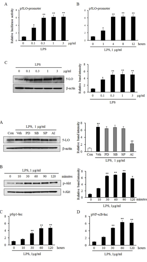
LPS increased 5-LO expression in monocytes To determine the potential role for LPS in the transcrip- tional regulation of 5-LO expression, we initially examined the promoter activity of 5-LO in LPS-treated monocytes.
As shown in Fig. 1A and 1B, the levels of 5-LO promoter activity in LPS-treated monocytes were significantly in- creased in a concentration- and time-dependent manners.
In addition, stimulation of monocytes with various concen- trations of LPS was found to increase 5-LO protein expression in a concentration-dependent manner (Fig. 1C). These re- sults suggest that LPS increases 5-LO expression via an enhanced up-regulation of 5-LO transcription in monocytes.
Involvement of Akt signaling pathway in LPS-induced 5-LO expression
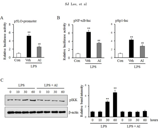
To determine the role played by various signaling pro- teins such as MAPKs and the Akt in LPS-induced 5-LO expression, we examined their pharmacological inhibitors on LPS-induced 5-LO expression in monocytes. In this study, monocytes were pretreated with various MAPK inhibitors including PD98059 (a ERK inhibitor), SB203580 (a p38 MAPK inhibitor), and SP600125 (a JNK inhibitor), and an inhibitor for Akt pathway (AI), and then stimulated with LPS. As shown in Fig. 2A, LPS-induced 5-LO expression was significantly inhibited by an Akt inhibitor, but not by MAPK inhibitors. In addition, LPS significantly and time-de- pendently increased the level of phosphorylated Akt, but not total Akt, indicating a pivotal role of LPS on the activation of Akt pathway in monocytes.
To identify transcription factors involved in LPS-induced 5-LO expression, this study determined whether Sp1 and NF-κB is involved in LPS-induced 5-LO transcription in monocytes. As shown in Fig. 2C and 2D, the luciferase re- porter activities of Sp1 and NF-κB in LPS-treated monocytes were significantly increased in a time-dependent manner. These results indicate that the Akt/Sp1 and Akt/NF-κB pathways mediate LPS-induced 5-LO transcription in monocytes.
Role of the Akt pathway on Sp1 and NF-κB activation in LPS-induced 5-LO expression
To determine the links between Akt pathway and the ac- tivities of Sp1 and NF-κB, the luciferase reporter activities of these transcription factors in LPS-treated monocytes were investigated in the presence or absence of an Akt
Fig. 2. Effects of various signal pathway inhibitors on LPS-induced 5-LO expression. (A) Monocytes we- re pre-treated with MAPK inhibitors including PD98059 (PD, 30 mM), SP600125 (SP, 30 mM), SB203580 (SB, 30 mM), or AI (3 mM) for 30 min, and then stimulated with 1 μg/
ml of LPS. 5-LO expressions were analyzed by immunoblotting, and data were presented as means±SEM from 4∼6 independent experiments.
**p<0.01 vs. value of control (Con),
##p<0.01 vs. value of vehicle (Veh).
(B) Monocytes were stimulated with 1 μg/ml of LPS for the indicated times. The cell lysates were analy- zed for phosphorylated (p-Akt) and total Akt (t-Akt) by Western blo- tting. Relative intensity of p-Akt to t-Akt was quantified, and data were presented as means±SEM from 6∼7 independent experiments. *p<0.05,
**p<0.01 vs. value at time 0. (C and D) Monocytes were transiently tran- sfected with the Sp1 and NF-κB luciferase reporter constructs for 36 h, and then stimulated with LPS for the indicated times. Sp1 and NF-κB activities were analyzed using luci- ferase reporter assays. Data were presented as means±SEM of 4-5 independent experiments performed in triplicate. **p<0.01 vs. value at time 0.
Fig. 1. Effects of LPS on 5-LO expre- ssion in monocytes. Monocytes were transiently cotransfected with the empty luciferase vector pRL CMV or 5-LO promoter constructs for 36 h, and then stimulated with the indicated concentrations of LPS for 4 h (A) or 1 μg/ml of LPS for the indicated time (B). The promoter activity of 5-LO was analyzed using a luciferase reporter assay. (C) Monocytes were stimulated with the indicated concentrations of LPS for 4 h, and the protein expression of 5-LO were analyzed by immuno- blotting. Relative band intensity of 5-LO to β-actin was quantified, and the results were presented as the mean±SEM of 4-5 independent expe- riments performed in triplicate. *p<
0.05, **p<0.01 vs. value at concen- tration 0 or time 0.
Fig. 3. Role of Akt on LPS-induced 5-LO expression mediated by Sp1 and NF-κB signaling pathways. (A) Monocytes were transiently cotransfected with the empty luciferase vector pRL CMV or 5-LO promoter constructs for 36 h, and then stimulated with LPS (1 μg/ml) in the presence of AI (3 μm). 5-LO promoter activities were determined using a luciferase reporter assay. The data is presented as the mean±SEM from 5∼6 independent experiments. (B) Monocytes were transiently transfected with the Sp1 and NF-κB luciferase reporter constructs for 36 h. After pretreatment with AI (3 μm) for 30 min, cells were treated with LPS (1 μg/ml). Sp1 and NF-κB activities were analyzed using luciferase reporter assays, and data were presented as the mean±SEM from 5∼6 independent experiments. **p<0.01 vs. control (Con), ##p<0.01 vs. vehicle (Veh). (C) Monocytes were stimulated with LPS (1 μg/ml) for the indicated time in the presence or absence of AI (3 μm). The cell lysates were analysed for the phosphorylated levels of IKK (pIKK). Relative intensity to β-actin was presented as the mean±SEM from 6∼7 independent experiments. **p<0.01 vs. value at time 0.
Fig. 4. Involvement of TLR4 pathway in Akt phosphorylation induced by LPS. Monocytes were pre-treated with a TLR4 functional blocking antibody (anti-TLR4) for 30 min, and then stimulated with LPS (1 μg/ml). Cell lysates were analyzed for the total (t-Akt) and phosphorylated levels of Akt (p-Akt) using Western blotting.
Relative band intensity of p-Akt to t-Akt was quantified, and data were presented as the mean±SEM from 5∼
6 independent experiments. **p<0.01 vs. control (Con), #p<0.05, ##p<0.01 vs. vehicle (Veh).
inhibitor. As shown in Fig. 3A, AI inhibited the increase in the LPS-induced 5-LO promoter activity, and also mark- edly attenuated the increase in the activities of Sp1 and NF-κB (Fig. 3B). Moreover, the increased phosphorylation of IKK by LPS was also attenuated by pre-treatment with AI in monocytes (Fig. 3C), suggesting an important role for Akt signaling pathway on the activation of Sp1 and NF-κB in LPS-stimulated monocytes.
Involvement of TLR4 on the regulation of Akt activity in monocytes stimulated by LPS
To determine whether the LPS-induced activation of Akt was mediated via the activation of TLR4, Akt phosphor- ylation was investigated in LPS-stimulated monocytes pre- treated with a TLR4 functional blocking antibody. As shown in Fig. 4, the increase in LPS-induced Akt phosphorylation was attenuated in a concentration-dependent manner in
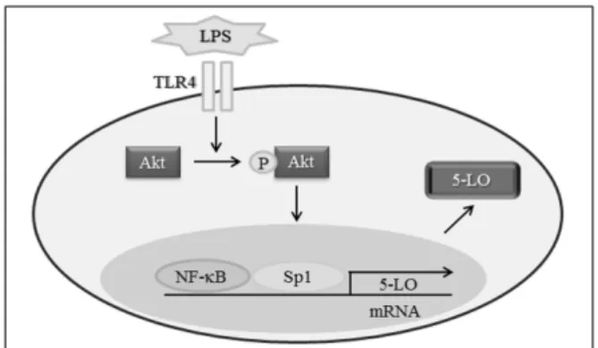
Fig. 5. Schematic diagram showing the signal pathways involved in LPS-induced 5-LO expression in Monocytes.
monocytes pretreated with an anti-TLR4 antibody. These results suggest that TLR4 on the surface of monocytes is a pivotal mediator linking LPS and Akt pathway in monocytes.
DISCUSSION
In this study, we examined molecular mechanisms that regulate LPS-induced 5-LO expression in monocytes. Our results show that LPS enhances the activities of Sp1 and NF-κB in monocytes, and that these activations increase 5-LO promoter activity and protein expression. We found that increased Sp1 and NF-κB activities and the sub- sequent expression of 5-LO by LPS were significantly atte- nuated by inhibiting the Akt pathway. Furthermore, LPS-en- hanced Akt phosphorylation was attenuated markedly by a TLR4 functional blocking antibody. These results support the hypothesis that LPS enhances 5-LO expression in mon- ocytes via the TLR4-mediated up-regulation of the activ- ities of Sp1 and NF-κB and activation of the Akt pathway.
Atherosclerosis is known to be a major contributor to car- diovascular disease [19,20]. Bacterial infection, such as lip- opolysaccharide (LPS) has also been shown to be an im- portant factor responsible for triggering atherosclerosis and the associated cardiovascular diseases [21]. LPS is a con- stituent of the cell wall of gram-negative bacteria and major cause of inflammatory response associated with bacterial infection. In fact, elevated serum LPS levels have been re- ported to increase the risk of atherosclerosis development in man [22] and in mouse models [23], and the modulatory effects of inflammatory stimuli on 5-lipoxygenase-activating protein (FLAP) gene expression in monocytes have at- tracted considerable interest [24]. To investigate these rela- tionship, we evaluated the effects of LPS on the regulation of 5-LO expression in monocytes. Although our results sug- gest that activated monocytes play an important role in atherosclerosis, the role played by 5-LO signaling in LPS-medi- ated pathophysiological processes that lead to the pro- gression of atherosclerosis has not been fully determined.
The present study provides insight of the mechanism whereby LPS modulates 5-LO gene expression in monocytes.
Previous studies have shown that LPS activates MAPKs and Akt pathways in several cell types. In the present study, the induction of 5-LO protein expression by LPS was atte- nuated by AI (an Akt inhibitor), but not by MAPK in- hibitors including PD98059 (an ERK inhibitor), SP600125 (a JNK inhibitor), or SB203580 (a p38 inhibitor), showing
that the Akt signaling pathway participates in LPS-induced 5-LO expression. In addition, it has been shown 5-LO ex- pression is regulated at the transcriptional level [25,26].
One of these studies showed that the promoter activity of LPS-stimulated p5LO-213 in monocytes was approximately 6.2 (±0.4) times higher than that in untreated controls. In our previous study [25], sequence analysis of the region be- tween nt −213 and +1 demonstrated the presence of con- sensus binding sites for Sp1 and NF-κB. This result was confirmed by observations of the reporter activities of Sp1 and NF-κB, as LPS significantly enhanced their reporter activities and 5-LO promoter activity, which showed Sp1 and NF-κB are essential transcription factors for LPS-in- duced 5-LO transcription. Furthermore, LPS-induced re- porter activities of Sp1 and NF-κB were significantly in- hibited by AI, an Akt inhibitor. Accordingly, we found that LPS-induced 5-LO expression occurs via Akt-mediated Sp1 and NF-κB pathways.
In the present study, 5-LO expression was significantly elevated in LPS-stimulated monocytes, and this elevation was associated with increased 5-LO promoter activity. The Sp1 and NF-κB transcription factors were found to be es- sential for LPS-induced 5-LO transcription, and increases in Sp1 and NF-κB activity induced by LPS were attenuated by inhibiting the Akt pathway. Furthermore, TLR4 func- tional blocking antibody attenuated Akt phosphorylation and 5-LO up-regulation in monocytes induced by LPS.
Collectively, these results suggest that LPS-induced 5-LO transcription is mediated via the TLR4 mediated activa- tions of Sp1 and NF-κB through the Akt signaling pathway (Fig. 5). This finding provides novel options for therapeutic interventions aimed at regulating 5-LO transcription in atherosclerosis.
DECLARATION OF INTEREST
The authors report no declarations of interest. The au- thors alone are responsible for the content and writing of the paper.
This study was supported by a 2-Year Research Grant of Pusan National University.
REFERENCES
1. Badimon L, Storey RF, Vilahur G. Update on lipids, inflammation and atherothrombosis. Thromb Haemost. 2011;105 Suppl 1:S34-42.
2. Hansson GK. Inflammatory mechanisms in atherosclerosis. J Thromb Haemost. 2009;7 Suppl 1:328-331.
3. Gupta H, Dai L, Datta G, Garber DW, Grenett H, Li Y, Mishra V, Palgunachari MN, Handattu S, Gianturco SH, Bradley WA, Anantharamaiah GM, White CR. Inhibition of lipopolysac- charide-induced inflammatory responses by an apolipoprotein AI mimetic peptide. Circ Res. 2005;97:236-243.
4. Kuhn AM, Tzieply N, Schmidt MV, von Knethen A, Namgaladze D, Yamamoto M, Brüne B. Antioxidant signaling via Nrf2 counteracts lipopolysaccharide-mediated inflammatory responses in foam cell macrophages. Free Radic Biol Med. 2011;50:1382- 1391.
5. Sikorski K, Chmielewski S, Przybyl L, Heemann U, Wesoly J, Baumann M, Bluyssen HA. STAT1-mediated signal integration between IFNγ and LPS leads to increased EC and SMC activation and monocyte adhesion. Am J Physiol Cell Physiol.
2011;300: C1337-1344.
6. Poeckel D, Funk CD. The 5-lipoxygenase/leukotriene pathway
2010;86:243-253.
7. Vila L. Cyclooxygenase and 5-lipoxygenase pathways in the vessel wall: role in atherosclerosis. Med Res Rev. 2004;24:399-424.
8. Mehrabian M, Allayee H. 5-lipoxygenase and atherosclerosis.
Curr Opin Lipidol. 2003;14:447-457.
9. Yonekawa K, Neidhart M, Altwegg LA, Wyss CA, Corti R, Vogl T, Grigorian M, Gay S, Lüscher TF, Maier W. Myeloid related proteins activate Toll-like receptor 4 in human acute coronary syndromes. Atherosclerosis. 2011;218:486-492.
10. Kawamoto T, Ii M, Kitazaki T, Iizawa Y, Kimura H. TAK-242 selectively suppresses Toll-like receptor 4-signaling mediated by the intracellular domain. Eur J Pharmacol. 2008;584:40-48.
11. Serio KJ, Reddy KV, Bigby TD. Lipopolysaccharide induces 5-lipoxygenase-activating protein gene expression in THP-1 cells via a NF-kappaB and C/EBP-mediated mechanism. Am J Physiol Cell Physiol. 2005;288:C1125-1133.
12. Zhao L, Moos MP, Gräbner R, Pédrono F, Fan J, Kaiser B, John N, Schmidt S, Spanbroek R, Lötzer K, Huang L, Cui J, Rader DJ, Evans JF, Habenicht AJ, Funk CD. The 5-lipoxygenase pathway promotes pathogenesis of hyperlipidemia-dependent aortic aneurysm. Nat Med. 2004;10:966-973.
13. De Caterina R, Zampolli A. From asthma to atherosclerosis-- 5-lipoxygenase, leukotrienes, and inflammation. N Engl J Med.
2004;350:4-7.
14. Jawień J. The putative role of leukotrienes in experimental atherogenesis. Pol Arch Med Wewn. 2009;119:90-93.
15. Rådmark O, Werz O, Steinhilber D, Samuelsson B. 5-Lipoxy- genase: regulation of expression and enzyme activity. Trends Biochem Sci. 2007;32:332-341.
16. Lötzer K, Funk CD, Habenicht AJ. The 5-lipoxygenase pathway in arterial wall biology and atherosclerosis. Biochim Biophys Acta. 2005;1736:30-37.
17. Yang HJ, Youn H, Seong KM, Yun YJ, Kim W, Kim YH, Lee JY, Kim CS, Jin YW, Youn B. Psoralidin, a dual inhibitor of
pulmonary inflammation. Biochem Pharmacol. 2011;82:524-534.
18. Sánchez-Galán E, Gómez-Hernández A, Vidal C, Martín- Ventura JL, Blanco-Colio LM, Muñoz-García B, Ortega L, Egido J, Tuñón J. Leukotriene B4 enhances the activity of nuclear factor- kappaB pathway through BLT1 and BLT2 receptors in atherosclerosis. Cardiovasc Res. 2009;81:216-225.
19. Hansson GK, Hermansson A. The immune system in atheros- clerosis. Nat Immunol. 2011;12:204-212.
20. Drüeke TB, Massy ZA. Atherosclerosis in CKD: differences from the general population. Nat Rev Nephrol. 2010;6:723-735.
21. Gitlin JM, Loftin CD. Cyclooxygenase-2 inhibition increases lipopolysaccharide-induced atherosclerosis in mice. Cardiovasc Res. 2009;81:400-407.
22. Szeto CC, Kwan BC, Chow KM, Lai KB, Chung KY, Leung CB, Li PK. Endotoxemia is related to systemic inflammation and atherosclerosis in peritoneal dialysis patients. Clin J Am Soc Nephrol. 2008;3:431-436.
23. Lalla E, Lamster IB, Hofmann MA, Bucciarelli L, Jerud AP, Tucker S, Lu Y, Papapanou PN, Schmidt AM. Oral infection with a periodontal pathogen accelerates early atherosclerosis in apolipoprotein E-null mice. Arterioscler Thromb Vasc Biol.
2003;23:1405-1411.
24. Serio KJ, Reddy KV, Bigby TD. Lipopolysaccharide induces 5-lipoxygenase-activating protein gene expression in THP-1 cells via a NF-kappaB and C/EBP-mediated mechanism. Am J Physiol Cell Physiol. 2005;288:C1125-1133.
25. Lee SJ, Kim CE, Seo KW, Kim CD. HNE-induced 5-LO expression is regulated by NF-{kappa}B/ERK and Sp1/p38 MAPK pathways via EGF receptor in murine macrophages. Cardiovasc Res.
2010;88:352-359.
26. Serezani CH, Lewis C, Jancar S, Peters-Golden M. Leukotriene B4 amplifies NF-κB activation in mouse macrophages by reducing SOCS1 inhibition of MyD88 expression. J Clin Invest. 2011;
121:671-682.