Vol. 20, No. 3, September, 2008 원 저
파골 전구세포에서 여러 마모입자에 의한 NF-κB와 c-Jun/AP-1 전사 인자의 활성화
김용식*∙권순용�∙한석구∙권재영∙최남용
가톨릭대학교 의과대학 강남성모병원�, 성모병원�, 성바오로병원 정형외과
목적: 파골 전구세포에서 다양한 조성과 입자의 크기를 갖는 여러 마모입자들이 NF-κB와 c-Jun/AP-1 활성화에 미치는 영향 을 관찰하고자 한다.
대상 및 방법: C57BL 쥐의 골수 대식세포로부터 파골 전구세포를 배양하여 PMMA (polymethylmethacrylate) 시멘트, polystyrene, titanium 입자 및 실패한 무시멘트형 인공 고관절 치환술 환자에서 채취한 마모입자에 대한 NF-κB와 c-Jun/AP- 1 전사 인자의 DNA binding activity를 electrophoretic mobility shift assays (EMSA)로 관찰하였다.
결과: 내독소 검사상 PMMA 입자와 polystyrene 입자는 내독소 (endotoxin) 음성이었으며, titanium 입자와 환자에서 채취한 마모입자는 양성이었다. PMMA 입자에 의해 NF-κB와 c-Jun/AP-1 활동도는 증가하였으며, 특히 자극 30분 후 0.6 mg/ml에 서 최적의 반응을 나타내었다. 또한 polystyrene, titanium 입자와 추출한 마모입자도 대조군에 비해 NF-κB와 c-Jun/AP-1의 활동도를 증가시켰으며 내독소 제거 시 약 40%의 활동도가 감소되었다.
결론: 파골 전구세포에서 다양한 조성과 크기의 PMMA 입자, titanium 입자, polystyrene 및 환자에서 추출한 마모입자 모두 파 골세포 분화 및 활성에 중요한 두 신호전달 체계인 NF-κB와 c-Jun/AP-1 전사 인자의 DNA binding activity를 증가시켜 인체 의 인공관절 주위 골용해 기전에 NF-κB와 c-Jun/AP-1 전사 인자는 중요한 역할을 할 것으로 사료된다.
색인단어: 전사 인자, NF-κB, c-Jun/AP-1, 마모 입자, 파골 전구세포, Electrophoretic mobility shift assay
서 론
마모입자에 의한 인공관절물 주위 골용해는 인공관절 전 치환술 후 장기추시상 인공관절물의 생존을 위협하는 가장 심각한 문제가 된다12). 이러한 병적 골소실(bone loss)은 인 공관절물에서 떨어져 나온 마모입자에 의해 파골세포의 생 성과 분화가 촉진되어 발생한다. 마모입자는 섬유세포, 조 골세포 및 기질세포(stromal cells)에서 파골세포의 분화를 활성화시키는 IL-1, IL-6, TNF-α등의 염증 전구 cytokine 단백질 (proinflammatory cytokines)과 RANKL (Receptor Activator of NF-κB Ligand)을 생성하여 간접적으로 파골 세포 형성(osteoclastogenesis)을 촉진시킬뿐만 아니라, 직
접적으로 파골 전구세포를 분화 및 활성화시킨다6,7,21). 최근 파골세포 발생에 관여하는 세포내 기전으로 전사 인자 NF-κ B와 c-Jun/AP-1이 중요하다고 하였다14). NF-κB 전사 인자 는 파골세포의 분화에 필수적이며, NF-κB의 활성화를 차단 하면 마모입자에 의한 파골세포 활성이 발생하지 않는다고 보고하였다5,8). AP-1 (Activator protein-1)은 dimeric complex로 구성되며 subfamily로는 Fos, Jun, ATF와 CREB 등이 있다. 이중 Fos계의 c-Fos와 Jun 계의 c-Jun 전 사 인자가 파골세포 형성에 중요하며, 실험적으로 c-Fos 또 는 c-Jun knockout mice는 파골세포를 형성하지 못해 골경 화증 (osteopetrosis)이 발생한다고 하였다28). 이에 저자들은 다양한 크기와 조성의 마모입자가 파골 전구세포의 NF-κB 와 c-Jun/AP-1 활성화에 미치는 영향을 비교 관찰하였다.
대상 및 방법
1. 마모입자
PMMA 입자 (Polysciences. Warrington, PA, U.S.A), 는 평균 6.2 μm의 직경을 갖는 구형으로 95% 이상이 10
투고일: 2008년 4월 15일 1차수정일: 2008년 5월 20일 2차수정일: 2008년 7월 14일 게재확정일: 2008년 7월 14일
※ 통신저자 : 한 석 구
서울특별시 동대문구 전농동 620-56
가톨릭대학교 의과대학 성바오로병원 정형외과 TEL: 82-2-958-2448
FAX: 82-2-965-1456
E-mail: [email protected]
μm이하의 크기였다. 이를 15분간 100% ethanol로 씻고 (x4) overnight로 담가 증발시킨 후, PBS로 씻어(x4) serum free MEM으로 resuspend하고 -20�C에 보관하였 다. Titanium 입자 (Johnson Matthey, Wayne, PA, U.S.A.)는 평균 2.1 μm 크기로 전자현미경상 모양은 구 형 , 입 방 형 또 는 불 규 칙 한 형 태 였 고 , polystyrene (Polysciences, Warrington, PA, U.S.A.)은 구형으로 평 균 직경 0.1 μm의 제품을 각각 사용하였다. 추출한 마모 입자는 8명의 마모와 골용해로 실패한 무시멘트형 고관절 재치환술 환자의 인공관절주위 조직을 Margevicius 등17) 의 방법으로 분리하여 얻었으며 Decking 등의 방법10)으 로 시행한 scanning electromicros copy (SEM)상 평균 0.87 μm 크기였고, energy dispersive X-ray anaylsis (EDX)와 (X-ray photoelectron microscopy (XPS) 검사 상 85%이상이 CoCr이었다.
2. 내독소 검사 및 제거
모든 마모입자는 Limulus Amebocyte Lysate (LAL) assay를 통해 내독소 검사를 하였고, PMMA 입자 및 polystyrene은 음성이었으나 titanium 입자와 환자에서 추출한 마모입자는 내독소 검사상 양성이었다. 내독소 제 거는 Ragab 등의 방법20)으로 제거하였다.
3. 파골 전구세포 채취
생후 4~6주의 C57BL 쥐의 골수로부터 골수 대식세포 (bone marrow macrophage)를 얻어 이미 발표된 방법7) 에 따라 파골 전구세포를 배양하였다. 약술하면 쥐의 대퇴 골 및 경골을 MEM으로 세척하여 얻은 골수세포에 M-CSF (1000 U/ml)를 넣고 37�C, 5% CO2에서 24시간 배양 후 100 mm2 세포 배양용기에 부착된 기질세포를 제거하고 부착하지 않은 세포들을 Ficoll-Hypaque gradient 용액에 넣고 원심 분리하여 monocyte/macrophage 계열의 세포 를 얻었다. 이를 α-MEM (heat inactivated 10% FBS)으로 분주한 후, M-CSF (10 ng/ml)을 넣고 37�C, 5% CO2에 배양하였다. M-CSF는 2일 간격으로 투여하면 세포들은 용기바닥에 부착하며 배양 7일째는 monocyte-specific α-napthyl acetyltransferase activity가 99%이상 보였고, M-CSF 제거시 72시간 내에 모든 세포는 사멸하였다. 이 러한 M-CSF dependent bone marrow macrophage는 배양 3일째 RANKL (Receptor Activator of NF-kB ligand)를 마모입자 투여 전 처치하고 배양 4일째 마모입 자를 투여하여 마모입자에 대한 파골 전구세포의 반응을 관찰하였다.
4. 핵 단백질 분리(Nuclear extraction)
파골 전구세포인 골수 대식세포에 마모입자를 투여 후, ice-cold PBS로 두 번 씻은 후, 5 mM EDTA와 5 mM EGTA PBS로 용기 바닥에 자란 세포들을 떼어내었다.
3000 rpm으로 10분간 4�C에서 원심 분리 후, hypotonic lysis buffer (HLB) A (10 mM HEPES, 10 mM KCL, 1.5 mM MgCl, 0.5 mM dithiothreitol, 0.5 mM AEBSF, 5 ug/ml pepstatin A, 5 ug/ml leupeptin)를 15분간 얼음 속에서 반응시켜 세포를 용해하고 10% NP-40를 넣어 0.64% 농도로 만들었다. HLB A와 nuclear extraction buffer A를 이용하여 핵과 세포질 단백질을 분리하였다.
원심 분리 후, 세포질 단백질을 제거하여 얻은 핵 단백질 pellet을 nuclear extraction buffer B (20 mM HEPES, 420 mM NaCl, 1.2 mM MgCl, 0.2 mM EDTA 25%
glycerol, 0.5 mM dithiothreitol, 0.5 mM AEBSF, 5 ug/ml pepstatin A, 5 ug/ml leupeptin)로 재분주하여 30분간 0�C에서 반응시켰다. 15000 rpm으로 5분간 원심 분리 후, 부양액을 새 튜브에 넣고 BCA kit (Pierce, Rockford, IL, U.S.A.)로 단백질 농도를 정량하였다. 모든 단백질은 EMSA 시행까지 -80�C에 보관하였다.
5. Electrophoretic mobility shift assays (EMSA)
각각의 샘플 중 10 ug의 핵단백질을 20 μl binding buffer (20 mM HEPES, 100 mM NaCl, 0.5 mM dithiothreitol, 1 μg poly dI-dC, 10% glycerol)에서 TNF promoter의 kB3 site에 서 얻은 5‘-AAA CAG GGG GCT TTC CCT CCT C-3’염기배 열의 NF-kB end-labeled double stranded oligonucleotide pobe와 c-Jun/AP-1 oligonucleotide (Santa Cruz, Santa Cruz, CA, U.S.A.)에 실온에서 60분간 반응시켰다. 이를 4�C, 4% polyacrylamide gel에서 전기 영동 후, gel을 말려 필름을 찍고 현상하였다. 현상된 필름은 Biospectrum densitometry (UVP, Upland, CA, U.S.A.)와 VisionWorks program (UVP, Upland, CA, U.S.A.)을 이용하여 정량적 분석을 하였다.
결 과
모든 마모입자를 이용한 실험 전, PMMA 입자를 이용 하여 파골 전구세포에서 NF-κB와 c-Jun/AP-1 전사 인자 의 DNA binding activity를 실험하였다. PMMA 0.6 mg/ml (4.74×10⁷particles per 15x106cells)을 파골 전 구세포에 투여 30분 후에 NF-κB (p50/p65)의 활동도는 대조군에 비해 크게 증가하였다(Fig. 1). 또한 PMMA 입 자(0.6 mg/ml)투여 후, NF-κB의 p50와 p65 subunit dimer의 DNA binding activity는 TNF 또는 RANKL과 같이 대조군에 비해 크게 증가하였으며 이는 투여된
PMMA 농도에 비례 (dose responsive)하였다. c- Jun/AP-1 전사 인자의 DNA binding activity는 TNF, RANKL 또는 NF-κB와 같이 PMMA 입자 투여 30분 후 대조군에 비해 크게 증가하였다(Fig. 2). 본 실험으로 0.6 mg/ml PMMA 입자, 1 mg/ml titanium 입자, 10 μl/ml polystyrene 및 0.2 mg/ml의 추출된 마모입자에 대한 각 각의 NF-κB의 DNA binding activity는 대조군에 비해 모두 증가하였으며, 특히 densitometry상 추출된 마모입 자는 내독소 제거 시 약 40%의 활동도가 감소하였다 (Fig. 3). 또한 c-Jun/AP-1 전사 인자의 활동도는 대조군 에 비해 추출된 마모입자, titanium 입자, PMMA 입자 및 polystyrene 모두 자극 30분 후 대조군에 비해 증가하였
고, 내독소 제거 시 c-Jun/AP-1의 DNA binding activity 는 약 40% 감소하였다(Fig. 4).
고 찰
인공관절 치환술 후 장기 추시상 인공관절 주위 골용해 는 가장 흔한 합병증으로 알려졌으며, 인공관절의 실패를 일으키는 가장 흔한 합병증인 해리(loosening)의 원인이 기도 하다11). 인공관절 주위 골용해는 관절면 또는 골-삽 입물간에서 발생하는 마모입자에 대해 반응하는 면역기 전의 결과이며, 분자생물학의 발달로 그 기전이 최근 계속 밝혀지고 있다. 특히 인체내에서 골흡수를 담당하는 파골
Fig. 1. PMMA particles (0.6 mg/ml) activate NF-κB (p50/p65) in osteoclast precursor cells for the times interval indicated.
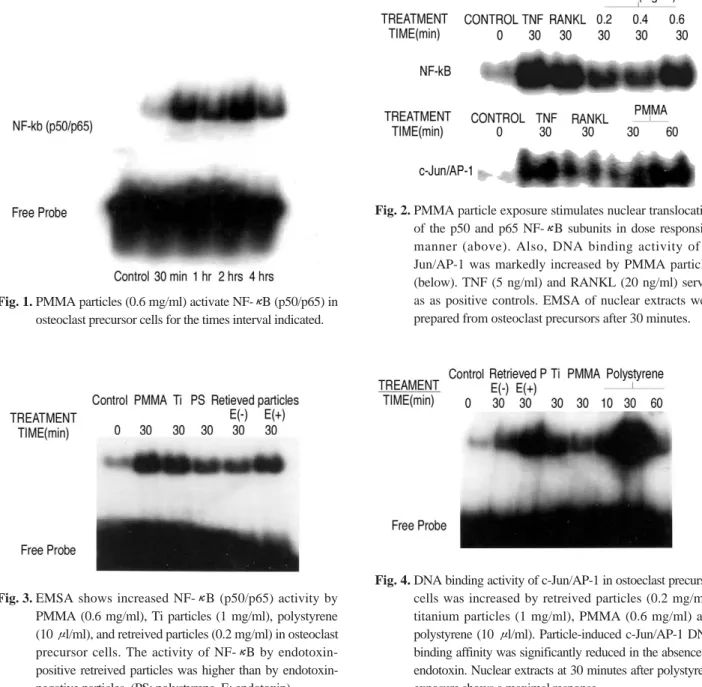
Fig. 2. PMMA particle exposure stimulates nuclear translocation of the p50 and p65 NF-κB subunits in dose responsive manner (above). Also, DNA binding activity of c- Jun/AP-1 was markedly increased by PMMA particles (below). TNF (5 ng/ml) and RANKL (20 ng/ml) served as as positive controls. EMSA of nuclear extracts were prepared from osteoclast precursors after 30 minutes.
Fig. 4. DNA binding activity of c-Jun/AP-1 in ostoeclast precursor cells was increased by retreived particles (0.2 mg/ml), titanium particles (1 mg/ml), PMMA (0.6 mg/ml) and polystyrene (10 μl/ml). Particle-induced c-Jun/AP-1 DNA binding affinity was significantly reduced in the absence of endotoxin. Nuclear extracts at 30 minutes after polystyrene exposure shows a maximal response.
Fig. 3. EMSA shows increased NF-κB (p50/p65) activity by PMMA (0.6 mg/ml), Ti particles (1 mg/ml), polystyrene (10 μl/ml), and retreived particles (0.2 mg/ml) in osteoclast precursor cells. The activity of NF-κB by endotoxin- positive retreived particles was higher than by endotoxin- negative particles. (PS: polystyrene, E: endotoxin).
세포의 분화 및 활성에 결정적인 역할을 하는 RANKL의 발견으로 골용해의 병리기전에 대한 실험적 연구는 크게 진척되었다. RANKL은 osteoprotegerin ligand (OPGL), osteoclast differentiation factor (ODF) 또는 TNF- related activation-induced cytokine (TRANCE)라고 불 리우며 TNF계의 cytokine으로 파골세포를 분화 및 활성 화시킨다. 골모세포, 기질세포 또는 T 세포가 분비하는 RANKL이 결 핍 된 실 험 쥐 는 골 경 화 증 이 발 생 하 며 , RANKL은 nuclear transcription factorκB (NF-κB)을 매개하여 파골세포를 분화시킨다13,15).
NF-κB는 TNF-α1), lipopolysacchride19) 및 마모입자2,25) 에 대해 초기에 반응하는 유전자 (early response gene)이 며, NF-κB1 (p50/p105), NF-κB2 (p52/p100), RelA (p65), RelB, c-Rel 등 Rel homology domain (RHD)을 공유하는 다섯 종류의 subunit으로 구성되어 있고 각종 면 역반응, 세포성장, 세포사멸 (apoptosis), 염증반응 뿐만 아니라 암 발생에도 관여한다고 보고되었다4,17). 세포 외 자 극이 없을 때는 세포질 내에 IκB (Inhibitor of NFκB)와 결합해 있으며, RANKL, TNF, lipopolysacchride 등의 세 포 외 자극이 있으면 IκB 인산효소의 활성화와 함께 IκB 의 tyrosine염기가 인산화되어 IκB는 26S proteosome에 의해 제거되며, 유리된 NF-κB는 세포핵 내로 이동하여 DNA와 결합하여 target gene발현에 관여한다1,19,27). 여러
보고들2,7,25)에 의하면 PMMA 입자, titanium 입자 및 추출
된 마모입자가 쥐의 골수 대식세포 또는 대식세포 세포주 (ANA-1, THP-1)에서 NF-κB를 활성화시킨다고 하였다.
인공관절의 가장 흔한 마모재질인 polyethylene12)은 섬유 세포를 분화시켜 염증 전구단백질을 생성하고23), 골모세 포에서 RANKL을 유리시켜 파골세포 형성을 촉진시킨다 고 알려졌다11).
하지만 polyethylene의 비중은 배양액보다 월등히 적어 세포배양액 위에 부양(floating)하며 세포 배양용기 바닥 에 서 자 라 는 대 식 세 포 와 접 촉 하 지 어 렵 기 때 문 에 polyethylene 마모입자를 이용한 실험은 근본적으로 한 계가 있다. 따라서 본 저자들은 polyethylene과 조성이 비 슷 하 지 만 보 다 비 중 이 크 며 액 상 형 태 로 사 용 되 는 polystyrene을 실험에 사용하였다. 또한 마모입자에 의한 NF-κB의 활성화를 증명하기 위해 여러 in vivo5) 및 in vitro 실험8)에서 약제를 이용한 NF-κB의 활성화 차단으 로 마모입자에 대한 파골세포의 분화 억제가 증명되었다.
본 연구에서는 NF-κB p50와 p65의 DNA binding activity를 관찰하여 그 활동도가 여러 종류의 마모입자에 의해 증가된 것으로 밝혀졌다. 여러 NF-κB subunit 중 p50와 p65를 관찰한 이유는 이미 밝혀진대로 titanium 입 자21)와 PMMA 입자7)에 의한 p50와 p65 subunit의 활성화 가 확인되었고, Soloviev 등25)은 titanium을 ANA-1세포에 투여하여 p105를 관찰하였는데 p105는 p50과 p65가 결
합된 전구 단백질이기 때문이다. 그러나 마모입자에 따른 다른 subunit을 포함한 subunit간의 활동도 차이에 대해 서는 아직 보고된 바 없으며, 각 subunit의 기능적 역할 차 이에 대해서는 향후 연구가 필요하다고 생각된다.
전사 인자 복합체 AP-1은 Fos, Jun, ATF와 CREB으로 구성되며 세포의 증식, 분화 및 사멸 등에 광범위하게 관 여하는 단백질이다 . NF-κB와 마찬가지로 dimer형태로 존재하며, 이 중 Jun (c-Jun, Jun B, Jun D) 단백질과 Fos (c-Fos, Fos B, Fra-1, Fra-2) 단백질이 파골세포의 분화 과정에 중요하다고 보고되었다14).
Growth factor, cytokines, polypeptide hormone, 세 균감염과 RANKL 등의 자극에 의해 MAPK (mitogen activated protein kinase)는 활성화되고 c-Jun은 핵 내로 이동하여 유전자 단백질과 결합한다9,24). c-Jun 또는 Fos가 knockout된 쥐는 파골세포를 형성하지 못해 골경화가 일 어나며, 최근 Ikeda 등14)에 의하면 RANKL-TRAF6- MAPKK7-JNK1을 통해 c-Jun이 Fos와 결합하고 이는 NFAT (nuclear factor of activated T cells)를 활성화시 킨다고 알려졌다. 본 실험에서는 여러 종류의 마모입자에 의해 NF-κB 뿐만 아니라 c-Jun/ AP-1도 활성화되어 두 신호전달체계가 마모입자에 의한 파골세포 분화과정에 관여함을 알 수 있었다.
Shanbhag 등22)은 마모입자의 크기, 조성과 표면적에 따라 대식세포의 IL-1, PGE2분비와 골흡수 효과가 다르 다고 보고하였다. 본 실험에서 내독소 포함 유무에 따른 추출된 마모입자외 다른 여러 마모입자간의 NF-κB와 c- Jun/AP-1 활성화의 정량적 차이를 비교하지 않았으며, 이 는 사용된 마모입자의 용량이 달라 활성 능력의 차이를 비 교가 불가능하기 때문이었다.
Titanium 입자와 환자에서 추출한 마모입자는 내독소 검사상 양성을 나타내었는데, 내독소는 자체만으로도 파 골 세포를 분화 및 활성화시킨다26). 인공관절 주위 골용해 에서 내독소의 역할에 대해서는 아직 확실하게 밝혀지지 않았으나, Cho 등6)은 내독소의 마모입자 부착은 대식세포 의 TNF-α, IL-1β와 IL-1α의 생성을 크게 증가시킨다고 하 였고, 본 실험에서 추출한 마모입자의 내독소 양성 소견과 이를 제거 시 NF-κB와 c-Jun/AP-1의 생물학적 활성도가 약 40% 감소하였다는 결과는 의미있는 것으로 볼 수 있 다. 다만, 인공관절 주위 골용해의 생물학적 발생기전을 밝히기 위해 NF-κB와 c-Jun/AP-1 이외의 다른 경로를 통 한 마모입자의 파골세포 분화과정과 최근 밝혀진 NFAT의 역할에 대해서는 추후 연구가 필요할 것으로 사료된다.
결 론
다양한 조성과 크기의 여러 마모입자들은 파골 전구세포 에서 NF-κB와 c-Jun/AP-1 신호전달체계(signaling
pathway)를 활성화시켰으며, 특히 실패한 인공관절 환자에 서 추출된 마모입자도 두 전사 인자의 DNA binding activity 를 증가시키는 것으로 나타나, 마모입자에 의한 인체의 인공 관절 주위 골용해 발생기전에 NF-κB와 c-Jun/AP-1 신호전 달체계는 중요한 역할을 할 것으로 사료된다.
REFERENCES
01) Abu-Amer Y, Ross FP, McHugh KP, Livolsi A, Peyron JF and Teitelbaum SL:Tumor necrosis factor-αactivation of Nuclear transcription factor-κB in marrow macrophages is mediated by c-src tyrosine phosphorylation of IκBα. J Biol Chem, 273: 29417-29423, 1998.
02) Akisue T, Bauer TW, Farver CF and Mochida Y: The effect of particle wear devris on NF-κB activation and pro-inflammatory cytokine release in differentiated THP-1 cells. J Biomed Mater Res, 59: 507-515, 2002.
03) Bi Y, Motter RV, Ragab AA, Goldring VM, Anderson JM and Greenfield EM: Titanium particles stimulate bone resorption by inducing differentiation of murine osteoclasts. J Bone Joint Surg, 83A: 501-508, 2001.
04) Chen F, Castranova V and Shi X: New insights into the role of nuclear factor-κB in cell growth regulation. Am J Pathol, 159: 387-397, 2001.
05) Childs LM, Paschalis EP, Xing L, et al.: In vivo RANK signaling blockade using the receptor activator of NF-κB:Fc effectively prevents and ameliorates wear debris-induced osteolysis via osteoclast depletion without inhibiting osteogenesis. J Bone Miner Res, 17: 192-199, 2002.
06) Cho DR, Shanbhag AS, Hong CY, Baran GR and Goldring SR: The role of absorbed endotoxin in particle- induced stimulation of cytokine release. J Orthop Res, 20:
704-713, 2002.
07) Clohisy JC, Teitelbaum S, Chen S, Erdmann JM and Abu-Amer Y: Tumor necrosis factor-α mediates polymethylmethacrylate particle-induced NF-κB activation in osteoclast precursor cells. J Orthop Res, 20:
174-181, 2002.
08) Clohisy JC, Hirayama T, Frazier E, Han SK and Abu- Amer Y: NF-κB signaling blockade abolishes implant particle-induced osteoclastogenesis. J Orthop Res, 22: 13- 20, 2004.
09) Curran T and Franza BRJ: Fos and Jun. The AP-1 connection. Cell, 55: 395-397, 1988.
10) Decking R, Reuter P, Hutter M, Puhl W, Claes E and Scharf P: Surface composition analysis of failed cementless CoCr- and Ti-base-alloy total hip implants. J Biomed Mater Res, 64B: 99-106, 2003.
11) Granchi D, Ciapetti G, Amato I et al.: The influence of alumina and ultra-high molecular weight polyethylene particles on osteoblast-osteoclast cooperation. Biomaterials, 25: 4037-4045, 2004.
12) Harris WH: The problem is osteolysis. Clin Orthop, 311:
46-53, 1995.
13) Howling GI, Barnett PI, Tipper JL, Stone MH, Fisher J and Ingham E: Quantitative characterization of polyethylene debris isolated from periprosthetic tissue in early failure knee implants and early and late failure Chanley hip implants. J Biomed Mater Res, 58: 415-420, 2001.
14) Ikeda F, Nishimura R, Matsubara T, et al.: Critical roles of c-Jun signaling in regulation of NFAT family and RANKL-regulated osteoclast differentiation. J Clin Invest, 114: 475-484, 2004.
15) Jacobs JJ, Roebuck KA, Archibeck MA, Hallab NJ and Glant TT: Osteolysis. Basic science. Clin Orthop, 393: 71-77, 2001.
16) Lacey DL, Timms E, Tan HL, et al.: Osteoprotegerin ligand is a cytokine that regulates osteoclast differentiation and activation. Cell, 93: 165-176, 1998.
17) Margevicius KJ, Bauer TW, McMahon JT, Brown SA and Merritt K: Isolation and characterization of debris in membranes around total joint prostheses. J Bone Joint Surg, 76A: 1664-1675, 1994.
18) Mercurio F and Manning AM: Multiple signals converging on NF-κB. Curr Opin Cell Biol, 11: 226-232, 1999.
19) Mordmuller B, Krappmann D, Wegener E and Scheidereit C: Lymphtoxin and lipopolysaccharide induce NF-κB p52 generation by a co-translational mechanism.
EMBO, 4: 82-97, 2003.
20) Ragab AA, Motter RV, Lavish SA, et al.: Measurement and removal of adherent endotoxin from titanium particles and implant surfaces. J Orthop Res, 17: 803-809, 1999.
21) Schwartz EM, Lu AP, Goater J, et al.: Tumor necrosis factor-α/Nuclear transcription factor-κB signaling in periprosthetic osteolysis. J Orthop Res, 18: 472-480, 2000.
22) Shanbhag AS, Jacobs JJ, Black J, Galante JO and Glant TT: Macrophage/particle interactions: Effect of size, composition and surface area. J Biomed Mater Res, 28: 81-90, 1994.
23) Shanbhag AS, Jacobs JJ, Black J, Galante JO and Glant TT: Effects of particles on fibroblast proliferation and bone resorption in vitro. Clin Orthop, 342: 205-217, 1997.
24) Shaulian E and Karin M: AP-1 as a regulator of cell life and death. Nature Cell Biol, 4: E131-136, 2002.
25) Soloviev A, Schwartz EM, Kuprash DV, et al.: The role of p105 protein in NF-κB activation in ANA-1 murine macrophages following stimulation with titanium particles. J Orthop Res, 20: 714-722, 2002.
26) Suda K, Woo JT, Takami M, Sexton PM and Nagai K:
Lipopolysaccharide supports survival and fusion of preosteoclasts independent of TNF-α, IL-1, and RANKL J Cell Physiol, 190: 101-108, 2002.
27) Verma IM, Stevenson JK, Schwarz EM, Antwerp DV and Miyamoto S: Rel/NF-κB/IκB family. Initmate tales of association and dissociation. Genes Deveolp, 9: 2723- 2735, 1995.
28) Wagner EF: Functions of AP1 (Fos/Jun) in bone development. Ann Rhem Dis, 61: 40-42, 2002.
Activation of Transcription Factor NF-κ κB and c-Jun/AP-1 by Various Compositions of Particles in Osteoclast Precursor Cells
Yong Sik Kim, M.D.*, Soon Yong Kwon, M.D.��, Suk Ku Han, M.D., Jae Young Kwon, M.D., and Nam Yong Choi, M.D.
Department of Orthopaedic surgery, Kangnam St. Mary’s Hospital*, St. Mary’s Hospital�, St. Paul’s Hospital, The Catholic university of Korea, Seoul, Korea
Purpose: This study was performed in order to investigate the effects of various particle preparations on NF-κB and c-Jun/AP-1 activity in osteoclast precursor cells.
Materials and Methods: Osteoclast precursor cells isolated from C57BL mice were treated with PMMA (polymethylmethacrylate) spheres, polystyrene, titanium particles, and retrieved metal particles from failed cementless total hip replacements. NF-κB and c-Jun/AP-1 DNA binding activities were analyzed using electrophoretic mobility shift assays (EMSA).
Results: Commercially available PMMA and polystyrene spheres routinely showed negativity on endotoxin assays, but titanium particles and retrieved metal particles consistently showed positivity. PMMA spheres, with a maximal response noted at 30 minutes with an optimal concentration of 0.6 mg/ml, were potent stimulator of NF-κB and c-Jun/AP-1 activity in osteoclast precursor cells. Other particles (polystyrene, titanium, metal retrievals) also activated transcription factor NF-κB and c-Jun/AP-1 compared to controls. Endotoxin removal from retrieved metal particles diminished the biologic effect by approximately 40%.
Conclusion: Particles of various compositions and sizes (PMMA, polystyrene, titanium, and retrieved metal particles) activated the NF-κB and c-Jun/AP-1 signaling pathways. This suggests that NF-κB and c-Jun/AP-1 may have important roles in the pathogenesis of periprosthetic osteolysis.
Key Words: Transcription factor, NF-κB, c-Jun/AP-1, Particle, Osteoclast precursor cell, Electrophoretic mobility shift assay A
ABBSSTTRRAACCTT