강화사자발쑥의 마크로파지 RAW 264.7 세포에 대한 Tumor Necrosis Factor -α, Prostaglandin E 2, Cyclooxygenase-2 및 LPS 유도 Nitric Oxide 생성 저해
윤준용*·최세영*·박표잠*·정해곤**·신흥묵***·석경호****·임병우*†
*건국대학교 의료생명대학 생명과학부, **강화농업기술연구센터,
***동국대학교 한의과대학 생리학 교실, ****경북대학교 의과대학 약리학교실
Extracts of Artemisia princeps Pampanini Inhibit Lipopolysaccharide-induced Nitric Oxide, Cyclooxygenase-2, Prostaglandin E 2 , and Tumor Necrosis Factor- α
Production from Murine Macrophage RAW 264.7 Cells
Jun-Yong Yun
*, Se-Yong Choi
*, Pyo Jam Park
*, Hae Gon Chung
**, Heung Mook Shin
***, Kyoungho Suk
****, and Beong Ou Lim
*†*College of Biomedical & Health Science, Department of Life Science, Konkuk University, Chungju 380-701, Korea.
**Ganghwa Agricultural Development & Technology Center, Department of Physiology
***College of Oriental Medicine, Dongguk University, Kyongju 780-714, Korea.
****Department of Pharmacology, Kyungpook National University School of Medicine, Daegu, Korea.
ABSTRACT : To search for immunoactive natural products exerting anti-inflammatory activity, we have evaluated the effects on the water extracts of Artemisia princeps Pampanini (APP) on lipopolysaccharide-induced nitric oxide (NO), tumor necrosis factor- α (TNF- α ), and prostaglandin E
2(PGE
2) production by RAW 264.7 macrophage cell line. Our data indicate that this extract is a potent inhibitor of NO production and it also significantly decreased PGE
2and TNF- α production. Con- sistent with these results, the protein and mRNA expression level of inducible NO synthase (iNOS) and cyclooxygenase-2 (COX-2) was inhibited by water extracts of APP in a dose-dependent manner. These results suggest that APP may exert anti-inflammatory and analgesic effects possibly by suppressing the inducible NO synthase and COX-2 expressions.
Key Words : Artemisia princeps Pampanini, Nitric Oxide, iNOS, TNF- α , IFN- γ , PGE 2
INTRODUCTION
Various in vivo and in vitro experimental models have been set up to assess inhibitory effects of natural products on the inflammatory mediators. Among these, RAW 264.7 mouse macrophage cells are an excellent model for the assessment of pro-inflammatory cytokines and reactive free radical mediators such as tumor necrosis factor (TNF- α ), inducible NO synthase (iNOS), and NO (Shin et al ., 2004; Jang et al ., 2004; Kwon et al ., 2007; Kwon et al ., 2008). These macrophage-derived inflammatory mediators are also reported to be involved in the development of inflammatory diseases (Freeman & Natanson, 2000).
Therefore, the inhibition of the excessive production of
TNF- α and/ or NO can be a critical point to evaluate anti-inflammatory effects of natural products (Pae et al., 2003).
The most conclusive evidence for NO as a mediator of tissue injury has been obtained from studies on an animal arthritis model, human osteoarthritism, and rheumatoid arthritis (Cochran et al ., 1996). In contrast to iNOS, the constitutive epithelial and neuronal forms of NOS are known to contribute relatively little to inflammation and carcinogenesis. Cyclooxygenase (COX) is the enzyme that converts arachidonic acid to prostaglandins (PGs). Like NOS, COX has been found in two isoforms, and COX-2 is an inducible form responsible for the production of large amounts of proinflammatory PGs at the inflammatory
†
Corresponding author: (Phone) +82-43-840-3570 (E-mail) [email protected]
Received July 23, 2008 / Revised September 2 / Accepted October 10, 2008
site (Weisz et al ., 1996). Furthermore, TNF- α is a potent proinflammatory cytokine that plays an important role in immunity and inflammation.
The genus Artemisia consists of more than 300 species, many of which have medicinal value. Among these plants, Artemisia princeps Pampanini (APP) has been used in traditional Oriental medicine for the treatment of microbial infections and inflammatory diseases. Recent studies revealed that the ethanol extract of Artemisia asiatica has anti-oxidative and anti-inflammatory activities, which contribute to its protective effects against gastric damage (Oh et al ., 2005), liver damage (Ryu et al ., 1998), experimental pancreatitis (Hahm et al., 1998), and tumor promotion (Seo et al ., 2002). Also, Genus Artemisia has been shown to exert the bactericidal activity in traditional medicine and proven to inhibit the cell growth (Lee et al ., 1996). But there are no data in the literature about anti-inflammatory effects. Thus, as a prelude to determining the underlying mechanisms of the anti-inflammatory effect of APP, lipopolysaccharide (LPS)-induced NO and PGE
2release, TNF- α production, and iNOS/COX-2 enzyme and mRNA expression levels in the macrophage cell line RAW 264.7 have been investigated. Considering the use of APP as an anti-inflammatory drug in the folk medicine, we evaluated the effects of distilled water extract of the APP on the production of TNF- α , NO, and PGE
2in RAW 264.7 cells which were stimulated by LPS. The effects of APP on the expressions of TNF- α , iNOS, and COX-2 genes in LPS-activated RAW 264.7 cells have been also studied to investigate the possible mechanisms of action in these studies.
MATERIAL AND METHODS
1. Preparation of the plant extract
Artemisia princeps Pampanini used in this study was collected at Ganghwa, Kyungki province, Korea, in August of 2004 . The air dried and pulverized Artemisia princeps Pampanini (20 g) was extracted with phosphate buffered saline for 3 hr. The extract was filtered, and the filtrate was concentrated under reduced pressure.
2. Reagents
LPS was obtained from Sigma Chemical Co. (St Louis, MO), DMEM medium, and 3-(4,5 dimethythiazol2-yl)-2,5-
diphenytetrazoleum (MTT) were obtained from Wako. Fetal bovine serum (FBS), oligo (dT) 18 primers, AMV reverse transcriptase, dNTP mixture, RNA inhibitor, Taq DNA polymerase were purchased from TaKaRa. The antibiotics were from Gibco-BRL (Rockville, MD). The pairs of polymerase chain reaction oligonucleotide primers were synthesized by Bioneer Co. (Korea).
3. RAW 264.7 cell line and sample treatment
The murine macrophage cell line (RAW 264.7) was obtained from the ATCC (Manassas, VA). The cells were cultured in 10
2㎜ dish (Falcon-Becton Dickinson Labwares, Franklin Lakes, NJ) and maintained in 37 ℃ DMEM containing 10% heat-inactivated FBS, penicillin (100 units/ ㎖ ), and streptomycin sulfate (100 units/ ㎖ ) in a humidified atmosphere of 5% CO
2. The extract was dissolved in PBS and applied to the cell cultures at final concentrations of 25, 50, 75 and 100 ㎍ / ㎖ alone or with 1 ㎍ / ㎖ of LPS.
4. Assessment of cell viability
Cytotoxicity studies were performed in 96-well plates.
RAW 264.7 cells were mechanically scraped and plated at 2 × 10
5/well in 96-well plates containing 100 ㎕ of DMEM with 10% heat-inactivated FBS and incubated overnight.
APP was dissolved in PBS. After overnight incubation, the test material was added, and the plates were incubated for 24 h. Cells were washed once before adding 50 ㎕ of FBS-free medium containing 5 ㎎ / ㎖ of MTT. After 4 h of incubation at 37 ℃ , the medium was discarded and the formazan blue that formed in the cells was dissolved in 100 ㎕ of DMSO. The optical density was measured at 540 ㎚ .
5. Nitric Oxide determination
The nitrite accumulated in culture medium was measured as an indicator of nitric oxide
(NO) production based on the Griess reaction. Briefly,
100 ㎕ of cell culture medium was mixed with 100 ㎕ of
Griess reagent [equal volumes of 1% (w/v) sulfanilamide
in 5% (v/v) phosphoric acid and 0.1% (w/v) naphtylethy-
lenediamine-HCl], incubated at room temperature for 10
min, and then the absorbance at 550 ㎚ was measured in
a microplate reader. Fresh culture medium was used as
the blank in all experiments. The amount of nitrite in the
samples was measured with the sodium nitrite serial dilution standard curve.
6. PGE
2assay
PGE
2levels in macrophage culture medium were quantified using EIA kits according to the manufacturer’s instructions (Cayman, USA).
7. RNA isolation and RT-PCR
To determine the expressions of iNOS and COX-2 mRNAs, RT-PCR was performed. Total RNA was isolated from RAW 264.7 cells using RNAzol
TMB (TEL-TEST, Friendswood, TX, USA). Two micrograms of RNA and 0.5 ㎕ of random 9 mers (TAKARA BIO INC, Japan), 2
㎕ of 25 mM MgCl
2, 1 ㎕ of 10X RT buffer, 1 ㎕ of 10 mM dNTP mixture, 0.25 ㎕ of AMV reverse transcriptase (1 unit/ ㎕ ) (TAKARA BIO INC, Japan) were added to the reaction mixture. And the final volume was brought up to 10 ㎕ with diethyl pyrocarbonate (DEPC)-treated water.
The reaction mixture was then incubated at 42 ℃ for 30 min. Then PCR analyses were performed on the aliquots of the cDNA preparations to detect iNOS, COX-2, TNF- α
and β -actin (as an internal standard) gene expression using a thermal cycler (TAKARA BIO INC, Japan). The reactions were carried out in a volume of 10 ㎕ containing 0.25 ㎕
of Taq DNA polymerase (1.25 units/50 ㎕ ) (TAKARA BIO INC, Japan), and 0.5 ㎕ of 5' and 3' primers (0.2 µ M). After initial denaturation for 2 min at 95, 30 amplification cycles were performed for iNOS (1 min of 95 ℃ denaturation, 1 min of 60 ℃ annealing, 1.5 min 72 ℃
extension), COX-2 (1 min of 94 ℃ denaturation, 1 min of 60 ℃ annealing, and 1 min of 72 ℃ extension), and TNF- α
(1 min 95 ℃ denaturation, 1 min 55 ℃ annealing, 1 min 72 ℃ extension). PCR primers used in this study are listed below and were purchased from Bioneer (Seoul, Korea):
TNF- α 5-'GCGACGTGGAACTGGCCAGAAG-3' (5'-primer) 5'-TCCATGCCGTTGGCCAGGAGG-3' (3'-primer); iNOS 5'- CCTTGTTCAGCTACGCCTTC-3' (5'-primer) 5'-CTGAGGGC TCTGTTGAGGT-3' (3'-primer); COX-2 5'-TGCATGTGGCTG TGGATGTCAT-3' (5'-primer), 5'-CACTAAGACACCCGTC ATCTCCA-3' (3'-primer); β -actin 5'-TACAGGCTTGTCAC TCGAANTT-3', 5' CCTAGAANGCATTTGCGGTGCACGA TG-3'. After amplification, portions of the PCR reactions were electrophoresed on 2% agarose gel and visualized by ethidium bromide staining and UV irradiation.
8. Western blot analysis
Cellular proteins were extracted from control and Artemisia princeps Pampanini -treated RAW264.7 cells.
Cells were collected by centrifugation and washed once with phosphate buffered saline. The washed cell pellets were resuspended in extraction lysis buffer (50 mM HEPES (pH 7.0), 250 mM NaCl, 5 mM EDTA, 0.1%
Nonidet P-40, 1 mM PMSF, 0.5 mM dithiothreitol (DTT), 5mM Sodium fluoride (NaF), 0.5mM Sodiium orthovanadate) containing 5 ㎍ / ㎖ each of leupeptin and aprotinin and incubated for 30 min at 4 ℃ . Cell debris was removed by microcentrifugation, followed by quick freezing of the supernatants. The protein concentration was determined using the Bio-Rad protein assay reagent according to the manufacturer’s instruction. Fifty micrograms of cellular protein from treated and untreated cell extracts electroblotted onto a nitrocellulose membrane following separation on 8- 12% SDS-polyacrylamide gel electrophoresis. The immu- noblot was incubated overnight with blocking solution (5%
skim milk) at 4 ℃ , followed by incubation for 1 h with a 1:500 dilution of polyclonal antibodies against iNOS, COX-2 and TNF- α (Santa Cruz Biotechnology Inc.). Blots were washed two times with Tween20/Tris-buffered saline (TTBS) and incubated with a 1 : 1000 dilution of horseradish peroxidase conjugated goat-anti rabbit IgG secondary antibody (Santa Cruz Biotechnology Inc.) for 1 h at room temperature. Blots were again washed three times with TTBS and then developed by enhanced chemiluminescence (Amersham Life Science, Arlington Heights, IL, USA).
9. Statistical analysis
Data are reported as mean ± S.D. values of three independent determinations. All experiments were performed at least three times, each time with three or more independent observations. Statistical analysis was performed using Student’s t-test with one-way analysis of variance.
RESULTS
1. Effects of Artemisia princeps Pampanini (APP) on LPS-Induced NO and PGE
2production
Treatment of RAW264.7 macrophages with APP alone
did not show any cytotoxicity (data not shown). The
endotoxin LPS at 1 ㎍ / ㎖ reduced the viability of
RAW264.7 macrophages by 22.24%. APP in the presence of LPS did not further affect the viability of RAW264.7 cells. To examine the effects of APP on the NO and PGE
2production in RAW264.7 cells, cell culture medium was harvested, and the production of nitrite or PGE
2was measured using the method of Griess reaction or EIA, respectively. APP reduced the LPS-induced NO and PGE
2production in a dose-dependent manner (Fig. 1).
2. Effects of Artemisia princeps Pampanini in LPS- induced TNF- α protein and mRNA expression
To investigate whether the APP could inhibit LPS-
stimulated TNF- α mRNA and protein expression, cells were preincubated with APP for 1 h, and then treated with 1 ㎍ / ㎖ of LPS for 4 h (for TNF- α mRNA) or 24 h (for TNF- α protein). Pretreatment of cells with APP decreased the LPS-induced mRNA expression and production of TNF- α in a dose-dependent manner (Fig. 2).
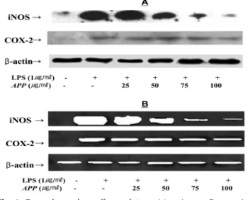
3. Effects of Artemisia princeps Pampanini on the iNOS and COX-2 expression
RT-PCR (Fig. 3A) or Western blot analysis (Fig. 3B) was conducted to determine whether the inhibitory effects of APP on NO and PGE
2production are related to the modulation of iNOS and COX-2 expression. In unstimulated RAW 264.7 cells, iNOS and COX-2 mRNA was not detectable. In response to LPS, the expression levels of iNOS and COX-2 were markedly increased.
Pretreatment of RAW 264.7 cells with APP significantly decreased iNOS and COX-2 mRNA expression in a dose- dependent manner (Fig. 3A). A similar inhibitory effect of APP was observed on the LPS-induced iNOS and COX-2
Fig. 1.
Dose dependent effects of Artemisia princeps Pampanini (APP) on NO and PGE
2production in RAW267.4 cells.
The cells were left untreated or treated with LPS (1
㎍/
㎖) and APP (25, 50, 75, 100
㎍/
㎖) for 24 h, and the production of NO (
A) and PGE
2(
B) was evaluated by Griess reaction and EIA, respectively. L -N
6-(1-iminoethyl) lysine ( L -NIL) (10
µM) or NS- 398 (10
µM) was used in the assay as a positive control (data not shown). The values are the mean ± S.D. from three independent experiments. * p < 0.05, ** p < 0.005 vs . LPS- treated group; significance of difference between treated groups in Student’s t -test.
Fig. 2.
Dose dependent effects of Artemisia princeps Pampanini on LPS-induced TNF-
αprotein and mRNA expression in RAW 264.7 cells.
(
A) Lysates were prepared from control or stimulated cells treated
for 24 h with LPS (1
㎍/
㎖) alone or in combination with
increasing concentrations (25, 50, 75, 100
㎍/
㎖) of APP. All
lanes contained 50
㎍of total proteins. The experiment was
repeated three times with similar results. (
B) Total RNA was
prepared for the RT-PCR analysis of gene expression. The cDNAs
(10 ng) of RAW 264.7 macrophages either untreated, stimulated
with LPS (1
㎍/
㎖), or co-treated with LPS (1
㎍/
㎖) and various
concentrations (25, 50, 75, 100
㎍/
㎖) of APP for 4 h were
subjected to PCR. TNF-
αspecific sequences (351 bp) were
detected by agarose gel electrophoresis. PCR of
β-actin was
performed to control the initial cDNA content of samples. The
experiment was repeated three times with similar results.
protein levels (Fig. 3B). The expression of housekeeping gene β -actin was not affected by APP. These results are consistent with the profile of the inhibitory effect of the APP on NO, PGE
2, and TNF- α production (Fig. 1 and 2).
DISCUSSION
Because the mechanism of the anti-inflammatory effects of APP has not been reported, we examined the effects of APP on the release of inflammatory mediators (NO, PGE
2, and TNF- α ) and the expression levels of iNOS and COX-2 in LPS-activated macrophages. The results of the present study indicate that APP inhibits the induction of iNOS in LPS-activated murine macrophages at the transcriptional level and NO production was reduced by APP in a concentration-dependent manner. Also, iNOS protein levels in the LPS-activated cells were markedly reduced by APP as compared with untreated cells. NO is considered as a proinflammatory product of iNOS. From these results, it is thought that APP inhibits the NO production and it may also repress the iNOS induction, which mediates inflammatory processes. NO synthase inhibitors used for the treatment of NO-mediated inflam- matory processes require high specificity for iNOS, and thus inhibitors of iNOS induction may function as
relatively safe modulators of NO for various pathologic conditions (Clancy & Abramson, 1995).
The mechanisms of various anti-inflammatory drug actions are related with the induction of PG synthesis, induced by COX (Vane, 1971). Among COX group, COX-2 is considered to be responsible for proinflammatory PG production (Seibert et al ., 1994: Masferer, 1994). In our results, APP significantly inhibited PGE
2production and COX-2 expression at mRNA as well as protein levels. From these results, it is suggested that APP may suppress the proinflammatory effect of PGE
2through inhibition of COX-2 expression.
In the resting murine macrophages, TNF- α expression is not detected at the transcription or translation levels (Park et al ., 1995). Inflammatory stimulus like LPS strongly induces both the transcription and the translation of TNF- α . Now, our current results indicate that APP may interfere with the transcriptional and/or translational activation of TNF- α gene, which results in the suppression of intracellular TNF- α
synthesis. These results clearly demonstrated the anti- inflammatory effects of APP as well as the possibility of therapeutic use of APP. However, further studies on the precise mechanism of action and the isolation/characterization of active chemical constituents are needed.
CONCLUSION
Considering the use of Artemisia princeps Pampanini (APP) as an anti-inflammatory drug in the folk medicine, we evaluated the effects of extract of the APP on the production of TNF- α , NO, iNOS, COX-2 and PGE
2in RAW 264.7 cells which were stimulated by LPS. Results show that the water extract of APP inhibits the production of NO, PGE
2, and TNF- α in the LPS-stimulated RAW 264.7 macrophages at least in part by interfering with the transcription and/or translation of iNOS, COX-2, and TNF- α genes.
ACKNOWLEDGEMENTS
This paper was supported by Konkuk University in 2005.
LITERATURE CITED
Chang SH, Mun SH, Ko NY, Lee JH, Jun MH, Seo JY, Kim
Fig. 3.