Korean J. Environ. Biol. 33(2) : 189~196(2015) http://dx.doi.org/10.11626/KJEB.2015.33.2.189
INTRODUCTION
To date, more than 240 ciliate species (including parasitic ciliates), mainly belonging to the class Spirotrichea, have been reported from Korea (The Korean Society of Systema
tic Zoology 1997; Kwon 2009; Jung et al. 2012; Shin 2012;
Jung 2013; Park and Min 2014; Lee et al. 2015; Nam et al.
2015 etc).
Most species of dysteriid ciliates (Ciliophora: Phyllopha
ryngea) are found in marine benthic habitats. Dysteria spe
cies are characterized by a laterally flattened body shape, oral structure with conspicuous cytopharynx, and restricted ciliature in the anterior and ventral regions (Kahl 1931;
Deroux 1965; Dragesco 1966; Wilbert 1971; Song and Wil
bert 2002; Chen et al. 2011). To date, more than 30 Dyste- ria morphospecies have been investigated (FauréFremiet 1965; Petz et al. 1995; Gong et al. 2002, 2003, 2007; Song and Wilbert 2002; Gong and Song 2003, 2004; Hu and Su
zuki 2005; Wilbert and Song 2005; Chen et al. 2011; Pan et al. 2011; Park and Min 2014). However, only one species
Dysteria nabia Park & Min, 2014 has previously been recorded in Korea.
MATERIALS AND METHODS Sample collection, observation, and identification The dysteriid ciliates were collected by filtering the wash
ed seawaters of mussels and seaweeds that were sampled from harbors. The obtained samples were maintained in Pe
tri dishes at room temperature. We observed the specimens by using brightfield and differential interference contrast microscopy, at magnifications of ×50 to ×1000. We used the protocol of Foissner (1991) to reveal the infraciliature.
We used the ChattonLwoff method to reveal the silverline systems for the three dysteriids (Corliss 1953). We counted and measured the stained specimens at a magnification of
× 1000 (DM2500; Leica, Wetzlar, Germany). Terminology follows that of Corliss (1979), Gong and Song (2004), and Chen et al. (2011).
All specimens were deposited in the National Institute of Biological Resources (NIBR), Korea.
RESULTS AND DISCUSSION Class Phyllopharyngea de Puytorac et al., 1974 Order Dysteriida Deroux, 1970
* Corresponding author: GiSik Min, Tel. 0328607692, Fax. 0328746737, Email. [email protected]
ⓒ2015. Korean Society of Environmental Biology.
New Records of Three Dysteriids (Ciliophora: Pyllopharyngea) from Korea
MiHyun Park and GiSik Min*
Department of Biological Sciences, Inha University, Incheon 402-751, Korea
Abstract - We verified three ciliates -
Dysteria brasiliensis Faria et al., 1922, Dysteria lanceolataClaparède and Lachmann, 1859, and Dysteria ovalis (Gourret and Roeser, 1886) Kahl, 1931 - as previously unrecorded species in Korea. These three ciliates were collected from Incheon Harbor and Geojin Harbor in Korea. We describe the three species based on live observations, protargol- impregnated specimens, and silver nitrate-impregnated specimens.
Key words: ciliates, Dysteria brasiliensis, Dysteria lanceolata, Dysteria ovalis, Korea, morphology
Family Dysteriidae Claparède & Lachmann, 1858
Genus Dysteria Huxley, 1857
납작충속1.
Dysteria brasiliensis Faria et al.,1922
꼬리납작충(Fig. 1A-K; Table 1)
Dysteria brasiliensis Faria et al. 1922, p. 196, Fig. 25;
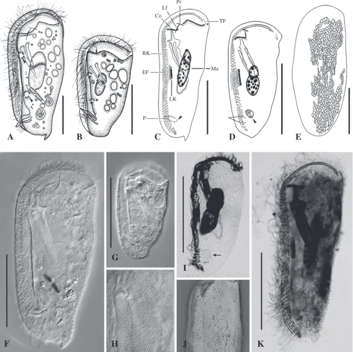
Fig. 1. Dysteria brasiliensis(normal and smallsized type) from life(A, B, FH), after protargol(C, D, I, K), and silver nitrate impregnation(E, J). A, B left side view of two individuals with differing body size and shape, normal type(A) and smallsized type(B), showing two contractile vacuoles(arrowheads); C, D left side view showing the infraciliature, arrowheads indicate the kinetosomelike granules at the base of the podite(C) and glandule(D); E left side view showing the silverline system; F, G left lateral view of two typical in
dividuals; H left side view showing ectosymbiotic bacteria; I, K left side view of two type of specimens, arrowheads indicate right kineties of smallsized type and arrow indicates glandule; J left lateral view showing the silverline system. Co circumoral kineties, EF equatorial fragment, Lf left frontal kineties, LK left kineties, Ma macronucleus, P podite, Pr preoral kineties, RK right kineties, TF terminal fragment. Scale bars: 40μm.
A B C D E
Lf Pr
Co TF
Ma RK
EF
P
LK
G I
F H J K
Gong et al. 2007, p. 157, Figs. 89.
Material examined: On September 27, 2011 at Incheon Harbor, the Yellow Sea, Korea (37˚26′N, 126˚35′E).
Diagnosis: (i) Normal type: Marine Dysteria, size approx
imately 105 μm×47 μm in vivo; dorsal spine present; six right kineties; macronucleus approximately 33 μm×15 μm in vivo; two ventral contractile vacuoles; ectosymbiotic bac
teria rodshaped.
(ii) Smallsized type: Marine Dysteria, size approximate
ly 59 μm×33 μm in vivo; dorsal spine absent; five right ki
neties; macronucleus approximately 23 μm×13 μm in vivo;
two ventral contractile vacuoles; ectosymbiotic bacteria rodshaped.
Remarks: This species was originally reported by Faria et al. (1922). Gong et al. (2007) redescribed the species as three types (normal type, smallsized type, and elongate type) according to size and body outline. In the present study,
we identified two types of D. brasiliensis, i.e., normal and smallsized type, in the same population; our normal and smallsized types correspond well with the two previously described types of Chinese isolate (Gong et al. 2007). How
ever, the normal type in our population differs from that of the Chinese isolate in terms of the cell size and number of right kineties cell size approximately 90~126 μm×44~52 μm in our population and 120~140 μm×46~60 μm in the Chinese isolate; six right kineties in our population and five right kineties in the Chinese isolate (Table 1, Fig. 1AK;
Gong et al. 2007). We considered the slightly different cell size and number of right kineties to be variations of D. bra- siliensis.
Habitat: Marine waters with mussels and seaweeds.
World distribution: Brazil, China, Korea.
Deposition: NIBRPR0000106074NIBRPR0000106075.
Identifiers: MiHyun Park and GiSik Min.
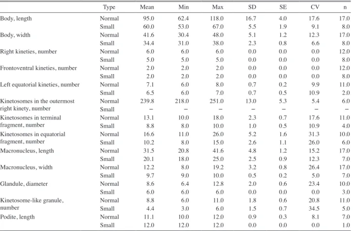
Table 1. Morphometric characteristics of Dysteria brasiliensis: normal type(upper row) and smallsized type(lower row) based on protar
golimpregnated specimens.
Type Mean Min Max SD SE CV n
Body, length Normal 95.0 62.4 118.0 16.7 4.0 17.6 17.0
Small 60.0 53.0 67.0 5.5 1.9 9.1 8.0
Body, width Normal 41.6 30.4 48.0 5.1 1.2 12.3 17.0
Small 34.4 31.0 38.0 2.3 0.8 6.6 8.0
Right kineties, number Normal 6.0 6.0 6.0 0.0 0.0 0.0 12.0
Small 5.0 5.0 5.0 0.0 0.0 0.0 8.0
Frontoventral kineties, number Normal 2.0 2.0 2.0 0.0 0.0 0.0 12.0
Small 2.0 2.0 2.0 0.0 0.0 0.0 8.0
Left equatorial kineties, number Normal 7.1 6.0 8.0 0.7 0.2 9.9 11.0
Small 6.5 6.0 7.0 0.7 0.5 10.9 2.0
Kinetosomes in the outermost
right kinety, number Normal 239.8 218.0 251.0 13.0 5.3 5.4 6.0
Small - - - -
Kinetosomes in terminal
fragment, number Normal 13.1 10.0 18.0 2.3 0.7 17.6 11.0
Small 8.8 8.0 10.0 1.0 0.5 10.9 4.0
Kinetosomes in equatorial
fragment, number Normal 16.6 11.0 26.0 5.2 1.6 31.3 10.0
Small 10.2 8.0 15.0 2.6 1.1 26.0 6.0
Macronucleus, length Normal 31.5 20.8 41.6 4.8 1.2 15.2 17.0
Small 20.1 18.0 25.0 2.5 0.9 12.3 7.0
Macronucleus, width Normal 12.2 8.0 19.2 3.2 0.8 26.4 17.0
Small 9.7 9.0 10.0 0.5 0.2 5.0 7.0
Glandule, diameter Normal 8.6 6.4 12.8 2.0 0.6 23.4 10.0
Small 6.0 6.0 6.0 0.0 0.0 0.0 3.0
Kinetosomelike granule,
number Normal 8.8 6.0 11.0 1.8 0.6 20.8 11.0
Small 4.4 3.0 6.0 1.5 0.7 34.5 5.0
Podite, length Normal 11.1 10.0 12.0 0.9 0.3 8.1 7.0
Small 12.0 12.0 12.0 0.0 0.0 0.0 1.0
CV - coefficient of variation in %; Max - maximum; Mean - arithmetic mean; Min - minimum; n - number of individuals examined; SD - standard deviation; SE
standard error of the mean. All measurements inμm.
2.
Dysteria lanceolata Claparède and Lachmann,1859
창납작충(
신칭) (Figs. 2A-H; Table 2)
Dysteria laceolata Chen et al. 2011, p. 106, Figs. 134.
Material examined: On February 9, 2011 at Incheon Har
bor, the Yellow Sea, Korea (37˚26′N, 126˚35′E).
Diagnosis: Cell size approximately 51 μm×37 μm in vivo;
body oval in outline; seven to eight right kineties, three out
ermost kineties extending to dorsoapically; macronucleus 23 μm×15 μm in vivo; two ventral contractile vacuoles.
Remarks: Dysteria lanceolata was recently collected from China and was redescribed by Chen et al. (2011) based on its body shape, size, and infraciliature. The D. lanceolata
isolate identified in our present study is slightly smaller than the Chinese isolate (49~53 μm×34~38 μm vs. 60~80 μm
× 30~60 μm) in vivo and has seven to eight right kineties (vs. six to seven right kineties in the Chinese isolate). How
ever, most other features correspond well with those of the Chinese isolate as follows: oval body shape; presence of groove and rodshaped ectosymbiotic bacteria on surface of plate; and three frontoventral kineties (cf. two to three in Chinese isolate) (Table 2, Fig. 2AH; Chen et al. 2011).
Habitat: Marine waters with mussels and seaweeds.
World distribution: France, China, Korea.
Deposition: NIBRPR0000106076NIBRPR0000106077.
Identifiers: MiHyun Park and GiSik Min.
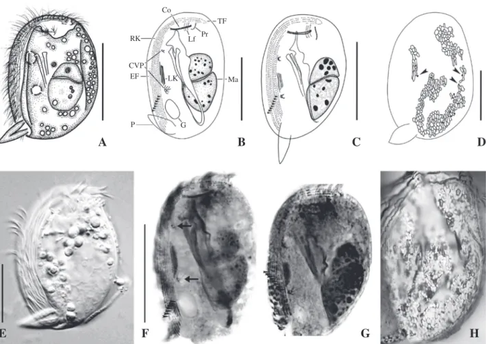
Fig. 2. Dysteria lanceolata from life(A, E), after protargol(B, C, F, G), and silver nitrate impregnation(D, H). A left side view; B, C left side view of two individuals with differing number of right kineties, arrowheads indicate rows of right kineties; D left side view showing the silverline system, arrowheads indicate the tiny argentophilic granules. E left lateral view of typical individual; F, G left side view of two specimens which have different number of right kineties, arrowheads indicate right kineties of having seven(F) and eight(G), arrows show two contractile vacuoles(F) and one short row under end of frontoventral kineties and terminal fragment(G);
H left lateral view showing the silverline system. Co circumoral kineties, CVP contractile vacuole pore; EF equatorial fragment, G glandule; Lf left frontal kineties, LK left kineties, Ma macronucleus, P podite, Pr preoral kineties, RK right kineties, TF terminal fragment. Scale bars: 30μm.
A B C D
RK Co
Lf Pr TF
EF Ma CVP
P G
LK
E F G H
3.
Dysteria nabia Park &Min, 2014
나비납작충(
신칭) Dysteria nabia Park & Min 2014, p. 258, Figs. 1AF, 2AI,
3AJ.
4.
Dysteria ovalis(Gourret and Roeser, 1886) Kahl, 1931
둥근납작충(
신칭) (Fig. 3A, D-H; Table 2) Dysteria ovalis Kahl 1931, p. 256, Figs. S 252, 7; Fauré
Fremiet 1965, p. 6681, Fig. 11.
Material examined: On September 25, 2008 at Geojin Har
bor, Gangwondo, the East Sea, Korea (salinity, 32.3 psu;
temperature, 22.6˚C; pH 8.0; 38˚26′N, 128˚27′E).
Diagnosis: Cell size approximately 58 μm×29 μm; body oval in outline; four right kineties, one innermost kinety conspicuously shortened; macronucleus 19 μm ×13 μm in vivo; two ventral contractile vacuoles. Silverlines show irregular and rather large reticulate, with equatorial trans
verse stripe.
Remarks: Dysteria ovalis was originally reported by Gour
ret and Roeser (1886) and was subsequently described by Kahl (1931) (Fig. 3B, C). After then, FauréFremiet (1965) described the infraciliature of D. ovalis. Our population corresponds well with the isolate reported by FauréFremiet (1965) in terms of the cell size, infraciliature, and silverline structure (Table 2, Fig. 3AH; Kahl 1931, FauréFremiet 1965).
Habitat: Marine waters with mussels and seaweeds.
World distribution: France, Korea.
Deposition: NIBRPR0000106078NIBRPR0000106079.
Identifiers: MiHyun Park and GiSik Min.
Key to the Korean species of the genus Dysteria 1. Four right kineties ···
···D. ovalis (Gourret and Roeser, 1886) Kahl, 1931
Table 2. Morphometric characteristics of Dysteria lanceolata(upper row) and D. ovalis(lower row) based on protargolimpregnated specimens.
Species Mean Min Max SD SE CV n
Body, length DL 56.8 48.0 68.0 6.3 1.4 11.2 20.0
DO 58.4 48.0 66.0 5.8 1.6 10.0 14.0
Body, width DL 36.6 31.0 47.0 4.5 1.0 12.2 20.0
DO 28.5 23.0 33.0 3.8 1.1 13.5 13.0
Right kineties, number DL 7.4 7.0 8.0 0.5 0.2 6.9 11.0
DO 4.0 4.0 4.0 0.0 0.0 0.0 13.0
Frontoventral kineties, number DL 3.0 3.0 3.0 0.0 0.0 0.0 20.0
DO 2.0 2.0 2.0 0.0 0.0 0.0 13.0
Left equatorial kineties, number DL 5.4 5.0 6.0 0.5 0.1 9.4 17.0
DO 5.7 4.0 9.0 1.5 0.4 27.2 13.0
Kinetiosomes in the outermost
right kinety, number DL - - - -
DO 112.8 106.0 118.0 5.0 2.2 4.4 5.0
Kinetiosomes in the innermost
right kinety, number DL - - - -
DO 11.4 9.0 13.0 1.4 0.5 12.4 8.0
Kinetosomes in terminal
fragment, number DL 18.5 14.0 23.0 3.2 1.3 17.3 6.0
DO 8.4 7.0 10.0 1.1 0.4 13.5 7.0
Kinetosomes in equatorial
fragment, number DL 15.3 9.0 24.0 5.0 1.6 32.5 10.0
DO 6.5 5.0 9.0 1.4 0.5 21.8 8.0
Macronucleus, length DL 24.5 19.0 31.0 2.9 0.6 11.7 20.0
DO 19.4 15.0 23.0 2.4 0.7 12.6 14.0
Macronucleus, width DL 13.5 9.0 17.0 2.4 0.5 17.4 20.0
DO 12.8 9.0 16.0 2.2 0.6 16.9 14.0
Glandule, diameter DL 7.2 6.0 9.0 1.0 0.2 13.4 20.0
DO - - - -
Podite, length DL 14.3 11.0 16.0 1.3 0.3 9.4 20.0
DO 13.4 12.0 15.0 1.1 0.5 8.5 5.0
CV - coefficient of variation in %; Max - maximum; Mean - arithmetic mean; Min - minimum; n - number of individuals examined; SD - standard deviation; SE
standard error of the mean. All measurements inμm.
- More than four right kineties ···2 2. Five or six right kineties ···3 - More than six right kineties ···
···D. lanceolata Claparède and Lachmann, 1859 3. Right kinety 5 terminates anteriorly in posterior 1/4 of
body···D. nabia Park & Min, 2014
Fig. 3. Dysteria ovalis after protargol(A, EG), and silver nitrate impregnation(D, H) and previously reported D. ovalis(Gourret and Roeser,1886) Kahl, 1931(B, C). A left side view, arrowhead indicates innermost shorten row of right kineties; B, C left side view of previ
ous reported two, B, from Kahl 1931 and C, from FauréFremiet 1965; D left side view showing the silverline system. E, F left lat
eral view of two individuals, arrowheads indicate ventral contractile vacuoles and arrow show innermost shorten row of right kineties;
G left side view showing equatorial fragment; H left lateral view showing the silverline system, arrowheads indicate the equatorial transverse stripe. Co circumoral kineties, CVP contractile vacuole pore; EF equatorial fragment, Lf left frontal kineties, LK left kineties, Ma macronucleus, P podite, Pr preoral kineties, RK right kineties, TF terminal fragment. Scale bars: 40μm.
B
A C D E
LfPr
Co Co
Pr
Lf TF
RK Ma EF
P
LK CVP
CVP
F G H
- Right kinety 5 or 6 terminates anteriorly at cytostome level or over equatorial transverse stripe ···
···D. brasiliensis Faria et al., 1922
ACKNOWLEDGEMENT
This work was supported by the grants from the National Institute of Biological Resources (NIBR), funded by the Ministry of Environment (MOE) of the Republic of Korea (NIBR NO. 201302001).
REFERENCES
Chen X, J Gong, KAS AlRasheid, SA Farraj and W Song.
2011. New contribution to the morphological taxonomy of three marine cyrtophorid ciliates from the Yellow Sea, Chi
na(Ciliophora: Cyrtophorida). Acta Protozool. 50:105119.
Corliss JO. 1953. Silver impregnation of ciliated protozoa by the ChattonLwoff technic. Stain Technol. 28:97100.
Corliss JO. 1979. The ciliated protozoa. Characterization, clas
sification and guide to the literature. Oxford: Pergamon Press.
Deroux G. 1965. Origine des cinéties antérieures, gauches et buccales dans le genre Dysteria Huxley. CR Acad. Sci.
Paris 260:66896691.
Dragesco J. 1966. Obsevation sur quelques Cilies libres. Arch.
Protistenkd. 109:155206.
Faria JG, AM da Cunha and C Pinto. 1922. Estudos sobre pro
tozoarios do mar. Mem. Inst. Oswaldo Cruz 15:186208.
FauréFremiet E. 1965. Morphologie des Dysteriidae(Ciliata Cyrtophorina). CR Acad. Sci. Paris 260: 66796684.
Foissner W. 1991. Basic light and scanning electron micro
scopic methods for taxonomic studies of ciliated protozoa.
Eur. J. Protistol. 27:313330.
Gong J and W Song. 2003. Morphology and infraciliature of two marine benthic ciliates, Dysteria procera Kahl, 1931 and Dysteria magna nov. spec.(Protozoa, Ciliophora, Cyr
tophorida), from China. Eur. J. Protistol. 39:301309.
Gong J and W Song. 2004. Description of a new marine cyrto
phorid ciliate, Dysteria derouxi nov spec., with an updated key to 12 wellinvestigated Dysteria species(Ciliophora, Cyrtophorida). Eur. J. Protistol. 40:1319.
Gong J, W Song and A Warren. 2002. Redescriptions of two marine cyrtophorid ciliates, Dysteria cristata(Gourret and Roeser, 1888) Kahl, 1931 and Dysteria monostyla(Ehren
berg, 1838) Kahl, 1931(Protozoa, Ciliophora, Cyrtophori
da), from China. Eur. J. Protistol. 38:213222.
Gong J, W Song, A Warren, X Lin and DM Roberts. 2007. Mi
croscopical observations on four marine Dysteria species (Ciliophora, Cyrtophorida). Eur. J. Protistol. 43:147161.
Gong J, XF Lin and W Song. 2003. Redescription of a poor
lyknown marine cyrtophorid ciliate, Dysteria pusilla (Claparède et Lachmann, 1859)(Protozoa: Ciliophora: Cyr
tophorida) from Qingdao, China. Acta Protozool. 42:215
Gourret P and P Roeser. 1886. Les protozoaires du VieuxPort 221.
de Marseille. Arch. Zool. Exp. Gen. 4:507511.
Hu X and T Suzuki. 2005. Light microscopical observations on two marine dysteriid ciliates from Japan, including a description of Dysteria yagiui nov. spec.(Ciliophora, Cyr
tophorida). Eur. J. Protistol. 41:2936.
Jung JH, KM Park and GS Min. 2012. Morphology, morpho
genesis, and molecular phylogeny of a new brackish water ciliate, Pseudourostyla cristatoides n. sp., from Songjiho lagoon on the coast of East Sea, South Korea. Zootaxa 3334:4254.
Jung JH. 2013. Systematics of Korean and Antarctic hypo
trichs(Protozoa: Ciliophora) and community analysis of Korean marine ciliates using pyrosequencing data. Ph.D.
Dissertation. University of Inha. Incheon.
Kahl A. 1931. Urtiere oder Protozoa I: Wimpertiere oder Cil
iata(Infusoria) 2. Holotricha außer den im 1. Teil behan
delten Prostomata. Tierwelt Dtl. 21:181398.
Kwon CB. 2009. Systematic study on estuarine littoral ciliates (Protozoa, Ciliophora) from Taehwagang River in Korea.
Ph.D. Dissertation. University of Ulsan, Ulsan.
Lee ES, YO Kim, S Agatha, JH Jung, D Xu and MK Shin.
2015. Revision of Strombidium paracalkinsi(Ciliophora:
Oligotrichea: Oligotrichia), with Comparison of Strom
bidiids Bearing Thigmotactic Membranelles. J. Eukaryot.
Microbiol. 62:400409.
Nam SW, W Shin, M Kang, W Yih and MG Park. 2015. Ultra
structure and Molecular Phylogeny of Mesodinium coatsi sp. nov., a Benthic Marine Ciliate. J. Eukaryot. Microbiol.
62:102120.
Pan H, X Hu, J Gong, X Lin, KAS AlRasheid, SA AlFarraj and A Warren. 2011. Morphological redescriptions of four marine ciliates(Ciliophora: Cyrtophorida: Dysteriidae) from Qingdao, China. Eur. J. Protistol. 47:197207.
Park MH and GS Min. 2014. A New Marine Cyrtophorid Cil
iate, Dysteria nabia nov. spec.(Ciliophora: Phyllopharyn
gea: Cyrtophorida: Dysteriidae), from South Korea. Acta Protozool. 53:257268.
Petz W, W Song and N Wilbert. 1995. Taxonomy and ecology
of the ciliate fauna(Protozoa, Ciliophora) in the endopagial and pelagial of the Weddell Sea, Antarctica. Stapfia 40:1- 223.
Shin MK. 2012. Invertebrate Fauna of Korea,Stichotrichs (Cil
iophora: Intramacronucleata: spirotrichea: Stichotrichia), 1(1). National Institute of Biological Resources. 193pp.
Song W and N Wilbert. 2002. Faunistic studies on marine cili
ates from the Antarctic benthic area, including descriptions of one epizoic form, 6 new species and, 2 new genera(Pro
tozoa: Ciliophora). Acta Protozool. 41:2361.
The Korean Society of Systematic Zoology. 1997. List of ani
mals in Korea(excluding insects). Academy, Seoul.
Wilbert N. 1971. Morphologie und Ökologie einiger neuer Ciliaten(Holotricha, Cyrtophorina) des Aufwuchses. Proti
stologica 7:357363.
Wilbert N and W Song. 2005. New contributions to the marine benthic ciliates from the Antarctic area, including descrip
tion of seven new species(Protozoa, Ciliophora). J. Nat.
Hist. 39:935973.
Received: 14 May 2015 Revised: 11 June 2015 Revision accepted: 11 June 2015