The Effects of Simultaneous Pulmonary
Rehabilitation during Thoracic Radiotherapy in the Treatment of Malignant Diseases
Myeong Geun Choi, M.D.
1, Hyang Yi Lee, B.P.T.
1, Si Yeol Song, M.D., Ph.D.
2, Su Ssan Kim, M.D., Ph.D.
2, Seung Hak Lee, M.D., Ph.D.
3, Won Kim, M.D., Ph.D.
3, Chang-Min Choi, M.D., Ph.D.
1,4and Sei Won Lee, M.D., Ph.D.
1Departments of
1Pulmonary and Critical Care Medicine,
2Radiation Oncology,
3Rehabilitation Medicine, and
4Oncology, Asan Medical Center, University of Ulsan College of Medicine, Seoul, Republic of Korea
Background: Radiotherapy is a common treatment option for lung or esophageal cancer, particularly when surgery is not feasible for patients with poor lung function. However, radiotherapy can affect pulmonary function and thereby induce pneumonitis or pneumonia, which can be fatal in patients with respiratory impairment. The purpose of this study is to evaluate if reductions in pulmonary function after radiotherapy can be minimized through simultaneous pulmonary rehabilitation (PR).
Methods: In this matched case control study, we retrospectively analyzed patients who had undergone radiotherapy for thoracic malignant disease between January 2018 and June 2019. We analyzed results from pulmonary function tests and 6-minute walking tests (6MWT) conducted within the six months before and after radiotherapy treatment.
Results: In total, results from 144 patients were analyzed, with 11 of the patients receiving PR and radiotherapy simultaneously. Of the 133 patients in the control group, 33 were matched with 11 patients in the PR group. Changes in forced expiratory volume in one second (FEV
1) and FEV
1/forced vital capacity were significantly different between the PR group and the matched control group (240 mL vs. –10 mL, p=0.017 and 5.5% vs. 1.0%, p=0.038, respectively).
The median distance of 6MWT in the PR group also increased significantly, from 407.5 m to 493.0 m after radiotherapy (p=0.017).
Conclusion: Simultaneous PR improved pulmonary function, particularly in measures of FEV
1, and exercise capacity for patients with lung or esophageal cancer even after radiotherapy treatment. These findings may provide an important base of knowledge for further large population studies with long-term follow-up analysis in the identification of the PR’s effects during thoracic radiotherapy.
Keywords: Pulmonary Rehabilitation; Radiotherapy; Pulmonary Function Tests
Address for correspondence: Sei Won Lee, M.D., Ph.D.
Department of Pulmonary and Critical Care Medicine, Asan Medical Center, University of Ulsan College of Medicine, 88 Olympic-ro 43-gil, Songpa-gu, Seoul 05505, Republic of Korea
Phone: 82-2-3010-3990, Fax: 82-2-3010-6968, E-mail: [email protected]
Received: Nov. 8, 2020, Revised: Jan. 1, 2021, Accepted: Feb. 10, 2021, Published online: Feb. 10, 2021
cc It is identical to the Creative Commons Attribution Non-Commercial License (http://creativecommons.org/licenses/by-nc/4.0/).
Copyright © 2021
The Korean Academy of Tuberculosis and Respiratory Diseases.
Introduction
Pulmonary rehabilitation (PR) is a broad, comprehensive treatment concept including exercise, education, and lifestyle modification. It improves both physiologic and psychologic condition by encouraging health-enhancing behaviors
1. PR is an essential non-pharmacologic treatment, which can im- prove quality of life, symptoms, and pulmonary functions
2.
The majority of evidence concerning PR focuses on chronic obstructive pulmonary disease (COPD); however, PR can also be performed for other respiratory diseases, such as interstitial lung disease, bronchiectasis, cystic fibrosis, asthma, pulmo- nary artery hypertension, lung cancer, and transplantation
3. Lung cancer is often accompanied by COPD and/or inter- stitial lung disease, which can limit treatment and increase treatment-related complications
4. PR has been suggested as means to minimize treatment-related complications. A previ- ous randomized controlled trial for lung cancer has shown that preoperative PR can increase exercise capacity
5, which is related to postoperative morbidity. In another study, PR was shown to significantly increase forced expiratory volume in one second (FEV
1) in lung cancer patients who undertook induction concurrent chemo-radiotherapy (CCRT), particu- larly among patients with respiratory impairment or history of smoking
6.
Radiotherapy can be a treatment option in patients with
thoracic malignancy, including lung and esophageal cancers.
However, thoracic radiotherapy can affect pulmonary func- tion especially with diminishing FEV
1and diffusing capacity for carbon monoxide (DLco). Moreover, it can also induce pneumonitis or pneumonia, which can be fatal in patients with respiratory impairment
7-9. Although some studies exist regarding PR in patients with thoracic malignancy, most of those studies were on the effect of preoperative and postoper- ative PR. Conversely, no study exists concerning the effects of simultaneous PR during radiotherapy with the control group.
The purpose of this study is to evaluate whether reduction in pulmonary function after radiotherapy can be minimized through simultaneous PR.
Materials and Methods
1. Study design and patients
This was a retrospective, matched case-control study. The inclusion criteria were as follows: patients >19 years who received thoracic radiotherapy for pathologically confirmed malignant diseases and underwent pulmonary function test (PFT) within 6 months before initiation and after the termina- tion of radiotherapy. The 180 patients who met these inclusion criteria were divided into two groups, PR group and control
7 Excluded
- 4 PR less than once a week during RT - 3 PR during other period than radiotherapy
28 Excluded
- 28 Thoracic surgery between pre- and post-PFT 1 Excluded
- Incomplete data in PFT 180 Patients with chest malignancy
who conducted radiotherapy and both pre- and post-PFT
179 Enrolled patients
161 Patients who did not conduct PR 18 Patients who
conducted PR
133 Control group 11 PR group
33 Matched control group 11 PR group
Matching
Figure 1. Consort flow diagram of the study. PFT: pulmonary function test; PR: pulmonary rehabilitation; RT: radiotherapy.
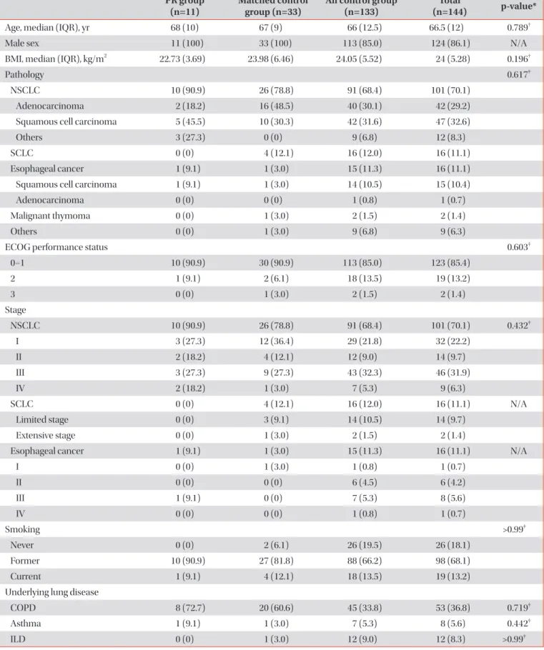
Table 1. Baseline characteristics of patients PR group
(n=11) Matched control
group (n=33) All control group
(n=133) Total
(n=144) p-value*
Age, median (IQR), yr 68 (10) 67 (9) 66 (12.5) 66.5 (12) 0.789
†Male sex 11 (100) 33 (100) 113 (85.0) 124 (86.1) N/A
BMI, median (IQR), kg/m
222.73 (3.69) 23.98 (6.46) 24.05 (5.52) 24 (5.28) 0.196
†Pathology 0.617
‡NSCLC 10 (90.9) 26 (78.8) 91 (68.4) 101 (70.1)
Adenocarcinoma 2 (18.2) 16 (48.5) 40 (30.1) 42 (29.2)
Squamous cell carcinoma 5 (45.5) 10 (30.3) 42 (31.6) 47 (32.6)
Others 3 (27.3) 0 (0) 9 (6.8) 12 (8.3)
SCLC 0 (0) 4 (12.1) 16 (12.0) 16 (11.1)
Esophageal cancer 1 (9.1) 1 (3.0) 15 (11.3) 16 (11.1)
Squamous cell carcinoma 1 (9.1) 1 (3.0) 14 (10.5) 15 (10.4)
Adenocarcinoma 0 (0) 0 (0) 1 (0.8) 1 (0.7)
Malignant thymoma 0 (0) 1 (3.0) 2 (1.5) 2 (1.4)
Others 0 (0) 1 (3.0) 9 (6.8) 9 (6.3)
ECOG performance status 0.603
‡0–1 10 (90.9) 30 (90.9) 113 (85.0) 123 (85.4)
2 1 (9.1) 2 (6.1) 18 (13.5) 19 (13.2)
3 0 (0) 1 (3.0) 2 (1.5) 2 (1.4)
Stage
NSCLC 10 (90.9) 26 (78.8) 91 (68.4) 101 (70.1) 0.432
‡I 3 (27.3) 12 (36.4) 29 (21.8) 32 (22.2)
II 2 (18.2) 4 (12.1) 12 (9.0) 14 (9.7)
III 3 (27.3) 9 (27.3) 43 (32.3) 46 (31.9)
IV 2 (18.2) 1 (3.0) 7 (5.3) 9 (6.3)
SCLC 0 (0) 4 (12.1) 16 (12.0) 16 (11.1) N/A
Limited stage 0 (0) 3 (9.1) 14 (10.5) 14 (9.7)
Extensive stage 0 (0) 1 (3.0) 2 (1.5) 2 (1.4)
Esophageal cancer 1 (9.1) 1 (3.0) 15 (11.3) 16 (11.1) N/A
I 0 (0) 1 (3.0) 1 (0.8) 1 (0.7)
II 0 (0) 0 (0) 6 (4.5) 6 (4.2)
III 1 (9.1) 0 (0) 7 (5.3) 8 (5.6)
IV 0 (0) 0 (0) 1 (0.8) 1 (0.7)
Smoking >0.99
‡Never 0 (0) 2 (6.1) 26 (19.5) 26 (18.1)
Former 10 (90.9) 27 (81.8) 88 (66.2) 98 (68.1)
Current 1 (9.1) 4 (12.1) 18 (13.5) 19 (13.2)
Underlying lung disease
COPD 8 (72.7) 20 (60.6) 45 (33.8) 53 (36.8) 0.719
‡Asthma 1 (9.1) 1 (3.0) 7 (5.3) 8 (5.6) 0.442
‡ILD 0 (0) 1 (3.0) 12 (9.0) 12 (8.3) >0.99
‡group, according to whether PR was performed or not. Pa- tients were excluded if they received any treatment (e.g., lung resection or initiation of new inhalers) between pre- and post- PFT that may affect study results. In addition, patients who received PR for less than once a week during radiotherapy or during the period when radiotherapy was not performed were also excluded. Finally, 11 patients performed PR simultane- ously more than once a week during radiotherapy, and 133 patients received only radiotherapy. Of the 133 patients in the control group, 33 patients were matched in a 1:3 ratio with re- spect to age, sex, and baseline PFT with 11 patients in the PR group (Figure 1).
This study was approved by the institutional review board of the Asan Medical Center (IRB No. 2019–0905) and was conducted in accordance with the ethical standards of the Declaration of Helsinki. Requirement for written informed consent was waived by the review board.
2. Pulmonary rehabilitation
All 11 patients of the PR group received PR when visiting the medical center for radiotherapy more than once a week.
The program included the following steps, which focused on supervised exercise (Supplementary Figure S1). First, we checked patient smoking status and provided smoking cessa- tion education and nutritional counseling. If the patient used an inhaler, inhaler operation was confirmed, and any mistakes were corrected. Then, patients practiced abdominal breath- ing and pursed-lip breathing, inhaling through the nose and slowly exhaling with lips closed. Next, patients performed
aerobic exercises such as walking and biking on treadmill to the degree assessed by 6-minute walking test (6MWT) or cardiopulmonary exercise test. The exercise was prescribed according to the frequency, intensity, time, and type principle based on their underlying disease and exercise capacity
10. Patients then performed muscle exercises of the upper and lower extremities, stretching and relaxation. Patients with pro- ductive sputum were educated about proper expectoration methods, such as forced expiration huffing. They were en- couraged to continue this exercise in their homes. All patients were required to fill out their exercise diary until next visit, with exercise intensity, duration and type being prescribed to maintain PR at home. The exercise prescription form and self- exercise diary are shown in Supplementary Table S1.
3. Outcomes
The primary outcome of this study was the change of PFT from baseline, including forced vital capacity (FVC), FEV
1, FEV
1/FVC, and DLco
11. Secondary outcomes were 6MWT, 6-month rate of pneumonia and radiation pneumonitis and 6-month overall survival. PFT results within 6 months before and after radiotherapy were analyzed and reported as base- line and post-PFT, respectively. Pneumonia and radiation pneumonitis were determined with chest computed tomog- raphy and chest X-ray images. Pneumonia was analyzed as an event when antibiotics were prescribed for pneumonia after referring to chest image and patient symptoms. Similarly, ra- diation pneumonitis was considered when steroids were pre- scribed for patient symptoms of grade 2 or higher, based on Table 1. Continued
PR group
(n=11) Matched control
group (n=33) All control group
(n=133) Total
(n=144) p-value*
Purpose of RT 0.366
‡Definitive 11 (100) 20 (60.6) 77 (57.9) 88 (61.1)
Palliative 0 (0) 1 (3.0) 4 (3.0) 4 (2.8)
Salvage 0 (0) 4 (12.1) 18 (13.5) 18 (12.5)
Preoperation 0 (0) 2 (6.1) 15 (11.3) 15 (10.4)
Postoperation 0 (0) 6 (18.2) 18 (13.5) 18 (12.5)
Others 0 (0) 0 (0) 1 (0.8) 1 (0.7)
PR duration, median (IQR), wk 4 (5) N/A N/A N/A
RT duration, median (IQR), day 29 (33) 41 (32.5) 41 (28.5) 40.5 (29.5) 0.487
†RT fraction, median (IQR), cGY 6,000 (400) 6,000 (745) 5,600 (1,000) 5,775 (1,000) 0.689
†Analysis period, median (IQR) 329 (326) 350 (216) 372 (222) 370 (225) 0.487
†Values are presented as number (%) unless otherwise indicated.
*Comparison between PR group and matched control group.
†Mann-Whitney test.
‡Fisher exact test.
PR: pulmonary rehabilitation; IQR: interquartile range; BMI: body mass index; NSCLC: non-small cell lung cancer; SCLC: small cell lung
cancer; ECOG: Eastern Cooperative Oncology Group; N/A: not available; COPD: chronic obstructive pulmonary disease; ILD: interstitial lung
disease; RT: radiotherapy.
chest image evidence. Survival was analyzed for the full study period.
4. Statistical analysis
Demographic characteristics at baseline were compared between enrolled patients. Wilcoxon signed-rank test or Mann-Whitney U test was used to compare continuous vari- ables. Fisher exact test was used to compare categorical vari- ables. Control group was extracted by baseline age, sex, FVC (%), and FEV
1(%) matching with the PR group at a ratio of 3:1.
Time to pneumonia, time to radiation pneumonitis, or overall survival analyses were performed using Kaplan-Meier curve and log-rank test. A p-value of less than 0.05 was considered statistically significant. Statistical analyses were performed us- ing IBM SPSS version 25.0 (IBM Corp., Armonk, NY, USA).
Results
1. Patients
From 144 total patients, 11 with simultaneous PR dur- ing radiotherapy were categorized as the PR group, and 33 were included as the matched control group. Patients in PR group underwent PR 1.5 times per week on average.
Baseline characteristics were similar between the PR group and the matched control group (Table 1). Median age of all patients was 66.5 years (68 years for the PR group and 67 for the matched control group), and 124 (86.1%) of all patients
were male. All patients in both the PR and matched control group were male. Ten patients (90.9%) in the PR group and 26 (78.8%) in matched control group had non-small cell lung cancer (NSCLC). The Eastern Cooperative Oncology Group scale of performance status values for most patients were be- tween 0 and 1. No difference was observed between the two groups regarding NSCLC stage and smoking status. Regarding underlying lung disease, eight (72.7%) of the PR group and 20 (60.6%) of the matched control group had COPD. Median fraction of radiotherapy was the same in the two groups (6,000 cGY). As baseline PFT was used for individual matching, FVC (76.0% vs. 79.0%) and FEV
1(57.0% vs. 59.0%) were similar be- tween the two groups.
2. Changes of PFT from baseline
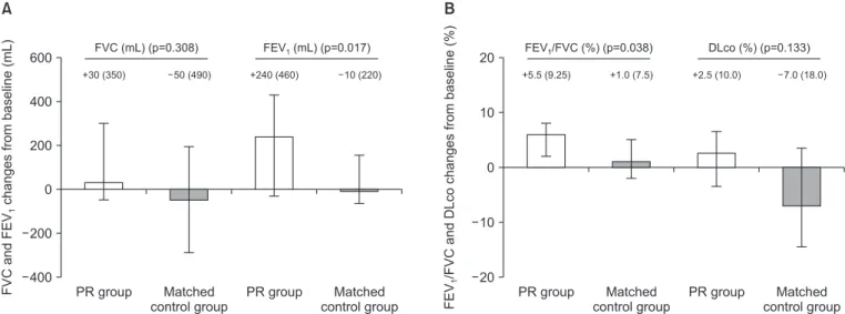
Change from baseline in FEV
1was significantly different be- tween PR group and matched control group (240 mL vs. –10 mL, respectively, p=0.017) (Figure 2). FEV
1/FVC was found to increase significantly more in the PR group (5.5% vs. 1.0%, p=0.038). Changes from baseline in FVC (30 mL vs. –50 mL) and DLco (2.5% vs. –7.0%) were also greater in the PR group than matched control group; however, these differences were not statistically significant (p=0.308 and p=0.133, respectively) (Figure 2).
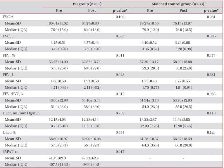
3. PFT, 6MWT, and safety in PR group
Comparing pre- and post-PFT in the PR group, FEV
1(L) and FEV
1/FVC (%) both increased significantly after PR dur-
600
400
200
0
200
400
PR group FVCandFEVchangesfrombaseline(mL)1
Matched control group
PR group Matched control group
+30 (350) 50 (490) +240 (460) 10 (220)
FVC (mL) (p=0.308) FEV (mL) (p=0.017)1
20
10
0
10
20
PR group FEV/1FVCandDLcochangesfrombaseline(%)
Matched control group
PR group Matched control group +5.5 (9.25) +1.0 (7.5) +2.5 (10.0) 7.0 (18.0)
FEV /FVC (%) (p=0.038)1 DLco (%) (p=0.133)
A B
Figure 2. Changes from baseline in pulmonary function test. (A) The forced vital capacity (FVC, mL) change from baseline was greater than
that of the pulmonary rehabilitation (PR) group (+30 mL vs. –50 mL, p=0.308), and the forced expiratory volume in 1 second (FEV
1, mL)
change from baseline was significantly greater than that of the PR group (+240 mL vs. –10 mL, p=0.017). (B) FEV
1/FVC (%) increased more in
the PR group (+5.5% vs. +1.0%, p=0.038), and the diffusing capacity for carbon monoxide (DLco, %) change from baseline was greater in the
PR group (+2.5% vs. –7.0%, p=0.133); Error bars represent the interquartile ranges (IQR) of the median. The text above each column indicates
the median (IQR).
ing radiotherapy (p=0.023 and p=0.012, respectively). Median distance of 6MWT also significantly increased from 407.5 m to 493.0 m after PR during radiotherapy (p=0.017). No signifi- cant differences were observed in FVC (L) or DLco (%). In the matched control group, no significant differences were observed between pre- and post-PFT values in any of these parameters (Table 2). No adverse events related to PR, such as musculoskeletal complications, extreme fatigue, dizziness or cardiac instability, were observed in PR group during the analysis period.
4. Sensitivity analysis
In 53 patients with COPD from the total patients, median changes in PFT and 6MWT from baseline improved more in patients who received PR during radiotherapy (n=8) than in
those who did not PR (n=45), although this was not statistically significant (FEV
1, 240 mL vs. 50 mL; p=0.075 and 6MWT, 93 m vs. –10 m; p=0.097). In 100 patients with NSCLC, median changes in FEV
1and 6MWT from baseline also improved significantly more in patients who received PR (n=10) than in those who did not (n=90) (FEV
1, 250 mL vs. –10 mL; p=0.023 and 6MWT, 93 m vs. –30 m; p=0.017). Among 107 patients receiving conventional radiotherapy, median changes in FEV
1and 6MWT from baseline were also greater in those who re- ceived PR (n=6) than in those who did not (n=101), although 6MWT changes did not reach statistical significance (FEV
1, 270 mL vs. –10 mL; p=0.023 and p=0.036, respectively) (Table 3).
5. Radiation pneumonitis, pneumonia, and survival The occurrences of radiation pneumonitis ≥grade 2 were
Table 2. Pulmonary function test before and after radiotherapy according to simultaneous pulmonary rehabilitation
PR group (n=11) Matched control group (n=33)
Pre Post p-value* Pre Post p-value*
FVC, % 0.196 0.281
Mean±SD 80.64±11.02 84.27±8.80 79.27±10.56 76.15±13.97
Median (IQR) 76.0 (15.0) 82.0 (13.0) 79.0 (12.0) 76.0 (18.5)
FVC, L 0.563 0.386
Mean±SD 3.45±0.51 3.57±0.43 3.40±0.52 3.29±0.68
Median (IQR) 3.42 (0.70) 3.39 (0.78) 3.36 (0.64) 3.28 (0.98)
FEV
1, % 0.011 0.474
Mean±SD 53.55±14.00 62.82±15.74 57.30±13.17 58.00±15.88
Median (IQR) 57.0 (26.0) 68.0 (27.0) 59.0 (20.5) 58.0 (25.0)
FEV
1, L 0.023 0.681
Mean±SD 1.66±0.50 1.93±0.58 1.75±0.48 1.77±0.55
Median (IQR) 1.71 (0.89) 2.11 (0.92) 1.70 (0.77) 1.81 (0.91)
FEV
1/FVC, % 0.012 0.085
Mean±SD 48.00±12.98 54.46±15.44 51.94±13.76 53.76±12.93
Median (IQR) 51.0 (23.0) 58.0 (30.0) 54.0 (23.0) 55.0 (20.5)
DLco, mL/mm Hg/min 0.759 0.110
Mean±SD 12.15±4.03 12.28±4.14 13.22±3.87 11.92±3.85
Median (IQR) 10.75 (5.40) 11.35 (5.78) 12.00 (7.25) 12.90 (5.45)
DLco, % 0.444 0.123
Mean±SD 58.60±16.97 60.00±16.08 61.78±18.67 56.67±18.59
Median (IQR) 57.5 (25.3) 56.5 (29.3) 64.0 (35.0) 66.0 (28.0)
6MWT, m 0.017 -
Mean±SD 419.9±89.9 478.3±62.4 - -
Median (IQR) 407.5 (116.3) 493.0 (80.5) - -
*Wilcoxon signed-rank test.
PR: pulmonary rehabilitation; FVC: forced vital capacity; SD: standard deviation; IQR: interquartile range; FEV
1: forced vital capacity in one
second; DL
CO: diffusing capacity for carbon monoxide; 6MWT: 6-minute walking test.
one (9.1%) and four (12.1%) in the PR and matched control group, respectively (p=0.789). The 6-month incidence of radiation pneumonitis was 0 in the PR group and 0.124 in the matched control group (Figure 3A). Four (36.4%) and nine (27.3%) cases of pneumonia were observed in the PR
and matched control group respectively during the analysis period (p=0.406). The median time to first pneumonia was 365 days for the PR group and could not be evaluated due to limited events in the matched control group. The 6-month incidence of pneumonia was 0.182 in the PR group and 0.216 Table 3. Subgroup analysis of median changes in PFT
PR group Control group Median difference p-value*
COPD (n=53) n=8 n=45
FVC, % 0.50 (7.75) –1.00 (12.50) 1.50 0.297
FVC, L 0.00 (0.37) –0.08 (0.53) 0.08 0.275
FEV
1, % 7.50 (15.00) 2.00 (10.00) 5.50 0.105
FEV
1, L 0.24 (0.44) 0.05 (0.32) 0.19 0.075
FEV
1/FVC, % 5.50 (5.75) 4.00 (6.50) 1.50 0.297
DLco, % 3.00 (11.00) –5.50 (12.50) 8.50 0.103
6MWT, m 93.0 (118.0) –10.0 (67.0) 103.0 0.097
NSCLC (n=100) n=10 n=90
FVC, % 1.50 (10.75) –1.00 (11.50) 2.50 0.110
FVC, L 0.00 (0.39) –0.08 (0.50) 0.07 0.204
FEV
1, % 9.00 (11.50) 0.00 (11.00) 9.00 0.011
FEV
1, L 0.25 (0.52) –0.01 (0.34) 0.25 0.023
FEV
1/FVC, % 6.00 (9.25) 3.00 (7.00) 3.00 0.094
DLco, % 3.00 (9.00) –3.00 (15.25) 6.00 0.053
6MWT, m 93.0 (88.5) –30.0 (64.25) 123.0 0.017
Conventional RT (n=107) n=6 n=101
FVC, % 0.50 (10.00) –1.00 (13.50) 1.50 0.336
FVC, L 0.00 (0.48) –0.09 (0.57) 0.09 0.361
FEV
1, % 9.50 (20.50) 0.00 (12.50) 9.50 0.038
FEV
1, L 0.27 (0.68) –0.01 (0.40) 0.28 0.036
FEV
1/FVC, % 7.50 (17.25) 2.00 (8.00) 5.50 0.032
DLco, % 2.50 (9.75) –9.00 (20.75) 11.50 0.080
6MWT, m 94.5 (92.0) –35.0 (40.0) 129.5 0.073
SBRT (n=37) n=5 n=32
FVC, % 2.00 (16.50) –1.00 (8.00) 3.00 0.079
FVC, L 0.03 (0.56) –0.05 (0.39) 0.09 0.213
FEV
1, % 8.00 (10.50) 0.00 (9.50) 8.00 0.071
FEV
1, L 0.24 (0.39) –0.02 (0.29) 0.22 0.117
FEV
1/FVC, % 5.00 (8.00) 1.50 (6.75) 3.50 0.714
DLco, % 2.50 (13.00) –2.00 (13.00) 4.50 0.357
6MWT, m 48.0 (101.75) 5.0 (82.0) 43.0 0.164
Values are presented as median (interquartile range).
*Mann-Whitney test.
PFT: pulmonary function test; PR: pulmonary rehabilitation; COPD: chronic obstructive pulmonary disease; FVC: forced vital capacity; FEV
1:
forced vital capacity in one second; DL
CO: diffusing capacity for carbon monoxide; 6MWT: 6-minute walking test; NSCLC: non-small cell lung
cancer; RT: radiotherapy; SBRT: stereotactic body radiation therapy.
in the matched control group (Figure 3B). One death (9.1%) and seven deaths (14.2%) were recorded in the PR group and matched control group, respectively, during the analysis pe- riod. The 6-month survival rate was 1.0 in the PR group and 0.938 in the matched control group; however, no difference was observed between the groups in terms of overall survival based on Kaplan-Meier survival analysis (p=0.662) (Figure 4).
Discussion
This study has showed several benefits of simultaneous PR during thoracic radiotherapy. All parameters of pulmonary function and exercise capacity improved despite radiotherapy, as combined PR, and FEV
1, FEV
1/FVC, and 6MWT showed
significant improvement. Meanwhile, pulmonary function, including FEV
1, FVC, and DL
COdid not change or decreased after radiotherapy without PR, although statistically not signifi- cant. Accordingly, FEV
1and FEV
1/FVC improved significantly in the PR group, compared with matched control. No adverse events related to PR were noted. Thus, these results suggest that simultaneous PR is a safe and effective treatment during radiotherapy, which could be an option for patients with poor lung function. As far as we know, this study was the first to confirm the effects of simultaneous PR during thoracic radio- therapy.
Most studies regarding PR in lung cancer patients have been conducted in preoperative or postoperative conditions
5,12-14. For radiotherapy, one previous study on PR for patients with CCRT demonstrated that PR significantly increases pulmo- nary function, particularly FEV
1, in smokers and those with respiratory impairment; however, this study included a single arm without a control group
6. Similar results were observed in our study. By comparing with a matched control, our study has provided more confirmative results that simultaneous PR can improve clinical performance after radiotherapy.
Among the changes of PFT from baseline, FEV
1was most prominent in the PR group. FEV
1generally decreases after ra- diotherapy
7; however, our study showed that FEV
1actually sig- nificantly increased in the PR group. This suggests that simul- taneous PR can attenuate potential declines in lung function associated with radiotherapy, especially in FEV
1. There are a few possible explanations for this. First, patients with COPD may have lung hyperinflation, and radiotherapy might reduce this
15,16. Simultaneous PR may additionally minimize the reduction of FVC, providing a synergistic effect
6,17,18. Second, increased regular physical activity through PR may delay FEV
1decline related with radiotherapy. It is known that FEV
1is
Overallsurvival(%)
0 100
90
80
200 Days
0
50 100 150
PR group
Matched control group
Figure 4. Kaplan-Meier curves. Overall survival (p=0.662 for log- rank test). PR: pulmonary rehabilitation.
0 30
20
10
CumulativeincidenceofRTpneumonitis(%) 200
Days 0
50 100 150
PR group
Matched control group
0 30
20
10
Cumulativeincidenceofpneumonia(%) 200
Days 0
50 100 150
PR group
Matched control group