The Effect of Integration Between Respiratory Muscle Training and Abdominal Drawing-in Maneuver on Decreased Pulmonary Function
in Young Subjects
Chang-yong Kim, B.H.Sc., P.T.
Dept. of Physical Therapy, The Graduate School, Daejeon University Jong-duk Choi, Ph.D., P.T.
Dept. of Physical Therapy, College of Natural Science, Daejeon University Dong-wook Byun, B.H.Sc., P.T.
Dept. of Physical Therapy, Yuseong Wellness Hospital Jin-seok Kim, B.H.Sc., P.T.
Dept. of Physical Therapy, Dunsan Oriental Hospital of Daejeon University Ji-yeol Lee, B.H.Sc., P.T.
Dept. of Physical Therapy, Cheongju Oriental Hospital of Daejeon University
Abstract
9)The aim of this study was to investigate the effects of respiratory muscle training (RMT) with abdominal drawing-in maneuver (ADIM) on pulmonary function. Twenty-two subjects with restrictive breathing participated in this study. All the subjects were randomly assigned to three groups (7 subjects in RMT group, 7 subjects in RMT with ADIM group, 8 subjects in control group). The first group performed the RMT by using incentive respiratory spirometer (IRS). The second group performed the RMT by using IRS and the ADIM by using a Stabilizer. The exercises were conducted over four days.
The pulmonary function was evaluated using the spirometer to measure the force exploratory volume in 1 second (FEV1) and forced vital capacity (FVC). Measurements were conducted on the first day and the last day. A paired-t test was used for pre-post changes and the change rates in FVC and FEV1 among each group were investigated by a one-way ANOVA. The findings of the the study were as follows: 1) There were significant differences of FVC and FEV1 between pre and post in the two training groups (p<.05) 2) There was no significant difference of the change ratio the FVC and FEV1 between the RMT group and RMT with ADIM group. Therefore, it is concluded that respiratory muscle and ADIM training, combined with two methods of treatment would suggest positive evidence for improving pulmonary function.
Key Words: Abdominal drawing-in maneuver training; Pulmonary function; Respiratory muscle training.
Introduction
Respiration is a process during which an organism exchanges carbon dioxide with oxygen in order to ob- tain the materials necessary to sustain life. The respi- ratory system supplies the blood with oxygen while removing carbon dioxide. During expiration, the sys- tem passes the air through the vocal cords, generating
sounds, regulating abdominal pressure, and aiding uri- nation, defecation, and parturition. Respiration consists of inspiration and expiration. Inspiration is an active movement that contracts the diaphragm and inter- costal muscles. The major inspiratory muscles are the diaphragm and external intercostal, and the auxiliary inspiratory muscles are the sternocleidomastoid, sca- lene, serratus anterior, pectoralis major, pectoralis mi-
Corresponding author: Jong-duk Choi [email protected]
nor, trapezius, and erector spinae. Contrary to inspira- tion, expiration is a passive process that occurs dur- ing relaxation of the muscles that were engaged in inspiration, and brings the thorax back to its original location. During expiration, the size of the thorax de- creases, increasing intrathoracic pressure. Pulmonary compliance contracts the lungs, resulting in the air pressure within them becoming greater than the at- mospheric pressure and thereby releasing the air from the lungs (Kim et al, 2009).
The thorax and abdominal muscles used for respira- tion are divided into the major and auxiliary muscles.
The auxiliary muscles include the scalene, sternoclei- domastoid, pectoralis major, and the abdominal. These aid the major muscles when the demand for air in- creases (Bach et al, 1993; Hanayama et al, 1997;
Jardins, 2002). Previous studies have noted that weak- ened expiratory muscles drastically lower the capacity for coughing and sputum drainage, and exercises to strengthen these expiratory muscles were conducive to reinforcing the muscular strength of the pectoralis and reducing residual volumes (Kim et al, 2008).
Consequently, the strengthened respiratory muscles af- ter exercises are linked with an enhanced capacity for coughing and sputum expectoration (Estenne et al, 1989). It is possible to utilize repetitive breathing re- sistance training to strengthen respiratory muscles us- ing simple instruments, and this can also improve car- diopulmonary functionality (Roth et al, 2010). One of the methods used to train respiratory muscles is one with an incentive respiratory spirometer (IRS). The method’s merits are that it is easy to learn how to use the instrument, it is economical, and patients are inspired to use it as it produces a visible achievement.
Its visual feedback helps trained patients to use the instrument independently and freely (Westwood et al, 2007), and maximizes their respiratory motivation (Bartlett et al, 1973; Houge, 2001).
10)The four muscles in the abdomen perform the important functions of trunk flexion and rotation, pull the abdominal wall medially, and increase the pres-
sure within the abdominal cavity. The elements of the abdomen are not actually pressurized, and there- fore these muscles induce the diaphragm to move in- wards toward the thoracic cavity. This movement is a result of increased pleural pressure and reduced lung capacity. These abdominal muscles are strong and play an important role in activities like coughing and deep respiration (De Troyer and Estenne, 1988).
Previous studies have presented conclusive evidence that abdominal muscles are the most crucial muscles in respiration (Suzuki et al, 1991). Most important, the activation of the upper and lower fibers of the transversus abdominis muscle contributes to changes in abdominal pressure and these are essential in in- creasing abdominal pressure. The diaphragm helps to enhance the spinal stability; its contraction, together with the abdominal muscles’ contraction, increases abdominal pressure (Bartelink, 1957; Cresswell et al, 1992; Grillner et al, 1978), and it continuously con- tributes to respiration and posture adjustment, along with the abdominal muscles (Hodge et al, 2000). The Stabilizer1) is a tool developed for abdominal draw- ing-in maneuver (ADIM) training that increases the abdominal pressure, stabilizing the lumbar spine. This Stabilizer provides visual feedback through a pressure sensor (Cairns et al, 2000).
Measuring forced vital capacity (FVC) is a method of measuring the vital capacity. The forced vital capacity refers to the total capacity of air that can be blown out by maximal forced expira- tion after maximal inspiration. The forced ex- piratory volume in 1 second (FEV1) means the ca- pacity of air that is blown out for a single second.
Most healthy adults can exhale 80% of their total capacity in 1 second during expiration (Westwood et al, 2007). Usually, in order to evaluate pulmo- nary functions after respiratory muscle training (RMT), measurements are taken of the pulmonary residual volume, the FVC and the FEV1 (Roth et al, 1995). A IRS may be also used to evaluate pul- monary functions and the respiratory muscles’ de-
1) STABILIZERTM Pressure Bio-Feedback, Chattanooga group Inc, Hixson, U.S.A.
Figure 1. Spirometer.
Variables RMTGb (n1=7) ITGc (n2=7) CGd (n3=8) F*
Age (yrs) 20.7±3.2a 19.1±.9 19.8±1.6 .99
Weight (㎏) 163.1±4.9 159.7±4.4 164.8±8.1 1.30
Height (㎝) 50.0±4.9 51.6±6.4 52.5±7.2 .30
FVCe (ℓ) 1.9±.6 2.0±.4 2.4±.6 1.94
FEV1f (ℓ) 1.8±.6 1.9±.4 2.3±.6 1.28
aMean±SD, brespiratory muscle training group, cintegrating training group, dcontrol group, eforced vital capacity,
fforced exploratory volume in 1 second, *p>.05.
Table 1. General characteristics of the subjects (N=22)
gree of weakness (Kim et al, 2009).
This study attempted to examine whether respira- tory muscle and ADIM training was conducive to enhancing pulmonary functions. This study estab- lished a hypothesis that RMT and the ADIM train- ing would increase abdominal pressure from the transversus abdominis muscle, improving pulmonary functions in the FVC and FEV1. We also predicted that a integrating exercise program of conducting respiratory muscle and ADIM training would be more conducive to enhancing pulmonary functions than either of them alone, and it also surmised that activation of deep abdominal muscles like trans- versus abdominis muscle would be effective in en- hancing respiratory capabilities. The purpose of this study was to examine the effects of a integrating exercise of respiratory muscle and ADIM training devised for internal stabilization of the spine on pul- monary functions.
Methods Subjects
Subjects were selected by taking a pre-measure- ment with a spirometer; the subjects selected com- prised 22 healthy male and female adults in their twenties with restricted pulmonary functions, with a vital capacity of less than 80%, or whose FEV1
was less than 70% compared to the norm (Kagaya et al, 2009). Those who had undergone orthopedic
or neurological surgeries, other thoracic surgeries, or those who received treatment due to neurological problems were excluded. Before this experiment was performed, this study was sufficiently explained to all the subjects and their consent to participate was obtained. 14 subjects were randomly allocated to ei- ther a respiratory muscle training group (RMTG, n1=7), a integrating training group of respiratory muscle and ADIM exercises (ITG, n2=7), or a con- trol group (n3=8) to which no exercise was prescribed. The three groups’ general characteristics were not statistically significantly different and their FVC and FEV1 were also not statistically sig- nificantly different (Table 1).
Instruments11)
A spirometer2) was used to measure the subjects’
FVC and FEV1 among pulmonary function indexes (Figure 1).
2) CHESTGRAPH HI-101, CHEST M. I. INC, Tokyo, Japan.
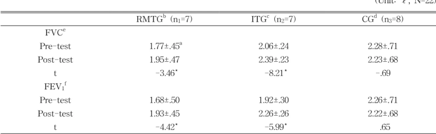
RMTGb (n1=7) ITGc (n2=7) CGd (n3=8) FVCe
Pre-test 1.77±.45a 2.06±.24 2.28±.71
Post-test 1.95±.47 2.39±.23 2.23±.68
t -3.46* -8.21* -.69
FEV1f
Pre-test 1.68±.50 1.92±.30 2.26±.71
Post-test 1.93±.45 2.26±.26 2.22±.68
t -4.42* -5.99* .65
aMean±SD, brespiratory muscle training group, cintegrating training group, dcontrol group, eforced vital capacity,
fforced exploratory volume in 1 second, *p<.05.
Table 2. Comparisons of the FVC and FEV1 before and after the intervention between groups (Unit: ℓ, N=22) Procedures
The RMTG received incentive spirometry training, the ITG of respiratory muscle and ADIM exercises received incentive spirometer and ADIM training, and the control group did not receive any intervention.
Each group’s training period spanned 4 days. For all the groups, the FVC and FEV1 were measured prior to the exercise and they were re-measured after the exercises were carried out for 4 days.
Respiratory Muscle Training12)
For the RMT method using an incentive spi- rometer3), a RMT method presented by Hall et al (1996) was modified and applied. The subject main- tained a maximal inspiration position for 2 to 3 sec- onds and then performed maximal expiration. This exercise was performed for a total of 5 sets, with 10 repetitions making up one set. After each set, a one-minute resting time was allowed. When the sub- ject complained of fatigue or felt dizzy during the respiratory exercise, they took a rest for a short while and then proceeded with the exercise. If these symp- toms were severe, the subjects stopped the exercise.
Abdominal Drawing-in Maneuver
For the ADIM training, a ADIM training method presented by Richardson et al. (2004) was modified and
applied. The subject lay in a prone position and the Stabilizer was placed horizontally, right under the ab- domen and the anterior superior iliac spine (ASIS) so that the instrument did not touch the ASIS. The pres- sure biofeedback unit was inflated to 70 ㎜Hg and the subject was instructed to perform an ADIM. The pres- sure biofeedback unit’s pressure was then lowered by 6~10 ㎜Hg, and this was maintained for 10 seconds.
The subject rested for 2 to 3 seconds after the 10-second maintenance. This exercise was conducted for a total of 5 sets with 10 repetitions making up one set. For each set, a one-minute rest was allowed. Prior to the exercise, the functions of the deep abdominal muscles were explained, and the subjects performed a preliminary exercise through feedback and reinforcement about whether the exercise was properly applied.
Statistical Analysis
Data were analyzed using Statistical Package for the Social Sciences software version 18.0 software.
Before the exercise, a one-way ANOVA was con- ducted in order to verify the statistical significance of the differences in the groups’ general character- istics and pulmonary functions. Then, a paired t-test was performed in order to verify the significance of the differences in each group between the measure- ments taken prior to the experiment and after the
3) Tri-ball Incentive Spirometer 600-1200cc, Ark Therapeutic, Lugoff, U.S.A.
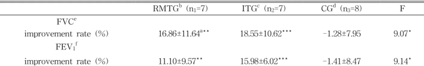
RMTGb (n1=7) ITGc (n2=7) CGd (n3=8) F FVCe
improvement rate (%) 16.86±11.64a** 18.55±10.62*** -1.28±7.95 9.07*
FEV1f
improvement rate (%) 11.10±9.57** 15.98±6.02*** -1.41±8.47 9.14*
aMean±SD, brespiratory muscle training group, cintegrating training group,dcontrol group, eforced vital capacity,
fforced exploratory volume in 1 second, *p<.05, **significant difference between RMGT and CG, ***significant difference between ITG and CG.
Table 3. Comparisons of improvement rate of the FVC and FEV1 after the intervention (N=22)
experiment. Furthermore, in order to examine the significance of the change ratio of FVC and FEV1, a one-way ANOVA was carried out. Tukey's honestly significant difference test was also applied as a post-hoc test. The significance level was set at .05 in order to analyze statistical significance.
Results
1. Comparison of pulmonary functions in accordance with the training methods
1.1. The Forced Vital Capacity
A comparison of the FVC prior to the exercise and after the exercise showed that this significantly increased in the RMTG and ITG (p<.05) (Table 2). FVC after the exercise was compared among the three groups; results from those in the RMTG and ITG were significantly dif- ferent from the results of the control group (p<.05) (Table 2), but there was no significant difference be- tween the RMTG and ITG (p>.05) (Table 3).
1.2. The Force Exploratory Volume in 1 second
The FEV1 prior to the exercise and after the ex- ercise were compared and results from participants in the RMTG and ITG significantly increased after the exercise (p<.05) (Table 2). Comparison of the FEV1
among the three groups after the exercise showed that the RMTG and ITG had a significantly larger volume than the control group (p<.05) (Table 2). However, there was no significant difference between the results
from the RMTG and ITG (p>.05) (Table 3).
1.3. Change ratio of forced vital capacity and forced expiratory volume in 1 second after the exercise
After the exercise, the FVC of the RMTG and ITG increased by an average of 16.86% and 18.55%, re- spectively; on the contrary, that of the control group decreased by an average of 1.28%. In addition, the FEV1 of the RMTG and ITG rose by an average of 11.10% and 15.98%, respectively, while that of the con- trol group reduced by an average of 1.41% (Table 3).
1.4. Comparison of the change ratio of forced vital capacity and forced expiratory volume in 1 second among the three groups
After the exercise, the RMTG and ITG’s FVC and FEV1 significantly increased in comparison with the control group (p<.05) (Table 3). After the 4day ex- ercise, the ITG and RMTG’s FVC and FEV1 increased further by an average of 1.69% and 4.88%. However, this was not statistically significant (p<.05) (Table 3).
Discussion
RMT using an incentive spirometer is conducive to enhancing pulmonary functions (Bartlett et al, 1973).
According to recent studies, deep abdominal muscles such as transversus abdominis muscle and multifidus muscle (Wilke et al, 1995) contribute not only to sta- bilization of the spine (Creewell et al, 1994) and ad- justment of posture (Hodge et al, 2000), but also to
significant improvement in pulmonary functions when ADIM training is applied (Kim et al, 2009). Studies have verified the respective effects of RMT and ADIM training and their relationships to improvement in pulmonary functions, but there has been no re- search examining the relationship between a integrat- ing training program of respiratory muscle and ADIM exercises and improvement in pulmonary functions.
Accordingly, this study attempted to investigate how a integrating exercise program of RMT and ADIM training affected improvement in pulmonary functions.
This study’s subjects are 22 males and females in their twenties; due to this limited sample, future re- search on patients with restrictive, obstructive, and mixed pulmonary functions is necessary. The subjects were randomly allocated to either a RMTG, ITG, or a control group. The tests on the subjects’ pulmonary functions were conducted with a spirometer. The test on their pulmonary functions was performed by the subjects putting on a nose clip. If the subjects coughed or made a mistake, their numerical value was not re- corded (Kim et al, 2001). The test results are affected by technological differences, biological differences un- related to diseases, differences in testing equipment, performance process, examiners and subjects, and dis- eases themselves. The most important concern when conducting and analyzing a pulmonary function test is to minimize technological differences, consider biological differences, and discover and analyze differences origi- nating from the diseases themselves. In determining differences in pulmonary functions, the most crucial el- ements are an subject’s gender, body size, and age;
these factors affect the results by 30%, 22%, and 8%
respectively. Race and technological differences affect the results by 20% and 3% respectively, and the re- maining 27% are individual differences that have yet to be explained (Becklake, 1986).
The rate of FVC is represented as a percentage of forced vital capacity; this is an index that approximately evaluates the degree of air obstruction. The FEV1 and FVC are indexes that are used to judge a relative re- duction in ventilation capacity in case the subject has
restrictive or obstructive ventilation disorders. Therefore, they are measured a lot in the clinical field (Jeon et al, 2010; Kreitzer et al, 1978). They have lower variability than other indexes and are often used to evaluate a prognosis and observe progression (Harber et al, 1985).
Based on prior studies, this study evaluated major res- piration indexes such as FVC and FEV1.
An incentive respiratory spirometer was used in or- der to apply RMT, for which a RMT method presented by Hall et al. (1996) was modified. For ADIM training, the Stabilizer was used to improve factors that were associated with decreased pulmonary function and the training was performed by modifying a ADIM training method presented by Richardson et al (2004).
FEV1 may be affected by variables that may influ- ence an expiration test such as expiratory effort, time of day, or whether the measurement was made before or after a meal (Kim et al, 2009). Both the RMTG’s and ITG’s pulmonary functions significantly improved after the respiratory muscle and ADIM training.
Measurements taken on the fourth day of the experi- ment showed significant enhancement. The increase in FEV1 on the fourth day after the experiment was judged to be because the subjects became accustomed to respiratory muscle and ADIM training and having their pulmonary functions measured (Kim et al, 2009).
However, the control group did not show a statisti- cally significant result, which means that there was no learning effect from repeated measurement and testing of pulmonary functions. The FVC and FEV1
increased significantly for the RMTG and ITG. The two groups’ pulmonary functions also improved sig- nificantly compared with the results from the control group. Although the integrating group’s change ratio was higher than the RMTG’s, their differences were not statistically significant.
Kim et al (2009) reported on a five-day period of ADIM training that was performed, and noted that the subjects’ pulmonary functions started to decline on the second to third day of the training, began to improve on the fourth day, and significantly improved from the fifth day. Therefore, they concluded that significant im-
provement in pulmonary functions required ADIM train- ing for at least five days. In this regard, the four-day exercise period of this study was insufficient to prove significant differences between the two groups.
This study has the following limitations. Firstly, the four-day exercise period applied in this study is judged to be insufficient to examine whether RMT and ADIM training are statistically significantly different in im- proving pulmonary functions. Therefore, future research considering this factor is necessary. Furthermore, addi- tional research to further investigate the relationship between pulmonary functions and deep abdominal mus- cles is needed. Secondly, a single measurement may be hard to obtain a reliable result. In the future, a more precise and reliable method will be necessary.
Conclusion
This study looked at the effects of a integrating ex- ercise program of respiratory muscle and ADIM train- ing on pulmonary functions by conducting experiments on 22 subjects with restricted pulmonary functions. For both the RMTG and ITG, pulmonary functions sig- nificantly improved in comparison with the results from the control group. The ITG’s average change ratio of pulmonary functions was larger than the RMTG’s, but they were not statistically significantly different. In conclusion, RMT alone, or a integrating exercise pro- gram of respiratory muscle and ADIM training, is con- ducive to enhancing the pulmonary functions of patients whose FVC and FEV1 have been reduced due to re- strictive pulmonary diseases.
References
Bach JR, Smith WH, Michaels J, et al. Airway se- cretion clearance by mechanical exsufflation for post-poliomyelitis ventilatior assisted individuals.
Arch Phys Med Rehabil. 1993;74(2):170-177.
Bartelink DL. The role of intra-abdominal pressure
in relieving the pressure on the lumbar verte- bral discs. J Bone Joint Surg Br.
1957;39-B(4):718-725.
Bartlett RH, Brennan ML, Gazaamiga AB, et al.
Studies on pathogenesis and prevention of post- operative pulmonary complications. Surg Gynecol Obstet. 1973;137(6):925-933.
Becklake MR. Concepts of normality applied to measurement of lung function. Am J Med.
1986;80(6);1158-1163.
Cairns MC, Harrison K, Wright C. Pressure biofeedback:
A useful tool in the quantification of abdominal muscular dysfunction? Phys Ther. 2000;86(3):
127-138.
Cresswell AG, Grundström H, Thorstensson A.
Observations on intra-abdominal pressure and patterns of abdominal intra-muscular activity in man. Acta Physiol Scand. 1992;144(4):409-418.
Cresswell AG, Oddsson L., Thorstensson A. The in- fluence of sudden perurbations on trunk muscles activity and intra-abdominal pressure while standing. Exp Brain Res. 1994;98(2):336-341.
De Troyer A, Estenne M. Functional anatomy of the res- piratory muscle. Clin Chest Med. 1988:9(2):175-193.
Estenne M, Knoop C, Vanvaerenbergh J, et al. The ef- fect of pectoralis muscle training in tetraplegic patients. Am Rev Respir Dis. 1989;139(5):
1218-1222.
Grillner S, Nilsson J, Thorstensson A. Intraabdominal pressure changes during natural movements in man. Acta Physiol Scand. 1978;103(3):275-283.
Hall LC, Tarala RA, Hall JL. A case-control study of postoperative pulmonary complications after laparoscopic and open cholecystectomy. J Lapaloendosc Surg. 1996;6(2):87-92.
Hanayama K, Ishikawa Y, Bach JR. Amyotrophic lateral sclerosis. Successful treatment of mu- cous plugging by mechanical insuf- flation-exsufflation. Am J Phy Med Rehabil.
1997:76(4):338-339.
Harber P, SooHoo K, Tashkin DP. Is the MVV:FEV1
ratio useful for assessing spirometer validity?
This article was received September 28, 2011, was reviewed October 13, 2011, and was accepted November 2, 2011.
Chest. 1985;88(1):52-57.
Hodges PW, Gadevia SC. Change in intra-abdominal pressure during postural and respiratory activation of the human diaphragm. J Appl Physiol. 2000;
89(3):967-976.
Houge A. Physiotherapy in Respiratory Care. 3rd ed.
Cheltenham, Nelson Thornes, 2001.
Jardins TD. Cardiopulmonary Anatomy and Physiology:
Essentials for respiratory care. 4th ed. Albany, Delmar Cengage learning, 2002.
Jeon YJ, Oh DW, Kim KM, et al. Comparison of the effect of inhalation and exhalation breathing ex- ercises on pulmonary function of patients with cer- vical cord injury. Physical Therapy Korea.
2010;17(1);9-16.
Kagaya H, Takahashi H, Sugawara K, et al.
Effective home-based pulmonary rehabilitation in patients with restrictive lung diseases. Tohoku J Exp Med. 2009;218(3):215-219.
Kim J, Davenport P, Sapienza C. Effect of expiratory muscle strength training on elderly cough function. Arch Gerontol Geriatr. 2008;48(3):
361-366.
Kim K, Kim BG, Kim SJ, et al. Introduction to
Cardiopulmonary Physical Therapy. Seoul, Terabooks, 2009:110-125.
Kim KS, Kwon OY, Yi CH. Effect of abdominal drawing-in maneuver on peak expiratory flow, forved expiratory volume in 1 second and pain during forced expiratory pulmonary function test in patients with chronic low back pain. Physical Therapy Korea. 2009;16(1):10-17.
Kim YS, Ahn AA, Kim SK, et al. Peak Expiratory Flow in Normal Healthy Korean Subjects Measured by Mini-Wright Peak Flow Meter. Tuberculosis and Respiratory Diseases. 2001;50(3):320-333.
Kreitzer SM, Saunders Na, Tyler HR, et al.
Respiratory muscle function in amyotrophic lateral sclerosis. Am Rev Respir Dis. 1978;117(3);437-447.
Richardson, CA, Hodges, PW, Hides, J. Therapeutic Exercises for Lumbopelvic Stabilization, 2nd ed.
Churchill Livingstone, Edinburgh, 2004.
Roth EJ, Nussbaum SB, Berkowitz M, et al.
Pulmonary function testing in spinal cord injury:
Correlation with vital capacity. Paraplegia.
1995;33(8):454-457.
Roth EJ, Stenson KW, Powley S, et al. Expiratory muscle training in spinal cord injury: A
randomized controlled trial. Arch Phys Med Rehabil.
2010;91(6):857-861.
Suzuki S, Juzuki J, Okubo T. Expiratory muscle fa- tigue in normal subject. J Appl Physiol.
1991;70(6):2632-2639.
Westwood K, Griffin M, Roberts K. Incentive spirometer decreases respiratory complications following major
abdominal surgery. Surgeon. 2007;5(6);339-342.