Received: May 11, 2020 Revised: August 26, 2020 Accepted: October 6, 2020 Journal of
Trauma and InJury
CASE REPORT
J Trauma Inj 2021;34(2):126-129 https://doi.org/10.20408/jti.2020.0007
Correspondence to Jae Guk Kim, M.D.
Department of Neurology, Daejeon Eulji Medical Center, Eulji University, 95 Dun- sanseo-ro, Seo-gu, Daejeon 35233, Korea Tel: +82-42-611-3429
Fax: +82-42-611-3858 E-mail: [email protected]
http://www.jtraumainj.org eISSN 2287-1683
pISSN 1738-8767
Copyright © 2021 The Korean Society of Traumatology
This is an Open Access article distributed under the terms of the Creative Commons Attribution Non-Commercial License (http://creativecommons.org/licenses/by-nc/4.0/) which permits unrestricted noncommercial use, distribution, and reproduction in any medium, provided the original work is properly cited.
Cerebral fat Embolism That Was Initially negative on diffusion- Weighted magnetic resonance Imaging
Seung Je Go, M.D.
1, Yun Su Mun, M.D.
1, Seung Ho Bang, M.D.
1,
Yong Han Cha, M.D., Ph.D.
2, Young Hoon Sul, M.D., Ph.D.
3, Jin Bong Ye, M.D.
3, Jae Guk Kim, M.D.
41
Department of Trauma Surgery, Trauma Center of Daejeon Eulji Medical Center, Eulji University, Daejeon, Korea
2
Department of Orthopedic Surgery, Trauma Center of Daejeon Eulji Medical Center, Eulji University, Daejeon, Korea
3
Department of Trauma Surgery, Trauma Center of Chungbuk National University Hospital, Cheongju, Korea
4
Department of Neurology, Daejeon Eulji Medical Center, Eulji University, Daejeon, Korea
Fat embolism syndrome is a rare, but serious condition that occurs in patients with fractures of the long bones or who undergo orthopedic surgery. The main clinical fea- tures of fat embolism syndrome are an altered mental status, hypoxia, and petechial rash. Cerebral fat embolism is the most severe manifestation of fat embolism syndrome because it can lead to an altered mental status. The diagnosis of cerebral fat embolism is clinical, but brain magnetic resonance image (MRI) is helpful. There is usually an interval until symptoms, such as an altered mental status, develop after trauma. We report a case of cerebral fat embolism in which the patient’s mental status deteriorated several hours after trauma and the initial findings were negative on diffusion-weighted MRI.
Keywords: Embolism, fat; Magnetic resonance
INTRODUCTION
Fat embolism syndrome (FES) is an uncommon, but potentially life-threatening con-
dition occurring in patients with fractures of the long bones or pelvis. The clinical
127
http://www.jtraumainj.org
Seung Je Go, et al. Cerebral Fat Embolism
Cerebral fat Embolism That Was Initially negative on diffusion- Weighted magnetic resonance Imaging
Seung Je Go, M.D.
1, Yun Su Mun, M.D.
1, Seung Ho Bang, M.D.
1,
Yong Han Cha, M.D., Ph.D.
2, Young Hoon Sul, M.D., Ph.D.
3, Jin Bong Ye, M.D.
3, Jae Guk Kim, M.D.
41
Department of Trauma Surgery, Trauma Center of Daejeon Eulji Medical Center, Eulji University, Daejeon, Korea
2
Department of Orthopedic Surgery, Trauma Center of Daejeon Eulji Medical Center, Eulji University, Daejeon, Korea
3
Department of Trauma Surgery, Trauma Center of Chungbuk National University Hospital, Cheongju, Korea
4
Department of Neurology, Daejeon Eulji Medical Center, Eulji University, Daejeon, Korea
Fat embolism syndrome is a rare, but serious condition that occurs in patients with fractures of the long bones or who undergo orthopedic surgery. The main clinical fea- tures of fat embolism syndrome are an altered mental status, hypoxia, and petechial rash. Cerebral fat embolism is the most severe manifestation of fat embolism syndrome because it can lead to an altered mental status. The diagnosis of cerebral fat embolism is clinical, but brain magnetic resonance image (MRI) is helpful. There is usually an interval until symptoms, such as an altered mental status, develop after trauma. We report a case of cerebral fat embolism in which the patient’s mental status deteriorated several hours after trauma and the initial findings were negative on diffusion-weighted MRI.
Keywords: Embolism, fat; Magnetic resonance
manifestations of FES include neurological symptoms, hypoxia, and gradual development of a petechial rash after trauma [1]. Among these manifestations, cerebral fat embolism (CFE) is particularly concerning because it can lead to an altered mental status. CFE is a clinical diagnosis, but specific findings on neuroimaging studies can be strongly supportive. Brain computed tomography (CT) is not diagnostic for CFE, while brain magnetic res- onance imaging (MRI) is more sensitive. However, initial diffusion-weighted imaging (DWI) on brain MRI may be negative because there is usually an interval until the symptoms develop after trauma. Herein, we report a case of CFE in which the patient’s mental status deteriorated within several hours after trauma and had initially nega- tive DWI.
CASE REPORT
A 59-year-old man visited the emergency room after a motorcycle accident. On arrival, he was alert and orient- ed without external evidence of head trauma. He com- plained of pain in the pelvic area and both thighs. The vital signs were stable and the results of the initial blood tests, including arterial blood gas analysis, were normal.
Brain and abdominal CT scans revealed no abnormalities.
Simple X-rays showed an inter-trochanteric fracture of the right femur and a distal shaft fracture of the left femur (Fig. 1).
While additional X-rays were being taken, the patient abruptly became stuporous and developed hypoxemia,
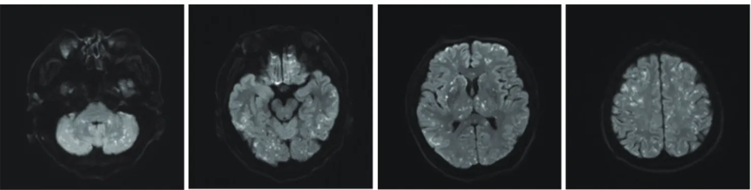
requiring endotracheal intubation and mechanical ven- tilation. A repeated brain CT scan showed no abnormal findings, and brain DWI also demonstrated no abnor- malities (Fig. 2). On the second day, chest CT showed no definite evidence of pulmonary thromboembolism and echocardiography showed no abnormalities in regional wall motion and valvular function. On the third day, a neurologist was consulted regarding the patient’s di- minished mental status. On a neurological examination, the patient had roving eye movements and decerebrate posturing. Electroencephalography was done to exclude non-convulsive status epilepticus, but there was no epi- leptiform discharge. Follow-up brain MRI was conduct- ed, and there were innumerable punctate foci of restricted diffusion, producing a “starfield” appearance on DWI (Fig.
3). After the diagnosis of CFE, he received steroid treat- ment, but his mental status did not change. Neuropsycho- logical testing was not possible because of mutism. After a month with no change of consciousness, he opened his eyes spontaneously, but could not make comprehensible sounds. Follow-up MRI performed 40 days after trauma showed slight resolution of the previous multifocal fat embolism and interval development of hypoxic-ischemic insults. He was discharged to another hospital for rehabil- itation with no change of consciousness.
DISCUSSION
FES is an uncommon, but potentially lethal condition that occurs in patients with long bone and pelvic frac- tures. FES, which was first described by Zenker in 1861, is characterized by major features such as altered mental status, hypoxemia, and petechial rash, as well as minor features such as tachycardia, pyrexia, retinal petechia, a sudden drop in hemoglobin levels, sudden thrombocyto- penia, oliguria, a high erythrocyte sedimentation rate, and fat globules in the sputum [2]. The most severe manifes- tation of FES is CFE, which can lead to an altered mental status on a spectrum from confusion to coma. The clini- cal symptoms of CFE usually develop gradually within 24 to 72 hours after trauma [3]. Hypoxemia can lead to the requirement for endotracheal intubation and mechanical ventilator use. A petechial rash is observed in the con-
Fig. 1. X-rays show fractures of the right femur intertrochanter and theleft femur shaft.
128
https://doi.org/10.20408/jti.2020.0007Journal of Trauma and Injury Volume 34, Number 2, June 2021
junctiva, around the neck, the axillary area, and the up- per body in 20–50% of FES patients [4]. In our case, the decline of the patient’s consciousness first occurred and deteriorated rapidly within a few hours after trauma, and then his mental status became comatose. We performed endotracheal intubation and applied a mechanical venti- lator because of the hypoxemia.
The pathophysiology of CFE involves high pressure in the marrow due to fracture or orthopedic surgery forc- ing marrow fat into the veins. The disruption of mar- row-containing long bone allows globules of marrow fat to pass into the venous sinuses that drain the marrow and then into the systemic circulation [5]. Having reached the circulation, fat may cause symptoms as a result of embolic occlusion of arteries in the lung, brain, skin, and elsewhere [6].
CFE occurs after fat emboli enter the arterial circula- tion. Fat globules may enter the arterial circulation by two mechanisms. First, fat globules can enter the left atrium
directly from the right heart through a shunt, such as a patent foramen ovale (PFO). Second, microglobules of fat may filter directly through the lung capillaries to reach the arterial system. These microemboli are small and malleable and may not lead to significant pulmonary in- jury. There is direct evidence of the passage of fat through a PFO, yet the absence of PFO in many patients with CFE supports the latter mechanism in some patients [7].
Therefore, PFO should be considered an additional risk factor for CFE, but is not necessary for CFE to occur [6].
In our case, echocardiography demonstrated no intra-car- diac shunt.
CFE is a clinical diagnosis, but specific findings on neuroimaging studies can be strongly supportive. Brain CT shows generally normal findings in most cases, but brain MRI is more sensitive. The most distinctive brain MRI finding is the starfield pattern, with scattered foci of high-intensity restricted diffusion on DWI. This is most apparent in the acute phase, from 4 hours to the first few
Fig. 3. Follow-up magnetic resonance diffusion-weighted images on the third day after injury demonstrate multiple hyperintense dots in the bilateral
cerebellum; both hemispheres; the cerebellar peduncle; pons; both basal ganglia; both thalami; both hippocampi; both frontal, temporal, occipital, and parietal cortices; and white matter area.
Fig. 2. Initial magnetic resonance diffusion-weighted images 3 hours after trauma display no abnormal findings.
129
http://www.jtraumainj.org