CASE REPORT
164 Copyright ⓒ 2008 Korean Neurological Association
Print ISSN 1738-6586 / On-line ISSN 2005-5013 10.3988/jcn.2008.4.4.164 J Clin Neurol 2008;4:164-166
Gradient-Echo MRI in Defining the Severity of Cerebral Fat Embolism
Jun Lee, MD
Department of Neurology, Yeungnam University School of Medicine, Daegu, Korea
Received April 21, 2008 Revised August 25, 2008 Accepted August 25, 2008 Correspondence Jun Lee, MD
Department of Neurology, Yeungnam University School of Medicine,
317-1 Daemyeong-dong, Nam-gu, Daegu 705-717, Korea
Tel +82-53-620-3680 Fax +82-53-627-1688 E-mail [email protected]
BackgroundᄏA few studies have found that abnormal findings on diffusion-weighted mag- netic resonance imaging (MRI) are useful for diagnosing cerebral fat embolism in the acute stage.
Case ReportᄏWe applied serial MRI to a case of cerebral fat embolism with cognitive im- pairment lasting for 2 months. Although marked resolution of the previous abnormal findings was demonstrated, T2*-weighted gradient-echo MRI revealed multiple tiny lesions.
ConclusionsᄏWe suggest that T2*-weighted gradient-echo MRI is useful in defining the cli- nical severity of patients with cerebral fat embolism. J Clin Neurol 2008;4:164-166 Key Wordsᄏmagnetic resonance imaging, embolism, fat, intracranial embolism.
Introduction
Cerebral fat embolism is an uncommon but serious compli- cation of long-bone fractures. Although such embolisms are diagnosed on the basis of clinical manifestations accompani- ed by hypoxemia, neurologic dysfunction, and petechiae, their neurologic symptoms are variable and often nonspecific.
There are several reports of magnetic resonance imaging (MRI) being a useful diagnostic tool for assessing cerebral fat embolism,1,2 with a few reports emphasizing that abnor- mal findings on diffusion-weighted MRI (DWI) have signi- ficant diagnostic value in acute cerebral fat embolism.3,4 The pathogenesis of disruption of the blood-brain barrier by ce- rebral fat embolism is still unclear, but it is thought that fat globules occlude the microvasculature, producing necrosis and hemorrhage in the surrounding parenchyma.5 Local in- flammatory and toxic reactions by free fatty acid causes breakdown of the blood-brain barrier.6 Considering that the pathologic findings in cerebral fat embolism are characteriz- ed by multiple petechiae and purpura, and that T2*-weighted gradient-echo MRI is a sensitive method for detecting resi- dual hemorrhage, this MRI sequence might aid the diagnosis of cerebral fat embolism.
To the best of our knowledge, no previous study has em- phasized the diagnostic usefulness of gradient-echo MRI in
patients with cerebral fat embolism. This report presents a patient with cerebral fat embolism documented by the pre- sence of multiple low signals on T2*-weighted gradient-echo MRI, in whom there was marked resolution of the previous abnormal findings.
Case Report
A 54-year-old male suffered from bilateral femoral fractures due to a traffic accident. He was alert and well oriented on admission, but became stuporous with extensor posturing 36 hours after the accident. The findings of basic blood tests, including arterial blood gas, were normal, and the findings of chest X-rays were also unremarkable. Brain computer tomo- graphy (CT) performed 3 hours after the deterioration showed normal findings. MRI was performed 10 hours later, with DWI, T2-weighted imaging (T2WI), and fluid-attenuated in- version recovery (FLAIR) images showing multiple high- signal lesions in the bilateral hemisphere.
Multiple lesions were prominent in the subcortical and deep white matter. Other lesions were observed in the thalamus, basal ganglia, corpus callosum, and occipital cortex (Fig. 1).
Electrocardiography and echocardiography showed normal findings. Three days later, multiple petechiae were detected on the skin of the anterior chest.
Lee J
www.thejcn.com 165 The patient received supportive therapy, and his condition
improved slowly. Neuropsychologic testing was not possible due to poor cooperation from the patient. His severe cogni- tive impairment, which included disorientation, apraxia, ac- alculia, and memory impairment, lasted for 2 months. Follow- up MRI performed 50 days after the onset of the cognitive impairment showed marked resolution of previous abnormal high-signal lesions and a few subtle remaining white-matter lesions (Fig. 2). In addition, T2*-weighted gradient-echo MRI revealed multiple tiny low-signal lesions distributed over the areas where the previous abnormal lesions had been located.
Discussion
Fat embolism syndrome has nonspecific and variable cere- bral manifestations, including headache, lethargy, irritability,
convulsions, and coma. Despite the marked neurologic dys- function in our patient, there were no obvious pulmonary symptoms or signs. There are several reports of isolated neu- rologic disorder occurring after cerebral fat embolism.7 The history of traumatic bone fractures, delayed onset of a non- focal neurologic dysfunction, the appearance of petechiae over the anterior chest, and marked abnormal signals over conspicuous sites on MRI supported the diagnosis of cere- bral fat embolism in this patient.
Small, scattered, hyperintense lesions on T2WI and FL- AIR imaging have been found previously.2-4 These lesions typically appear in the periventricular, subcortical, and deep white matter, as well as in the deep gray matter, including the corpus callosum.8-10 The lesions are usually not detected by CT and are more prominent on DWI than T2WI and FLAIR images, especially during the acute stage. These abnormalities
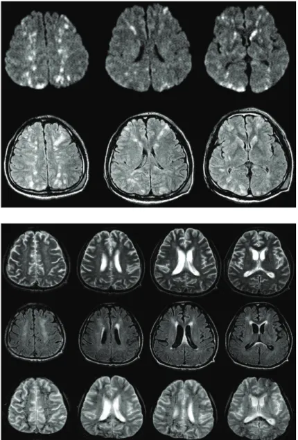
Fig. 1. Multiple scattered high signals were evident on DWI and FLAIR images obtained 10 hours after the on- set of drowsiness. DWI: diffusion-weighted MRI, FLAIR:
fluid-attenuated inversion recovery.
Fig. 2. T2WI, FLAIR, and T2*-weighted gradient-echo MRI was performed on day 50. Most of the lesions had disappeared, but ill-defined high-signal lesions remained in the periventricular and subcortical white matter and in the cortex. T2*-weighted gradient-echo MRI showed nu- merous punctate low-signal lesions in the centrum se- miovale, subcortical white matter, periventricular white matter, and corpus callosum. T2WI: T2-weighted imag- ing, FLAIR: fluid-attenuated inversion recovery.
Gradient-Echo MRI in Cerebral Fat Embolism
166 J Clin Neurol 2008;4:164-166
in the signal intensity on DWI presumably reflect the foci of cytotoxic edema. Clinical studies by Simon et al. and But- teriss et al. suggested that focal parenchymal hemorrhage is not a predominant feature in T2*-weighted gradient-echo MRI.8,9 However, their cases might not accurately reflect the full spectrum of patients with cerebral fat embolism because they showed relatively good clinical improvement. The num- ber of white-matter lesions on MRI is correlated with the patient’s score on the Glasgow Coma Scale.10 Simon et al.
considered that gradient-echo imaging might be useful in determining the severity of cerebral fat embolism, although hemorrhage was not a predominant feature of the multiple white-matter lesions in their reported case.9 Neuropsycholo- gical testing has revealed obvious cognitive deficits in pa- tients with stenosis of the internal carotid artery and lesions on the periventricular, subcortical, and deep white matter.11 Periventricular and subcortical white-matter lesions have been associated with worse performance on all cognitive measures tested.12 The patient in the present study exhibited continuous neurologic impairment, and T2*-weighted gradient-echo MRI showed that the lesions were distributed throughout the white matter of the cerebrum.
Reports of serial MRI and gradient-echo MRI in cases of cerebral fat embolism are rare. Previous studies of this con- dition found improvement of the neurologic impairment and the gradually disappearance of lesions on MRI within a few weeks to a few months. These findings might reflect imme- diate cytotoxic edema secondary to ischemia and subsequent vasogenic edema developing at a later stage.4 The patient in the present study presented with persistent cognitive impair- ment during the follow-up period. Follow-up MRI (T2WI and FLAIR) showed that some lesions disappeared, with only subtle ill-defined signal changes remaining. These findings did not explain the persistent neurologic impairment. A T2*- weighted gradient-echo MRI sequence is sensitive to hemo- siderin deposits in the brain parenchyma and is very useful for assessing the presence of old and new hemorrhage.13,14 Gradient-echo MRI sequences were not obtained during the initial MRI examination in this patient. However, T2*-wei- ghted gradient-echo MRI in the subacute and chronic stages of the follow-up MRI study in this patient revealed promi- nent multiple low signals, which might explain the residual neurologic impairment.
The findings of the present study suggest that T2*-weight- ed gradient-echo MRI can be used to analyze the neurologic impairment in patients with cerebral fat embolism, with the
findings also closely reflecting the clinical severity. Gradient- echo imaging is useful for detecting the petechial hemorr- hage that is the characteristic pathologic finding of cerebral fat embolism. Gradient-echo MRI might be useful in defin- ing the clinical severity of cerebral fat embolism with diffuse petechial lesions in the subacute or chronic stage, especially when abnormal signals are not detected in other forms of MRI.
In conclusion, MRI can be used for the early diagnosis of cerebral fat embolism with high sensitivity. We suggest that T2*-weighted gradient-echo MRI provides useful informat- ion for determining the clinical severity of patients with cere- bral fat embolism.
REFERENCES
1. Marshall GB, Heale VR, Herx L, Abdeen A, Mrkonjic L, Powell J, et al. Magnetic resonance diffusion weighted imaging in cerebral fat embolism. Can J Neurol Sci 2004;31:417-421.
2. Parizel PM, Demey HE, Veeckmans G, Verstreken F, Cras P, Jorens PG, et al. Early diagnosis of cerebral fat embolism syndrome by dif- fusion-weighted MRI (starfield pattern). Stroke 2001;32:2942-2944.
3. Ryu CW, Lee DH, Kim TK, Kim SJ, Kim HS, Lee JH, et al. Cerebral fat embolism: diffusion-weighted magnetic resonance imaging find- ings. Acta Radiol 2005;46:528-533.
4. Eguia P, Medina A, Garcia-Monco JC, Martin V, Monton FI. The value of diffusion-weighted MRI in the diagnosis of cerebral fat embolism.
J Neuroimaging 2007;17:78-80.
5. Kamenar E, Burger PC. Cerebral fat embolism: a neuropathological study of a microembolic state. Stroke 1980;11:477-484.
6. Kim HJ, Lee CH, Kim HG, Lee SD, Son SM, Kim YW, et al. Re- versible MR changes in the cat brain after cerebral fat embolism induc- ed by triolein emulsion. AJNR Am J Neuroradiol 2004;25:958-963.
7. Kamano M, Honda Y, Kitaguchi M, Kazuki K. Cerebral fat embolism after a nondisplaced tibial fracture: case report. Clin Orthop Relat Res 2001;206-209.
8. Butteriss DJ, Mahad D, Soh C, Walls T, Weir D, Birchall D. Reversi- ble cytotoxic cerebral edema in cerebral fat embolism. AJNR Am J Neuroradiol 2006;27:620-623.
9. Simon AD, Ulmer JL, Strottmann JM. Contrast-enhanced MR imag- ing of cerebral fat embolism: case report and review of the literature.
AJNR Am J Neuroradiol 2003;24:97-101.
10. Takahashi M, Suzuki R, Osakabe Y, Asai JI, Miyo T, Nagashima G, et al. Magnetic resonance imaging findings in cerebral fat embolism:
correlation with clinical manifestations. J Trauma 1999;46:324-327.
11. Kim JE, Lee BR, Chun JE, Lee SJ, Lee BH, Yu IK, et al. Cognitive dysfunction in 16 patients with carotid stenosis: detailed neuropsy- chological findings. J Clin Neurol 2007;3:9-17.
12. Desmond DW. Cognition and white matter lesions. Cerebrovasc Dis 2002;13 Suppl 2:53-57.
13. Parizel PM, Van Goethem JW, Ozsarlak O, Maes M, Phillips CD.
New developments in the neuroradiological diagnosis of craniocere- bral trauma. Eur Radiol 2005;15:569-581.
14. Kim BJ, Lee SH. Silent microbleeds and hemorrhagic conversion of an embolic infarction. J Clin Neurol 2007;3:147-149.