Immunomodulatory Effects of Fucoidan on NK Cells in Ovariectomized Rats
Seung Namkoong
1, Ye-Jin Kim
2, Taeseong Kim
2and Eun-Hwa Sohn
2*
1
Department of Physical Therapy, Kangwon National University, Samcheok 245-710, Korea
2
Department of Herbal Medicine Resource, Kangwon National University, Samcheok 245-710, Korea
Abstract - In this study we investigated the effects of supplementation with fucoidan from brown alga on the function of natural-killer (NK) cells to evaluate the possibility as an immunomodulator in ovariectomized (OVX) rats. A total of 18 female Wistar rats (six weeks) were used this study and 12 rats were OVX, and the rest of rats were sham-operated. The sham and one OVX group were fed standard diet, and the remaining OVX group received fucoidan (0.05% supplemented diet). After 12 weeks of supplementation, rats were sacrificed to assess the tumoricidal activity of the NK cells and the NO-iNOS regulation from splenocytes. The mass of body and the immune organs such as spleen and thymus were also studied. In OVX rats, body and thymus weights increased, however fucoidan supplementation did not change the body mass and organs weight compared to OVX group. Fucoidan supplementation increased NK cell activity and reduced NO-iNOS production in OVX rats. Ex vivo treatment of fucoidan increased NK cell activity in splenocytes from shame and OVX rats.
Ex vivo, we confirmed that fucoidan partially reduced the NK cell activity in the presence of iNOS inhibitors in OVX-splenocytes. These results indicate fucoidan supplementation has a NK cell tumoricidal activity, which are regulated by the iNOS production in OVX rats. This suggests that fucoidan is useful for potential therapeutic strategies as a nutrient in regulating the NK cells in postmenopausal osteoporosis patients.
Key words - Antitumor effect, In vivo, iNOS, Osteoporosis
*Corresponding author. E-mail : [email protected]
Introduction
Aging plays an important role in many biological functions within a body, and it is the main predictor for osteoporosis.
With advancing aging and particularly with post-menopausal conditions, the activity of osteoclasts, cells that break down bone through bone resorption, is greater than that of osteoblasts, cells responsible for bone formation, in women. This ultimately gives rise to osteoporosis (Yamaguchi, 2006). Aging is also associated with a decline in immune function, known as im- munosenescence, that contributes to the increased susceptibility to infection, cancer, and autoimmune diseases observed in humans (Mocchegiani and Malavolta, 2004; Sohal and Allen, 1990). Aging-related alterations of the cellular components of innate immunity might therefore be involved in the impair- ment of adaptive immunity observed in the elderly (Mocchegiani and Malavolta, 2004; Jang et al., 2010).
Ovariectomy induced bone loss in rats and postmenopausal bone loss in humans share many similar characteristics, and similar skeletal response to therapy with 17 β-estradiol (Kalu, 1991; Kalu et al., 1991). These similarities are strong evidence that the OVX rat bone loss model is suitable for studying the prevention and treatment of postmenopausal bone loss.
NK cells are an important component of the innate immune system and mediate cytolytic activities against tumors and virus-infected targets. Recently increasing evidences indicate that NK cells may play a pivotal role in some female pre- dominant diseases or in normal physiological conditions. Some studies suggest that estrogen may be involved in the regulation of immunocompetence in immune system and it modulates activities of NK cells (Nakaya et al., 2006). Baral et al. also found that estrogen reduced NK cell cytotoxicity against tumor targets but this effect was dependent on duration of exposure to estrogen (Baral et al., 1995).
The brown seaweeds, which have been consumed as a food
or relish to provide a dietary fiber source, are the most important
economic seaweeds cultured in Asia. Fucoidan, which is found in extracts from brown seaweeds (e.g., Ascophyllumnodosum and Fucus vesiculosus), is a polyanionic macromolecule com- posed predominantly of sulfated fucose moieties. In recent years, fucoidan has been extensively studied due to its numerous biological activities including its anticoagulant, antithrombotic, antitumor, antiviral, anti-complement and anti-inflammatory effects (Patel et al., 2002; O’Leary et al., 2004; Mahony et al.,1991; Baba et al., 1988).
Although these favorable effects of fucoidan have been reported and evaluated, the effects of fucoidan on activity of NK cells in osteoporosis have not been evaluated. The estrogen- deficient ovariectomized (OVX) osteoporosis model is useful for evaluation of osteoporotic drugs because several bone- related parameters are clearly decreased by ovariectomy within 10 to 14 weeks after the operation. In this model, we examined the effects of fucoidan on the NK cell function in OVX rats and identify the possible involvement of iNOS that may mediate the regulatory effect of NK cell.
Materials and Methods
Experimental animals and diets
Female Wistar rats (six weeks) were used for the experiment.
The animals were kept for 5 days before the onset of the experiment to acclimatize to our laboratory conditions (the room temperature was 25℃ with a 12 h/12 h light/dark cycle), then 12 rats were OVX and 6 rats were sham-operated under anesthesia with intraperitoneal (IP) injection of sodium pento- barbital at a dose of 30 mg/kg body weight. After operation, rats were kept in separate cages and fed standard diet before being sacrificed. To prevent hyperphagia associated with ovariectomy, the OVX rats were pair-fed to the mean intake of those in the sham group (Wronski et al., 1987). All rats were allowed free access to drinking ion-exchanged distilled water for the duration of the whole experiment. The sham and one OVX group were fed standard diet, and the remaining OVX group received fucoidan (0.05% fucoidan modified AIN- 93 M diet) supplemented diet. We confirmed that the ovar- iectomy significantly decreased (t-test, p<0.05) bone mineral density at 10 weeks after surgery. The animals were sacri- ficed after ether anesthesia at 12 weeks. Spleen samples were
collected aseptically for the NK cell assays. The rats were observed daily for clinical signs. None of the rats showed any abnormal growth and behavior and no sign of illness. Animal care and all experimental protocols were performed following the Institute for Laboratory Animal Research (ILAR) guideline.
Preparation of fucoidan and Reagents
Unless stated otherwise, all chemicals were purchased from the Sigma Chemical Co. (St Louis, MO). Fucoidan ex- tracted from Fucus vesiculosus was also purchased from Sigma-Aldrich (St. Louis, MO) and dissolved in distilled water. The RPMI 1640 medium and fetal bovine serum (FBS) were purchased from GIBCO (Grand Island, NY). The XTT {2,3-Bis(2-methoxy-4-nitro-5-sulfophenyl-2H-tetrazolium- 5-carboxanilide inner salt} cell viability assay kit was purchased from WelGENE (Daegu, South Korea). All the tissue culture reagents and fucoidan were assayed for any endotoxin con- tamination using the Limulus lysate test (E-Toxate, Sigma) for in vitro assay, and the endotoxin levels were found to be <
10 pg/ml.
NK cell-mediated tumoricidal activity
A modification of the method reported by Mosmann et al.
was used (Mosmann, 1983; Namkoong et al., 2011). The
spleens were aseptically removed and dissociated into a single-
cell suspension in a culture medium. The concentration was
adjusted to 2×10
5cells/ml. The culture medium was RPMI
1640 containing 10% heat-inactivated FBS, penicillin (100
IU/ml) and streptomycin (100 μg/ml). Spleen cells from the
shame and OVX rats were tested as effector cells and YAC-1
mouse lymphoma cells (ATCC, Rockville, MD) were used as
the target cells. The NK cell assays were carried out in 96-
well v-bottom plates at effector/target (E:T) cells ratios 50:1
with 1×10
4of the target cells in a final well volume of 200 μl
at 37 ℃ for 4 h in a 5% CO
2humidified incubator. After
incubation of cells for 4 h, 20 µl of phenazine methosulphate
(PMS); electron-coupling reagent and 25 µl of XTT were
added to each well. The cells were further incubated for 3h to
allow XTT formazan production. The absorbance were deter-
mined with a microplate reader at a test wavelength of 450 nm
and a reference wavelength of 690 nm.
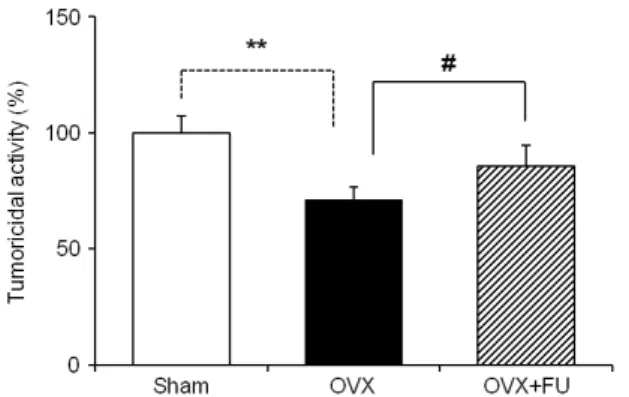
Fig. 1. Effect of the fucoidan (FU) supplementation on the NK cells tumoricidal activity from experimental rats. Splenocytes were cultured with Yac-1 tumor cells for 4 h. The NK cells tumoricidal activity examined at effecter/target cells ratio of 50:l by XTT assay. The data is expressed as a means ± S.E.M of quadruplicates of a representative experiment. ** P<0.01:
significantly different from the sham group.
#P<0.05: signifi- cantly different from the OVX group.
Fig. 2. The effects of the fucoidan (FU) supplementation on the production of NO in splenocytes from experimental rats. These results are means ± S.E.M of quintuplicates from a repre- sentative experiment. ** P<0.01: significantly different from the sham group.
##P<0.01: significantly different from the OVX group.
Western blot analysis
The amount of iNOS was measured by Western blot analysis.
Rats were immediately sacrificed and isolate spleen. After homogenized of spleen tissue and lysed in RIPA buffer.
Protein concentrations were measured using the DC Protein Assay (Bio-Rad Laboratories, Hercules, CA). Twenty micrograms of each sample was electrophoresed on 10% sodium dode- cylsulfate polyacrylamide gels and transferred to Hybond-ECL nitrocellulose membranes (Amersham Biosciences, Piscataway, NJ). The membranes were blocked with 5% skim milk in TBS-T for 1 h. The membranes were incubated with primary antibody against iNOS for 24 h. They were then washed with TBS-T once for 15 min and three times for 5 min and incubated with secondary HRP-conjugated anti-rabbit antibody for 1 h.
The membranes were washed again as described above. Auto- radiography was carried out using an enhanced chemilumi- nescence kit.
Statistical analysis
Each experiment was repeated at least two times, and the results of one representative experiment are shown. The results were expressed as means ± S.E.M. and analyzed via Student’s t test and ANOVA. The significant values are re- presented by an asterisk (*
, #p < 0.05 and **
, ##p < 0.01).
Results and Discussion
The most characteristic function of NK cells is non-MHC restricted killing of target cells, such as tumor, and thus NK cells are important for maintaining health in body (Borrego et al., 1999). It has been reported that aging attributed to various factors of NK cells. For example, NK cells exhibited decreased responsiveness to its positive modulator, IL-2, in aged group (Miller, 1991) and this explains the high frequency of cancer and infectious diseases with aging. Osteoporosis is one of the aging-related diseases (Yamaguchi, 2006). With advancing aging and particularly after menopause in women, the activity of osteoclasts, cells that break down bone, is greater than that of osteoblasts, cells that build bone. In this study, we deter- mined the tumoricidal activity of the NK cells in OVX and the regulatory effects of the supplementation with edible seaweed fucoidan. We showed that the increasing effects on NO-iNOS
production and the suppressive effect of NK cells tumoricidal activity in OVX rats compared to sham group. Furthermore, fucoidan supplementation in OVX group recovered all these effects such as NO-iNOS production and NK cells tumoricidal activity (Fig. 1-3).
Recently it has been reported that NK cell tumoricidal activity could be regulated by sex hormones. Yovel et al.
found that there was an inhibition of specific lysis of Yac-1
Fig. 3. The effects of the fucoidan (FU) supplementation on the expression iNOS in splenocytes and thymocytes from experi- mental rats. Spleen and thymus tissue lysate were determined by Western Blot assay.
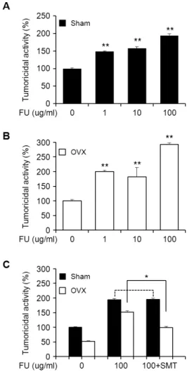
Fig. 4. The effects of fucoidan (FU) on the NK cell tumoricidal activity in the splenocytes from sham (A), OVX group (B) and the NK cell tumoricidal activity of FU in the absence or presences of s-methylisothiourea (SMT; 100 µM), iNOS in- hibitors (C). Splenocytes (5×10
4cells/well) were treated with various FU (1-l00 μg/ml or l00 μg/ml) or in the presence of SMT 4 h. The turmoridical activity was examined at E:T ratio of 50:1. The data represents the means ± S.E.M. of quadrupli- cate experiments. ** p <0.01, *p <0.05: significantly different from the control or FU alone treated cells.
target after a long-term exposure of the NK cells to 17β -estradiol (Yovel et al., 2001). In addition, several data in animal model studies also provided direct evidence that chronic administration of high doses of 17β-estradiol to mice resulted in a decreased activity of NK cells (Yovel et al., 2001). Thus, estrogen imbalance level caused change of NK cell cytotoxicity and the low NK activity induced by IL-2 was found to be a predictor of morbidity for various infection and cancer in osteoporosis (Levy et al., 1991; Kim et al., 2009).
To examine whether the effect of fucoidan on NK cell tumoricidal activity is mediated by NO production which is involved in iNOS expression, the splenocytes from sham and OVX rats were treated with fucoidan (100 μg/ml) in the absence or presence of s-methylisothiourea, preferential in- hibitor of iNOS. As shown in Fig. 4, the treatment of fucoidan resulted in a significant enhancement of NK cell activity in splenocyte from sham and OVX rats in dose dependant manners.
Moreover it appears that iNOS expression was involved in the regulation of NK cell activity in fucoidan-treated OVX splenocytes, whereas inhibition of iNOS has no effects of the NK cells activity in fucoidan-treated sham cells. These data imply that the estrogen-deficient stress activates innate immune cells such as NK cells and changed intracellular NO levels.
Thus it is speculated that fucoidan and cytokines produced by these cells could exert combinatory effects on NK cell activities in OVX state.
In previous study, red ginseng acidic polysaccharide (RGAP) alone had no effect on the function of macrophages for killing tumor cells. However, a combination of RGAP and recom- binant IFN- γ (rIFN-γ) enhanced their killing of tumor cells (Choi et al., 2008). There was a report that IFN-γ was a major product of murine NK cells during pregnancy and its pro- duction was enhanced by a long-term exposure of cells to estradiol. Therefore, we could postulate that hormonal im- balance such as estrogen-deficiency changed the secretion of various cytokines of NK cells. Moreover, the fucoidan exerted combinatory influence on such cytokines regulation for NK cell activities, and it was regulated by secondary signals such as NO-iNOS production.
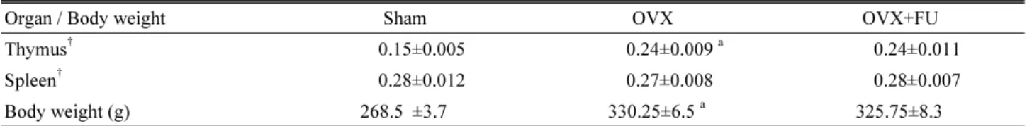
In our experimental systems using OVX rat model, as
shown in Table 1, ovariectomy significantly increased the
body mass and thymus weight index but not spleen compared
Table 1. Effects of fucoidan supplement on the body mass and index of immune organs in ovariectomized (OVX) rats
Organ / Body weight Sham OVX OVX+FU
Thymus
†0.15±0.005 0.24±0.009
a0.24±0.011
Spleen
†0.28±0.012 0.27±0.008 0.28±0.007
Body weight (g) 268.5 ±3.7 330.25±6.5
a325.75±8.3
†