CASE REPORT eISSN 2384-0293 https://doi.org/10.12701/yujm.2017.34.2.275
Yeungnam Univ J Med 2017;34(2):275-278YUJM VOLUME 34, NUMBER 2, DECEMBER 2017 275
신경계 증상을 동반한 부분적으로 자연완화된 소세포폐암
윤경현1, 송성헌2, 김충현1, 황찬희1, 이준호1, 최재형1, 김선영3
1동강병원 내과, 2제주한라병원 내과, 3동강병원 병리과
Partial spontaneous remission of small cell lung carcinoma with neurologic symptom
Kyung Hyun Yun1, Sung Heon Song2, Chung Hyoun Kim1, Chan Hee Hwang1, Jun Ho Lee1, Je Hyoung Choi1, Sun Young Kim3
1
Department of Internal Medicine, DongKang Medical Center, Ulsan;
2Department of Internal Medicine, Cheju Halla General Hospital, Jeju;
3
Department of Pathology, DongKang Medical Center, Ulsan, Korea
Small cell lung carcinoma (SCLC) is a cancer that shows aggressive behavior, early spread to distant sites, and frequent association with distinct paraneoplastic syndromes. Spontaneous remission of cancer, parti- cularly of SCLC, is a rare biological event. Cases involving spontaneous regression of SCLC were reported, and were associated with paraneoplastic syndromes of the nervous system. This article reports on a 78-year- old man with SCLC in remission, with neurological symptoms. The patient visited the hospital because of generalized weakness, and imaging studies revealed a mass in the lower lobe of the left lung, pathological evaluation showed SCLC. The patient refused oncologic treatment and was treated only with conservative care. In follow-up study the diameter of the mass had decreased from initial 32 mm, 9 months after admission to 20 mm, 17 months after admission to 13 mm. The patient kept complaining of generalized weakness, dizzi- ness, and paresthesia of limbs. We assumed that, in this case, the spontaneous remission of lung cancer was related to the immunologic response directed against the tumor, which is believed to be an important factor in the pathogenesis of paraneoplastic neurologic syndromes.
Keywords: Small cell lung carcinoma; Paraneoplastic syndrome; Spontaneous neoplasm regression
Copyright ©2017 Yeungnam University College of Medicine
This is an Open Access article distributed under the terms of the Creative Commons Attribution Non-Commercial License (http://creative- commons.org/licenses/by-nc/4.0/) which permits unrestricted non-commercial use, distribution, and reproduction in any medium, provided the original work is properly cited.
Received: March 29, 2016, Revised: September 25, 2016 Accepted: September 26, 2016
Corresponding Author: Sung Heon Song, Department of Internal Medicine, Cheju Halla General Hospital, 65 Doreongno, Jeju 63172, Korea
Tel: +82-64-740-5009, Fax: +82-64-743-3110 E-mail: [email protected]
서 론
폐암은 전 세계적으로 가장 흔한 암사망 원인으로 암사망 원인의 19.4%를 차지하며[1], 국내의 경우도 폐암에 의한 사망률은 남, 녀 모두 1위로 예후가 매우 불량하다. 특히 소세
포폐암의 경우 임상경과가 공격적인 특성이 있어 폐암 중에 서도 예후가 가장 나쁘다고 알려져 있다.
악성종양이 자연완화 되는 것은 극히 드문 일이다[2]. 국내 에서 폐암이 부분 또는 완전 자연완화는 비소세포폐암에서 보고된 예는 다수 있으나, 소세포폐암이 자연완화 되는 것은 최근까지 2건의 증례 보고가 있을 정도로 매우 희귀하다[3,4].
소세포폐암의 자연완화는 신경계 증상을 호소하는 환자에서 신생물딸림신경병증에 의해 유발된 면역반응이 자연완화와 관련성이 있다고 추측하고 있으며, 이런 환자에서는 전신위 약, 이상 감각 등의 증상이 두드러졌다고 보고되었다[5-7].
저자들은 잘 설명되지 않았던 전신위약과 이상 감각을 호
Kyung Hyun Yun et al.
276 YUJM VOLUME 34, NUMBER 2, DECEMBER 2017
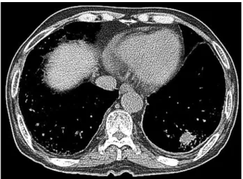
Fig. 1. Irregular margined mass (32 mm) in the left lower lobe.
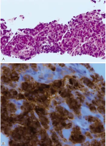
Fig. 2. Histologic finding. (A) The tumor cells have round or ovoid-shaped hyperchromatic nuclei and small amount of eosino- philic cytoplasm (H&E stain, ×200). (B) The tumor cells are posi- tive for synaptophysin (immunohistochemical stain, ×400).
소하였던 소세포폐암이 부분적으로 자연완화된 1예를 경험 하였기에 이를 문헌고찰과 함께 보고한다.
증 례
환 자: 남자, 78세
주증상: 전신 위약감과 사지 말단의 저림 증상
현병력: 당뇨병과 뇌경색으로 신경과에서 경과 관찰 중, 수개월 전부터 전신 위약감이 있었으며, 내원 1개월 전부터 증상이 악화되고 어지럼증도 동반되어 신경과에 입원하였 다. 뇌자기공명영상(magnetic resonance imaging)에서 새롭 게 발생된 병변은 관찰되지 않았으나, 흉부엑스선에서 좌하 엽에 음영이 증가된 소견이 관찰되어 흉부 컴퓨터단층촬영 (computed tomography, CT)을 하였고, 좌하엽에서 폐종괴 가 관찰되어 호흡기내과로 전과되었다.
과거력: 당뇨병 및 뇌경색
사회력: 흡연력은 50갑년이었으나, 7년 전 금연을 하였으 며, 음주는 하지 않음.
가족력: 특이사항이 없음.
신체검사: 혈압 110/70 mmHg, 맥박 112회/분, 호흡수 20 회, 체온 37.3℃였다. 양측 폐하부에 수포음이 들렸으며, 복 부 진찰에서 이상소견은 없었다.
검사실 소견: 말초혈액검사에서 백혈구 7,300/mm3, 혈색 소 10.8 g/dL, 혈소판 142,000/mm3였다. 혈청생화학검사는 알부민 3.5 g/dL, 총단백 5.6 g/dL, C-반응단백질 0.85 mg/dL, 적혈구침강속도 60 mm/hr, 감마-글루타밀트란스펩티데이즈 (gamma glutamyltranspeptidase) 64 IU/L, 칼슘 8.0 mg/dL, 인 3.0 mg/dL였으며, 그 이외 다른 혈액 검사는 정상이었다.
흉수검사에서 색깔은 볏짚색이었으며, 총 단백 3.4 g/dL, 젖산탈수소효소 287 IU/L로 삼출액 소견이었고, 포도당 162 mg/dL, 비중 1.029, pH 7.5, 백혈구 480/mm3였다. 백혈구는 호중구 11%, 림프구 71%, 호산구 8%, 대식세포 9%, 중피세 포 1%의 분획을 보였다. 아데노신아미노기제거효소(adeno- sine deaminase) 22.5 IU/L, 항산균도말검사 음성, 배양검사에 서 동정되는 미생물은 없었으며, 세포검사에서 악성세포는 관찰되지 않았다.
영상 소견: 처음 촬영한 흉부 CT에서 좌측하엽으로 32 mm 크기의 폐종괴가 관찰되었으며(Fig. 1), 폐는 전반적으로 폐기종이 있었고, 양측으로 흉수도 관찰되었다. 양전자방출 컴퓨터단층촬영(positron emission tomography-computed to- mography)에서 폐종괴의 포도당최대표준화섭취계수(maxi- mum standardized uptake value) 값은 19였고, 림프절이나 다른 장기로 전이된 소견은 관찰되지 않았다. 뇌 자기공명영 상에서 새롭게 발생된 뇌경색소견이나 전이암은 관찰되지
Spontaneous remission of SCLC
YUJM VOLUME 34, NUMBER 2, DECEMBER 2017 277
Fig. 3. Decreasing size of the LLL mass lesion (13 mm).
않아 임상적으로 T2aN0M0 병기의 제한병기로 확인되었다.
조직 소견: 현미경 소견에서 종양세포는 원형 혹은 난원형 의 과다염색 핵과 적은 양의 호산성 세포질을 보였다(Fig.
2A). 면역조직화학염색상 종양세포는 CD56와 synaptophysin 에 양성소견을 보였고(Fig. 2B), cytokeratin 7에는 일부 종양 세포가 양성이었다. TTF-1와 napsin A에서는 음성이었다.
조직소견과 면역조직화학염색소견은 소세포폐암에 잘 맞는 소견이었다. 추가로 시행된 흉수세포검사에서 악성세포는 관찰되지 않았다.
치료 및 경과: 환자와 보호자는 환자가 고령 및 뇌졸중의 과거력이 있어 항암 치료 및 방사선 치료를 하지 않고 증상 조절만 원하여 대증적인 치료를 하였다. 혈당조절은 잘 되었 으나, 전신 위약은 지속되었고, 어지럼증과 사지말단의 저림 감각도 간헐적으로 있었다. 그러나 뇌경색이 재발한 소견은 없었다. 외래에서 경과관찰 중 종괴 크기는 9개월째 20 mm 로 감소되었고, 17개월째 흉부 CT에서 좌측하엽의 종괴는 13 mm로 감소되었다(Fig. 3). 종양의 크기는 59.4%가 감소되 어 고형암에 적용되는 반응평가도구인 response evaluation criteria in solid tumor (RECIST) 기준에 따라 부분자연완화로 판정하였다. 환자는 외래에서 계속 추적 관찰 중이다.
고 찰
악성종양의 자연완화는 암에 대한 특정한 치료 없이 악성 종양이 소실 혹은 위축되는 것으로 정의한다. 악성종양이 수술, 방사선 치료, 항암제 등을 이용한 치료 없이 자연적으로 완화하는 것은 60,000-100,000명당 1명 꼴로 발생할 정도로 매운 드문 일이다[2,8]. 자연완화가 가능한 것으로 알려져
있는 종양은 신장암, 신경모세포종, 악성흑색종, 융모막암종 등이다.
악성종양의 자연완화기전은 확실히 입증된 바는 없지만 어떤 요인들로 인하여 면역반응이 유도되면서 자연완화를 일으키지 않을까 추정하고 있다. 이러한 기전은 1) 싸이토카 인이 분비되고 세포매개면역반응이 발생하여 종양세포와 조 직에 염증반응을 일으키고 T 세포에 의해 매개된 종양세포 세포자멸사를 유도하거나, 2) 종양에서 발현된 항원에 의해 활성화된 anti-idiotypic antibodies이 중요한 역할을 한다고 설명하였다[4]. 이 외에 가능한 기전으로 분화, 호르몬의 영향, 신생혈관생성방해, 끝분절효소방해, 정신신경면역학적인요 소, 그리고 조직 검사나 외과 외상에 의하여 면역학적 반응이 발생하여 자연완화가 발생한다고 보고되었다[8-10]. 그러나, 현재까지는 암의 자연완화에 대한 명확한 기전이 확립되어 있지 않다.
국내에서 보고된 폐암의 자연완화는 부분적 자연완화를 포함하여 최근까지 5건이 보고되었으며, 소세포폐암의 자연 완화는 2건이 보고되었다[3,4]. 국내에서 보고된 증례는 소세 포폐암의 자연완화 기전으로 신생물딸림증후군으로 인해 면 역체계가 변화되어 자연완화된 것으로 추측하기도 하였다 [3]. 이러한 가설은 소세포폐암에서 신경계 이상증상을 호소 하는 신생물딸림신경학증후군(paraneoplastic neurological syn- drome)이 나타난 환자들에서 항 Hu 항체 양성 소견을 확인하 였고, 이 항체로 인한 면역반응이 발생되어 소세포폐암이 자 연완화하였다고 설명하였다[5,11]. 신생물딸림신경학증후 군은 원인을 알 수 없는 신경학적 증상이 종양과 동반된 경우 를 이르는 것으로, 종양의 전이, 성장, 대사성 독소와 직접적인 관계가 없이 원격 효과에 의해 발생하는 신경학적 증후군으 로 정의할 수 있다[12]. 신생물딸림신경학증후군 환자들은 항암 치료에 대한 반응이 양호한 것으로 보고되기도 하였다 [13]. 본 증례는 뇌경색이 재발한 소견이 없고, 혈당 조절은 비교적 양호하였으나, 전신위약감이 지속되고 저림과 이상 감각을 호소하였다는 점에서 신생물딸림신경학증후군으로 판단되며, 확인하지는 못했지만 항 Hu 항체의 항종양면역매 개반응(anti-tumor immune mediated response)이 자연완화 의 기전으로 추측해 볼 수 있다. 그러나 현재까지 이러한 가설은 증례 보고 수준에 불과하여 명확하게 그 기전이 설명 되었다고 하기는 어렵다. 소세포폐암의 자연완화의 다른 기 전으로 CD8-positive T 세포에 매개된 세포매개면역반응이 중요한 역할을 하지 않을까 제시되기도 하였다[14]. 소세포 폐암과는 달리 비소세포폐암은 NY-ESO-1 특이면역반응이
Kyung Hyun Yun et al.
278 YUJM VOLUME 34, NUMBER 2, DECEMBER 2017
나타난 선암종에서 폐암의 자연완화가 보고되었다[15].
소세포폐암의 5년 생존율은 제한병기일 때 10-13%이며, 확장병기일 때 1-2%로 예후가 좋지 않으며, 제한병기라 하더 라도 치료받지 않을 경우에는 중앙 생존 기간이 12주로 보고 될 정도로 공격적인 암이다. 치료는 제한병기일 경우 항암 치료와 방사선 치료를 동시에 진행하게 되며, 확장병기에는 항암치료를 하는 것이 일반적이다. 본 증례는 제한병기였으 나 환자가 고령이며, 뇌경색과 당뇨병을 앓고 있어 항암 치료 와 방사선 치료를 하지 않고 보존적 치료를 하며 경과관찰만 하였다.
본 증례는 예후가 나쁜 소세포폐암의 경우에도 부분 완화 이기는 하지만 자연완화가 일어날 수 있음을 보여주었으며, 폐암치료의 최근 진전과 함께 자연적으로도 완화가 일어날 수 있는 폐암의 임상적 특징과 그 기전이 확인된다면 폐암을 치료하는데 더 도움이 될 수 있을 것이다.
CONFLICT OF INTEREST
No potential conflict of interest relevant to this article was reported.
REFERENCES