Changes in the Concentration and Localization of Accumulated Mercury in Kidney, Liver, and Spleen of Mice over Time
Yu Seon Kim, Young Eun Kim and Hyun Wook Cho*
Department of Biology, College of Life Science and Natural Resources, Sunchon National University, Jeonnam 57922, Korea Received April 23, 2019 /Revised July 9, 2019 /Accepted August 7, 2019
This study investigated the localization and changes in the concentration of injected mercury in the kidney, liver, and spleen of mice. To evaluate changes in the concentration of mercury over time, the mice were euthanized 10, 150, and 300 days post-treatment. Localization of accumulated mercury was identified by the autometallography method. Mercury was densely located in the supranuclear cyto- plasm of epithelial cells of proximal tubules of the kidney but was not detected in the glomerulus 10 days post-treatment. In the liver, mercury was mainly found in hepatocytes around the portal vein and in sinusoidal Kupffer cells 10 days post-treatment. Mercury was scattered throughout both white and red pulp of the spleen 10 days post-treatment. In terms of changes in the concentration of mer- cury, the levels were lower in the renal cortex and medulla 150 and 300 days post-treatment as com- pared with those 10 days post-treatment. Mercury was found at low concentrations in liver hep- atocytes 150 and 300 days post-treatment. The mercury concentration was also low in both the white and red pulp of the spleen 150 and 300 days post-treatment. Therefore, the concentrations of accumu- lated mercury in the kidney, liver, and spleen 150 and 300 days post-treatment were lower than those 10 days post-treatment. We identified the localization of mercury in cells and tissues of several organs and observed that accumulated mercury in organs decreased naturally over time.
Key words : Autometallography, kidney, liver, mercury, spleen
*Corresponding author
*Tel : +82-61-750-3614, Fax : +82-61-750-3208
*E-mail : [email protected]
This is an Open-Access article distributed under the terms of the Creative Commons Attribution Non-Commercial License (http://creativecommons.org/licenses/by-nc/3.0) which permits unrestricted non-commercial use, distribution, and reproduction in any medium, provided the original work is properly cited.
Journal of Life Science 2019 Vol. 29. No. 8. 879~887 DOI : https://doi.org/10.5352/JLS.2019.29.8.879
Introduction
Mercury is a well-known toxic metal that tends to be re- leased into the environment via various industrial activities.
Reports have suggested that people can be exposed to mer- cury through whitening creams applied to the skin [3], amal- gam fillings [5, 33] used as dental materials, and by the in- gestion of certain types of fish [23, 37]. In humans, the main route of exposure to methylmercury is through ingestion of certain types of seafood and freshwater fish [12].
The major organs affected by mercury toxicity in mam- mals are the kidneys and the nervous system [15]. Mercury is also an environmental contaminant that has toxic effects on the immune system and the lungs [26]. Mercury can in- duce various metabolic changes, particularly in the nervous system, kidneys, and liver [28]. It has been reported that high concentrations of mercury can accumulate in the liver,
kidney, and brain [1]. Moreover, the concentration of mer- cury in the kidney has been reported to be up to five times higher than that in the liver [6]. It has also been observed that administration of mercury can bring about changes in the epithelial cells constituting the tubules in the kidneys in mice [32], along with changes in the hepatocytes in the liver [26]. In rats, various changes caused by mercury were observed in the kidneys [27, 32, 36] and liver [36]. In the spleen, although it was seen that the accumulation of mer- cury differs according to the mouse strain, the concentration of mercury accumulated in the spleen increased in pro- portion from the time of mercury administration [24]. As a result, the lymphocytes of the spleen have been reported to decrease in response to the accumulation of mercury [30].
In this study, we administered compounds containing mercury into pubertal mice once a week for a total of 3 weeks, after which the administration was stopped and the levels and the localization of mercury in the kidney, liver, and spleen were measured at various time points by auto- metallography [16, 17].
Materials and Methods
Experimental animals, mercury administration and perfusion
Six-week-old pubertal female mice (n=36) belonging to the ICR (Damul Sciences) strain were used for this experi- ment. Adolescent mice were established according to the cri- teria reported by Emanuele et al [20]. The average weight of the mice was 24 g. During the experimental period, solid feed and distilled water were supplied ad libitum. In addi- tion, the environment of the feeding room was maintained at a temperature of 22±2℃, with a relative humidity of 50±5%, and an alternating light-dark cycle of 12 hr each.
The experiment was conducted with the approval of the Ethical Committee of Animal Experimentation at Sunchon National University (Approval No. SCNU IACUC-2014-05).
For the administration of mercury into the mice, 0.01 mg of methylmercuric chloride (CH3HgCl) was dissolved in 0.1 ml of physiological saline solution, then subcutaneously in- jected into the back of the neck of the mice for 3 weeks at a dose of 0.01 mg of mercury per week. For the control group, 0.1 ml of physiological saline was injected subcuta- neously in the same manner.
The mice in the control and the mercury-treated groups were randomly divided into three groups after receiving the mercury doses for 3 weeks and were killed at 10, 150, and 300 days post treatment. After the euthanasia, 12 mice were used in the control (n=6) and the mercury treated (n=6) groups. Before perfusion, heparin was administered into each mouse intraperitoneally, followed by inhalation anes- thesia with diethyl ether. The anesthetized mice were then weighed and perfused. For perfusion, the needle was in- serted into the left ventricle, and the animals were perfused with a peristaltic pump (Spectra/Chrom MP-1) for 2 min using Ringer's solution. This was subsequently replaced with 4% glutaraldehyde (pH 7.4) solution and perfused for 20 min.
Tissue preparation and mercury staining
The kidneys, livers, and spleens were excised from the perfused mouse, placed on filter paper to remove the water, and then weighed and averaged. A p value of <0.05 was considered statistically significant in terms of organ weight.
Each organ was weighed and subsequently stored in 4% glu- taraldehyde (pH 7.4) at 4℃ until further use. The tissues of these organs were subsequently trimmed to the appro- priate size, rinsed with 0.1 M phosphate buffer (pH 7.4), de- hydrated with 70%(twice), 80%(once), 95%(twice) and 100%(thrice) ethanol at 1 hr intervals, followed by infiltration solution treatment, and then embedded in JB-4 solution
(Polysciences, Inc.).
After the embedding process, the tissues were sectioned at a thickness of 2.5 μm using a microtome (RMC Rotary Microtome, MT 990) and stained using an autometallog- raphy-based method [16, 17]. Briefly, tissue sections were treated with distilled water for 10 min and developed for 2 hr at 26℃. The samples were then processed for 55 min in distilled water, 12 min in 5% sodium thiosulfate, 10 min in distilled water, and 5 s in 0.5% toluidine blue at room temperature. Next, the sections were washed with distilled water for 10 min, dried in a slide warmer at 35℃ for about 1 hr, sealed with permount (Fisher Chemical), and observed under an optical microscope.
Measurement of mercury concentration
A SPOT camera (Model No. 11.2 Color Mosaic) attached to an optical microscope (Olympus BX 50) was used to exam- ine the localization of the accumulated mercury and the changes in the mercury concentration in the kidney, liver, and spleen. To measure the concentration ratio of the accu- mulated mercury, the color range was adjusted so that the area with the accumulated mercury could be seen clearly using the color range designation module of the Adobe Photoshop CS 5 program, after which the pixel value of the area with accumulated mercury was obtained. For calculat- ing the ratio, the pixel value was divided by the total pixel value of the photograph, then multiplied by 100 to obtain the ratio of the accumulated mercury in the tissue.
Statistical analysis
Statistical differences between the control and the mer- cury treated groups were analyzed via one-way ANOVA us- ing Microsoft Office Excel, and a differences with a p value of <0.05 were considered to be statistically significant.
Results
Localization of mercury compound in organs Compared to the control group, the mean weights of the kidneys, livers, and spleens in mice that were euthanized 10~150 days post treatment, were found to be similar or in- creased in the mercury-treated group. When the mice were euthanized at 300 days, the weight of each organ was found to be lower in all the mercury-treated mice than in the con- trol mice (Table 1). Microscopic observations of the renal tis- sue showed that the mercury accumulation was in the form
Fig. 1. Microscopic photographs of the renal cortex region be- fore and after mercury treatment. Images of the renal cortex of the control mice at (A) 10 days, and mer- cury-treated mice at 10 (B), 150 (C) and 300 (D) days post treatment. Mercury granules (indicated by arrows) were found to be accumulated in the epithelial cells of the proximal tubule. The accumulated mercury (indicat- ed by arrows) was mainly distributed around the nu- cleus of the proximal tubule epithelium, or from the nu- cleus to the free surface of the cell (B, inset). As shown in the enlarged magnification, mercury was distributed on the free surfaces of proximal tubular epithelial cells, which was mainly in contact the lumen (C and D, inset).
DT, distal tubule; GL, glomerulus; PT, proximal tubule.
Bars = 50 μm in A-D and 20 μm in B-D insets.
Table 1. The average organ weight of the control and mercury-treated mice taken each day post treatment
Organ Group Day post treatment
10 150 300
Kidney (g) Control
Mercury
0.25±0.01 0.29±0.02*
0.27±0.05 0.28±0.04
0.32±0.03 0.25±0.01*
Liver (g) Control
Mercury
1.82±0.18 2.58±0.23*
2.33±0.73 2.54±0.62
2.61±0.66 2.01±0.33
Spleen (g) Control
Mercury
0.14±0.02 0.22±0.05*
0.15±0.05 0.15±0.06
0.18±0.06 0.12±0.02 Values represent the means ± SD.
For obtaining the organ weights each day post treatment, 12 mice were used as control (n=6) and as mercury treated (n=6) specimens.
*p<0.05 compared with the control group of the same day post treatment.
Fig. 2. Microscopic photographs of the renal medulla region before and after mercury treatment. Images showing the renal medulla in the control mice at (A) 10 days post treatment, and mercury treated groups at (B) 10, (C) 150 and (D) 300 days post treatment. The accumulated mer- cury (indicated by the arrows) was seen in granular form in the collecting duct and Henle's loop epithelial cells. The amount of mercury was decreased at 150 days, and there was very little or no mercury observed at 300 days post treatment. CD, collecting duct; HL, Henle's loop. Bars = 20 μm in A-D.
of granules, mainly in the epithelial cells constituting the proximal convoluted tubule in the cortical area at 10 days post treatment (Fig. 1). In the epithelial cells, mercury was located in the cytoplasm near the nucleus or in the supra- nuclear cytoplasm, rather than in the basal lamina (Fig. 1B).
Mercury was also accumulated in the epithelial cells of the distal convoluted tubule albeit in relatively small amounts as compared to that in the proximal convoluted tubule. We
did not find any mercury accumulation in the glomerulus.
When the mice were euthanized at 150 days post treat- ment, the glomeruli were found to be atrophied. Additional- ly, we observed that the mercury granules were mainly accu- mulated in the free surface of proximal tubular epithelium (Fig. 1C). We also found mercury granules in the cytoplasm of the distal tubule epithelial cells, as well as in the epithelial cells constituting the collecting ducts and Henle’s loop tu- bules in the medulla region (Fig. 2). When the mice were euthanized after 300 days of treatment, a small amount of mercury was found to be accumulated in the epithelial cells
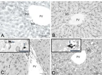
Fig. 3. Microscopic photographs of the liver before and after mercury treatment. Images of the livers in the control mice at (A) 10 days, and mercury treated groups at (B) 10, (C) 150 and (D) 300 days post treatment. The mer- cury was distributed in the form of granules in hep- atocytes around the portal vein and bile ductile at 10 days post treatment. A small amount of mercury was found to be accumulated in the hepatocytes, but many mercury granules (arrows) were still distributed in Kupffer cells in the sinusoid at 150 and 300 days post treatment (C and D, inset). BD, bile ductile; PV, portal vein; SI, sinusoid. Bars = 50 μm in A-D, and 20 μm in C and D, inset.
Fig. 4. Microscopic photographs of the spleen before and after mercury treatment. Images showing the spleen in con- trol mice at (A) 10 days, and mercury treated groups at (B) 10, (C) 150 and (D) 300 days post treatment. The mercury grains (arrows) as in the form of agglomer- ations were mainly distributed in the white pulp around the central arteriole and red pulp at 10 days post treat- ment. The amount of mercury accumulated in white pulp and red pulp was decreased at 150 and 300 days post treatment. CA, central arteriole; RP, red pulp; WP, white pulp. Bars = 50 μm in A and 100 μm in B–D.
of the proximal and distal convoluted tubular portion of the kidney cortex (Fig. 1D). In case of the renal medulla (Fig.
2), although we found accumulated mercury in the epi- thelium constituting the Henle’s loop and collecting duct, the amount was much lesser than that observed in the prox- imal convoluted tubule (Fig. 2B). The epithelial cells of the collecting ducts and Henle’s loop tubules in the medulla re- gion contained very little or no mercury at 150 and 300 days post treatment (Fig. 2C, Fig. 2D).
In the liver, mercury was mainly distributed in the form of granules in the hepatocytes around the portal vein at 10 days post treatment (Fig. 3). In addition, mercury was accu- mulated in the hepatocytes along the hepatocyte plate from the portal vein to the central vein and the mercury deposi- tion was observed to be linear (Fig. 3B). The mercury accu- mulation in the hepatocytes decreased toward the central vein as the hepatocytes surrounding the central vein showed little or no mercury accumulation. Mercury was also found to be accumulated in the Kupffer cells in the sinusoid. When mice were euthanized at 150 days post treatment, there was either no accumulation of mercury or very low concen- trations of mercury were observed in the hepatocytes.
However, Kupffer cells in the sinusoid contained mercury granules (Fig. 3C). When the mice were euthanized at 300 days post treatment it was revealed that though mercury accumulation did not appear in the hepatocytes, but it was still observed in Kupffer cells in sinusoid (Fig. 3D). In addi- tion, vacuoles were observed in the hepatocytes.
In the spleens of (Fig. 4) mice that were euthanized at 10 days post treatment, there was relatively higher amount of accumulated mercury in the white pulp than in the red pulp (Fig. 4B). Compared to the granular form of mercury observed in the liver and kidney, the mercury granules in the spleen were observed in the form of aggregations. When the mice were euthanized at 150 days post treatment, the distribution of black color in the white and red pulp portions of the spleen clearly indicated the aggregation of mercury granules (Fig. 4C). The bulk of the mercury was found to be mainly accumulated in the red pulp of the spleen at 300 days post treatment (Fig. 4D).
The concentration of accumulated mercury in the organs
The amount of mercury accumulated in the tubular epi- thelial cells of renal cortex gradually decreased at 150 and
Table 2. Mercury concentration rates (%) of kidney, liver and spleen in the mercury-treated group at each day post treatment
Organ Day post treatment
10 150 300
Kidney Liver Spleen
1.06±0.354 1.38±0.346 2.37±0.395
0.25±0.051 0.62±0.156 1.61±0.404
0.06±0.003 0.22±0.074 0.69±0.232 Values represent the mean ± SD.
For obtaining the mercury concentration rates each day post treatment, 12 mice were used as control (n=6) and as mercury treated (n=6) specimens.
300 days post treatment compared to the levels observed at 10 days post treatment. The concentration of mercury in the renal medulla region was decreased at 150 days, and there was very little or no mercury observed at 300 days post treatment. The ratio of the accumulated mercury con- centration in the kidneys was 1.06% when the mice were killed at 10 days post treatment and it went down to 0.25%
and 0.06% at 150 days and 300 days post treatment, re- spectively (Table 2).
The hepatocytes of the liver showed lower concentrations of mercury at 150 and 300 days post treatment compared to those at 10 days post treatment. The mercury concen- tration of the Kupffer cells in the sinusoid at 150 and 300 days post treatment was lower than that observed in the livers of mice that were euthanized at 10 days. The mercury concentration ratio in the liver decreased gradually over time from 1.38% on day 10 to 0.62% on day 150, and 0.22%
on day 300 post treatment (Table 2).
A small amount of mercury was observed in the white and red pulps at 150 and 300 days post treatment compared to that 10 days post treatment. The red pulps showed a de- crease in mass of mercury at 300 days post treatment com- pared to 150 days. The ratio of the concentration of accumu- lated mercury in the spleen gradually decreased over time from 2.37% at day 10 after the mice were killed to 1.61%
at day 150 and 0.69% at day 300 (Table 2).
Discussion
In this study, we investigated the distribution of mercury in various organ and tissues and the changes in the concen- trations of accumulated mercury over time. At 6 weeks of age, female mice were treated with a compound containing mercury (methylmercuric chloride) and then killed at 10, 150
and 300 days post treatment. We observed that the mean weights of the kidney, liver, and spleen in the group treated with mercury were either similar or higher at 10 days and 150 days post treatment compared to the control group, while they were lower at 300 days. In a previous study, methylmercury was orally administered into adult male mice daily for 35 days, after which the weight of their livers was found to be significantly lower (0.99 g) in the mer- cury-treated group compared to the control group (1.90 g) [18]. However, another study found that oral administration of mercuric chloride into male mice for 14 days led to a significant increase in the kidney and spleen weights in the mercury-treated group compared to the control group [30].
In the present study, the weights of the kidney, liver, and spleen of the mice in the mercury-treated group decreased significantly, especially at 300 days post treatment, which is considered to be the result of accumulation of mercury for a long period of time.
Based on several reports, it has been observed that more mercury accumulates in the kidney than in the liver [1, 3, 41, 42]. However, a lethal dose of mercury compounds killed mice within 2-4 min of intraperitoneal injection and the con- centration of mercury accumulated in the liver was found to be 10 times that of the mercury concentration in the kid- ney and 50 times that of the mercury concentration in the spleen [14]. In this case, the mercury accumulated in a very short period of time, which is extremely unusual.
In the present study, the kidneys of mice that were eu- thanized 10 days post treatment were found to have mercury accumulation in the epithelial cells constituting the proximal and distal convoluted tubules of the cortex, as well as in the collecting ducts and the Henle’s loop of the medulla.
These results are consistent with reports that have shown that mercury accumulates in the proximal tubular epithelial cells of the kidneys in mice [10] and rats [21]. In the kidneys of the mice that were euthanized 10 days post treatment, mercury was mainly distributed around the nuclei of the epithelial cells in the proximal convoluted tubules, or from the nucleus to the free surface of the cell. In comparison, mercury was mainly located on the free surface side of epi- thelial cells in the proximal convoluted tubules of mice killed at 150 or 300 days post treatment. This phenomenon might be attributed to the fact that the mercury accumulated in the cytoplasm of the epithelial cells might migrate towards the free surface of the cells and eventually appear in the filtrate of the proximal convoluted tubule lumen. It has been
reported that cytoplasmic vacuole formation [32, 34], py- knotic nucleus, and cell degeneration occurred in the epi- thelial cells of the proximal convoluted tubules following the accumulation of mercury [35]. In the present study, glomer- ular atrophy and vacuole formation was noted in response to mercury treatment, which is consistent with the results of previous studies [35, 36].
The effects of mercury also depends on the sex and the strain of the animal models. In the A.SW mouse strain, the accumulation of mercury is 2-5 times higher in the kidneys of the males than in those of females. The accumulation of mercury was also reported to be higher in the kidneys of the mice belonging to the A.SW strain than in the B10.S strain [19]. These findings indicate that the concentration of mercury that accumulates in the kidney differs from strain to strain, which means that the accumulation of mercury in the body is controlled or released depending on the genetic background of the animal. Higher accumulation of mercury was found in the A.SW, DBA/2, and BALB/C strains than in the B10.S strain, which might indicate that Pprc1 is an important regulator of mercury excretion in the kidney [2].
In this experiment, although the mercury concentrations in the kidney, liver, and spleen were not measured directly, the relative concentration of mercury was analyzed by com- paring the degree of mercury accumulation in the tissues of each organ under a microscope.
Generally, the blood in the hepatic portal vein flows to- wards the central vein. In this study, mercury was mainly found to be distributed in the hepatocytes around the portal vein after the initial contact with mercury in the blood and Kupffer cells in the sinusoid capillary. However, this was not the case in the hepatocytes around the central vein at 10 days post treatment. This suggests that mercury dis- tribution might be related to the flow of blood. These results were confirmed by the autometallography method using sections frozen and embedded in Epon [10]. Mercuric chlor- ide was also found in the Kupffer cells of the sinusoid capil- lary [11]. In the livers of mice killed on 150 and 300 days post treatment, although the amount of mercury accumu- lated in the hepatocytes was reduced to near zero, the mer- cury accumulated in the Kupffer cells still remained. There- fore, the effects of mercury on the hepatocytes is expected to decrease naturally over time. When the mercuric chloride was subcutaneously administered into adult male mice or rats, degeneration of hepatocytes and destruction of normal liver tissue structure was observed [26, 27]. The mechanism
of action behind the damage caused by mercury to living tissue is due to its ability to increase the concentration of reactive oxygen species in cells such as superoxide and hy- drogen peroxide, which leads to oxidative stress and sub- sequent tissue damage [22, 38]. In this experiment, vacuoles were still observed in hepatocytes of the mice killed at 150 and 300 days post treatment, which may be related to the damage to the hepatocytes.
According to previous reports, mercuric chloride was in- jected intraperitoneally into mice and autometallography confirmed that the mercury was localized in the macro- phages of the spleen, lymph nodes, and thymus [11]. The results of studies conducted on various types of fish col- lected from eight national parks in the United States suggest that the aggregation of macrophages in the spleen increases in response to mercury, which implies tissue damage to the spleen [40]. In the present study, the mercury that accumu- lated in the kidney tubular epithelial cells or hepatocytes appeared in the form of small granules, whereas mercury that accumulated in the spleen appeared in the form of ag- glomerated clusters and was distinctly different from that seen in the kidneys and liver. This may be explained by the fact that mercury is predominantly found in the macro- phages that are concentrated in the splenic tissues and ap- pear to be in the form of lumps. Previous reports have shown that the concentration of mercury accumulating in the C57BL/6 strain mouse spleen increased based on the du- ration of the subcutaneous administration of mercury into the female mice. However, the concentration of mercury in the spleen of the DBA/2 mouse did not show a significant increase with time [24]. Similar to the kidneys, these findings indicate that mercury accumulation in the spleen differs de- pending on the mouse strain.
In this study, the ratio of the concentration of mercury in the organs of the mercury-treated mice and the control mice decreased over time, which is consistent with the re- sults of a previous study [25]. Although mercury levels were seen to decline naturally over time, the pathways regulating the emission of mercury are not yet fully understood [9, 43].
One mechanism that might account for mercury excretion is via the binding of mercury to glutathione (GSH), which is present in almost all cells, to form GSH-Hg complexes [29, 39, 43]. This complex, which is the initial form of mer- cury that can be transported out of the cell [13], might be transported through the organic anion transporter to the proximal tubular cells, which are eventually transported into
the urine by multidrug resistant proteins (MRPs) [8]. These GSHs are induced by metallothionein (MT), which is a cys- teine-rich protein present in various animal tissues; it has four isomers (MT-I, II, III, and IV). Metallothionein also has the ability to remove heavy metals [42].
A widely recommended method of detoxifying mercury is the conversion of mercury to a chelate complex [31]. In animals and humans, chelating compounds such as 2,3-di- mercapto-1-propanesulfonic acid are known to be effective at removing mercury [4]. However, 2,3-dimercapto-1-pro- panesulfonic acid compounds have recently been reported to induce toxicity in the kidneys of mice treated with mer- cury [7]. The GSH-Hg complexes are transported into the urine by MRPs, meso-2,3-dimercaptosuccinic acid (DMSA), and 2,3-dimercaptopropane-1-sulfonic acid (DMPS) [4, 8].
The MRPs play a functional role in the DMSA- and DMPS-mediated extraction of mercuric ions from proximal tubular epithelial cells of kidney and resulting in the release of mercury through the urine.
Methylmercuric chloride was subcutaneously injected weekly into pubescent female mice for 3 weeks in the pres- ent study. Localization of accumulated mercury was identi- fied by autometallography. When mice were euthanized at 10 days post treatment, mercury was found to be distributed in the renal tubules and collecting ducts, especially in the proximal convoluted tubules. In the liver, mercury accumu- lated in the hepatocytes around the portal vein and Kupffer cells in the sinusoid capillary. In addition, vacuoles with ac- cumulated mercury were observed in hepatocytes. In the spleen, an unusual mass of lumpy mercury was distributed in the white and red pulp. When animals were euthanized at 150 and 300 days post treatment, the relative concen- trations of mercury accumulated in the kidney, liver, and spleen reduced naturally.
In conclusion, the present findings suggest that the endog- enous glutathione protein can form a complex with mercury, and this complex can be excreted out via urine. As a result, the concentration of mercury accumulated in cells and tis- sues naturally reduces over time.
References
1. Agarwal, R. and Behari, J. R. 2007. Role of selenium in mer- cury intoxication in mice. Ind. Health 45, 388-395.
2. Alkaissi, H., Ekstrand, J., Jawad, A., Nielsen, J. B., Havarin- asab, S., Soderkvist, P. and Hultman, P. 2016. Genome-wide association study to identify genes related to renal mercury
concentrations in mice. Environ. Health Perspect. 124, 920-926.
3. Al-Saleh, I., El-Doush, I., Shinwari, N., Al-Baradei, R., Kho- gali, F. and Al-Amodi, M. 2005. Does low mercury contain- ing skin-lightening cream (fair & lovely) affect the kidney, liver, and brain of female mice? Cutan. Ocul. Toxicol. 24, 11-29.
4. Aposhian, H. V., Maiorino, R. M., Gonzalez-Ramirez, D., Zuniga-Charles, M., Xu, Z., Hurlbut, K. M., Junco-Munoz, P., Dart, R. C. and Aposhian, M. M. 1995. Mobilization of heavy metals by newer, therapeutically useful chelating agents. Toxicology 97, 23-38.
5. Bates, M. N. 2006. Mercury amalgam dental fillings: an epi- demiologic assessment. Int. J. Hyg. Environ. Health 209, 309- 316.
6. Brandão, R., Moresco, R. N., Bellé, L. P., Leite, M. R., de Freitas, M. L., Bianchini, A. and Nogueira, C. W. 2011.
Diphenyl diselenide potentiates nephrotoxicity induced by mercuric chloride in mice. J. Appl. Toxicol. 31, 773-782.
7. Brandão, R., Santos, F. W., Zeni, G., Rocha, J. B. and Nogueira, C. W. 2006. DMPS and N-acetylcysteine induced renal tox- icity in mice exposed to mercury. Biometals 19, 389-398.
8. Bridges, C. C., Joshee, L. and Zalups, R. K. 2011. MRP2 and the handling of mercuric ions in rats exposed acutely to in- organic and organic species of mercury. Toxicol. Appl. Phar- macol. 251, 50-58.
9. Bridges, C. C., Joshee, L. and Zalups, R. K. 2014. Aging and the disposition and toxicity of mercury in rats. Exp. Gerontol.
53, 31-39.
10. Cho, H. W., Kim, M. H., Hwang, K. Y. and Yee, S. T. 1997.
Detection of mercury in kidney, liver, spleen and cerebellum of the mouse by autometallography. Kor. J. Toxicol. 13, 401-408.
11. Christensen, M. M. 1996. Histochemical localization of auto- metallographically detectable mercury in tissues of the im- mune system from mice exposed to mercuric chloride. Histo- chem. J. 28, 217-225.
12. Clarkson, T. W., Magos, L. and Myers, G. J. 2003. The toxicol- ogy of mercury-current exposures and clinical manifestations.
N. Engl. J. Med. 349, 1731-1737.
13. Clarkson, T. W., Vyas, J. B. and Ballatori, N. 2007. Mechan- isms of mercury disposition in the body. Am. J. Ind. Med.
50, 757-764.
14. Cunha, E. M., Cherdwongcharoensuk, D. and Aguas, A. P.
2003. Quantification of particles of lethal mercury in mouse viscera: high-resolution study of mercury in cells and tissues. Toxicol. Ind. Health 19, 55-61.
15. Cunha, E. M., Silva, D. P. and Aguas, A. P. 2003. High-reso- lution identification of mercury in particles in mouse kidney after acute lethal exposure. Biometals 16, 583-590.
16. Danscher, G. and Montagnese, C. 1994. Autometallographic localization of synaptic vesicular zinc and lysosomal gold, silver, and mercury. J. Histotechnol. 17, 15-22.
17. Danscher, G., Stoltenberg, M. and Juhl, S. 1994. How to de- tect gold, silver and mercury in human brain and other tis- sues by autometallographic silver amplification. Neuropathol.
Appl. Neurobiol. 20, 454-467.
18. de Freitas, A. S., Funck, V. R., Rotta Mdos, S., Bohrer, D., Mörschbächer, V., Puntel, R. L., Nogueira, C. W., Farina,
M., Aschner, M. and Rocha, J. B. 2009. Diphenyl diselenide, a simple organoselenium compound, decreases methyl- mercury-induced cerebral, hepatic and renal oxidative stress and mercury deposition in adult mice. Brain Res. Bull. 79, 77-84.
19. Ekstrand, J., Nielsen, J. B., Havarinasab, S., Zalups, R. K., Söderkvist, P. and Hultman, P. 2010. Mercury toxicoki- netics-dependency on strain and gender. Toxicol. Appl.
Pharmacol. 243, 283-291.
20. Emanuele, M. A., LaPaglia, N., Steiner, J., Jabamoni, K., Hansen, M., Kirsteins, L. and Emanuele, N. V. 1998 Reversal of ethanol-induced testosterone suppression in peripubertal male by opiate blockade. Alcoholism: Clin. Exp. Res. 22, 1199- 1204.
21. Eto, K., Yasutake, A., Miyamoto, K., Tokunaga, H. and Otsuka, Y. 1997. Chronic effects of methylmercury in rats.
II. Pathological aspects. Tohoku J. Exp. Med. 182, 197-205.
22. Farina, M., Brandão, R., de Lara, F. S., Pagliosa, L. B., Soares, F. A., Souza, D. O. and Rocha, J. B. 2003. Profile of non- protein thiols, lipid peroxidation and delta-aminolevulinate dehydratase activity in mouse kidney and liver in response to acute exposure to mercuric chloride and sodium selenite.
Toxicology 184, 179-187.
23. Grandjean, P., Budtz-Jørgensen, E., Steuerwald, U., Heinzow, B., Needham, L. L., Jørgensen, P. J. and Weihe, P. 2003.
Attenuated growth of breast-fed children exposed to in- creased concentrations of methylmercury and polychlorinated biphenyls. FASEB J. 17, 699-701.
24. Griem, P., Scholz, E., Turfeld, M., Zander, D., Wiesner, U., Dunemann, L. and Gleichmann, E. 1997. Strain differences in tissue concentrations of mercury in inbred mice treated with mercuric chloride. Toxicol. Appl. Pharmacol. 144, 163- 170.
25. Havarinasab, S., Björn, E., Nielsen, J. B. and Hultman, P.
2007. Mercury species in lymphoid and non-lymphoid tis- sues after exposure to methyl mercury: correlation with au- toimmune parameters during and after treatment in suscep- tible mice. Toxicol. Appl. Pharmacol. 221, 21-28.
26. Jin, G. B., Inoue, S., Urano, T., Cho, S., Ouchi, Y. and Cyong, J. C. 2002. Induction of anti-metallothionein antibody and mercury treatment decreases bone mineral density in mice.
Toxicol. Appl. Pharmacol. 185, 98-110.
27. Joshi, D., Mittal, D. K., Shukla, S., Srivastav, A. K. and Srivastav, S. K. 2014. N-acetyl cysteine and selenium pro- tects mercuric chloride-induced oxidative stress and anti- oxidant defense system in liver and kidney of rats: a histo- pathological approach. J. Trace Elem. Med. Biol. 28, 218-226.
28. Karapehlivan, M., Ogun, M., Kaya, I., Ozen, H., Deveci, H.
A. and Karaman, M. 2014. Protective effect of omega-3 fatty acid against mercury chloride intoxication in mice. J. Trace Elem. Med. Biol. 28, 94-99.
29. Khan, H., Khan, M. F., Jan, S. U., Mukhtiar, M., Ullah, N.
and Anwar, N. 2012. Role of glutathione in protection against mercury induced poisoning. Pak. J. Pharm. Sci. 25, 395-400.
30. Kim, S. H., Johnson, V. J. and Sharma, R. P. 2003. Oral ex-
posure to inorganic mercury alters T lymphocyte pheno- types and cytokine expression in BALB/c mice. Arch.
Toxicol. 77, 613-620.
31. Kostial, K., Kargacin, B., Blanusa, M., Piasek, M., Jones, M.
M. and Singh, P. K. 1994. Monoisoamyl meso-2,3-dimercap- tosuccinate as a delayed treatment for mercury removal in rats. Environ. Health Perspect. 102 Suppl 3, 309-311.
32. Liu, J., Lu, Y. F., Li, W. K., Zhou, Z. P., Li, Y. Y., Yang, X., Li, C., Du, Y. Z. and Wei, L. X. 2016. Mercury sulfides are much less nephrotoxic than mercury chloride and meth- ylmercury in mice. Toxicol. Lett. 262, 153-160.
33. Martin, M. D. and Woods, J. S. 2006. The safety of dental amalgam in children. Expert Opin. Drug Saf. 5, 773-781.
34. Moreira, E. L., de Oliveira, J., Dutra, M. F., Santos, D. B., Gonçalves, C. A., Goldfeder, E. M., de Bem, A. F., Prediger, R. D., Aschner, M. and Farina, M. 2012. Does methyl- mercury-induced hypercholesterolemia play a causal role in its neurotoxicity and cardiovascular disease? Toxicol. Sci.
130, 373-382.
35. Oliveira, C., Joshee, L., George, H., Nijhara, S. and Bridges, C. 2017. Oral exposure of pregnant rats to toxic doses of methylmercury alters fetal accumulation. Reprod. Toxicol. 69, 265-275.
36. Othman, M. S., Safwat, G., Aboulkhair, M. and Abdel Moneim, A. E. 2014. The potential effect of berberine in mer- cury-induced hepatorenal toxicity in albino rats. Food Chem.
Toxicol. 69, 175-181.
37. Passos, C. J., Mergler, D., Lemire, M., Fillion, M. and Gui- marães, J. R. 2007. Fish consumption and bioindicators of inorganic mercury exposure. Sci. Total Environ. 373, 68-76.
38. Perottoni, J., Rodrigues, O. E., Paixão, M. W., Zeni, G., Lobato, L. P., Braga, A. L., Rocha, J. B. and Emanuelli, T.
2004. Renal and hepatic ALA-D activity and selected oxida- tive stress parameters of rats exposed to inorganic mercury and organoselenium compounds. Food Chem. Toxicol. 42, 17- 28.
39. Schläwicke Engström, K., Strömberg, U., Lundh, T., Johans- son, I., Vessby, B., Hallmans, G., Skerfving, S. and Broberg, K. 2008. Genetic variation in glutathione-related genes and body burden of methylmercury. Environ. Health Perspect.
116, 734-739.
40. Schwindt, A. R., Fournie, J. W., Landers, D. H., Schreck, C. B. and Kent, M. L. 2008. Mercury concentrations in sal- monids from western U.S. National Parks and relationships with age and macrophage aggregates. Environ. Sci. Technol.
42, 1365-1370.
41. Shimojo, N., Kumagai, Y. and Nagafune, J. 2002. Difference between kidney and liver in decreased manganese super- oxide dismutase activity caused by exposure of mice to mer- curic chloride. Arch. Toxicol. 76, 383-387.
42. Yasutake, A. and Nakamura, M. 2011. Induction by mercury compounds of metallothioneins in mouse tissues: inorganic mercury accumulation is not a dominant factor for metal-
lothionein induction in the liver. J. Toxicol. Sci. 36, 365-372.
43. Zalups, R. K. 2000. Molecular interactions with mercury in the kidney. Pharmacol. Rev. 52, 113-143.
초록:생쥐 신장, 간, 비장 내 시간에 따른 수은 농도 변화와 수은 화합물의 위치
김유선․김영은․조현욱*
(순천대학교 생명산업과학대학 생물학과)
본 연구에서는 생쥐 신장, 간, 비장 내 축적된 수은의 위치와 아울러서 시간에 따른 수은 농도 변화를 조사하였 다. 수은 투여 종료 후 10일, 150일, 300일에 생쥐를 희생하여 수은 농도변화를 분석하였다. 10일에 희생시킨 생쥐 신장의 경우, 근위세뇨관 상피세포의 핵 위쪽 세포질에 수은이 다량으로 분포하였으나, 사구체에는 분포하지 않았 다. 간의 경우, 수은이 주로 간문맥 주위에 있는 간세포와 굴모세혈관에 있는 Kupffer 세포에 분포하였다. 10일에 희생시킨 비장의 경우, 백색 수질과 적색 수질에 수은이 흩어져 분포하였다. 수은의 농도 변화에 있어서, 150일과 300일에 희생시킨 신장의 피질과 수질에 축적되어 있던 수은이 낮은 농도로 나타났다. 역시 간세포에 축적되어 있던 수은도 150일과 300일의 경우, 낮은 농도로 나타났다. 비장의 경우, 백수와 적수 조직에 있던 수은 농도가 감소되었다. 이런 결과를 통해 세포나 조직에 축적되어 있던 수은의 위치가 확인되었으며, 또한 이 결과는 기관에 축적되어 있던 수은 농도가 시간이 지남에 따라 자연스럽게 감소된다는 사실을 확인해 주고 있다.