대한외과학회지:제 69 권 제 2 호
□ Case Report □
Vol. 69, No. 2, August, 2005
186
Primary Splenic Tuberculosis Presenting as a Large Solitary Mass
Department of Surgery, University of Ulsan Medical College, Seoul Asan Hospital, Seoul, Departments of 1Surgery, 2Diagnostic Pathology and 3Diagnostic Radiology, University of Ulsan Medical College, Gangneung Asan Hospital, Gangneung, Korea Ji Hoon Kim, M.D., Myoung Sik Han, M.D.1, Gil Hyun Kang, M.D.2, Seung Mun Jung, M.D.3, Yong Pil Cho, M.D.1, Hyuk Jai Jang, M.D.1, Yong Ho Kim, M.D.1, Jin Ho Kwak, M.D.1 and Youn Baik Choi, M.D.1
거대 단일 종괴의 형태로 나타난 원발성 비장 결핵
김지훈․한명식1․강길현2․정승문3․조용필1․장혁재1 김용호1․곽진호1․최윤백1
Tuberculosis may be difficult to diagnose when it presents in an uncommon extrapulmonary site. Although there has been a resurgence of abdominal tuberculosis in immunocom- promised patients, which is largely due to the extensive use of immunosuppressive drugs and the increasing incidence of a human immunodeficiency virus infection, splenic tuber- culosis is rare, particularly in the immunocompetent patients.
Almost all cases of splenic tuberculosis present as multiple hypoechoic foci on sonography or multiple focal hypodense lesions on contrast enhanced computed tomographic scan.
To our knowledge, splenic tuberculosis is an extremely rare condition. An 80-year-old man was found to have a large solitary splenic mass mimicking a splenic neoplasm on sonography and contrast enhanced computed tomographic scan. A diagnostic splenectomy revealed a large solitary mass in the spleen, which was consistent with splenic tuberculosis microscopically. We report a rare case of splenic tuberculosis in an elderly man presenting as a large solitary splenic mass on sonography and contrast enhanced computed tomographic scan. (J Korean Surg Soc 2005;
69:186-188)
Key Words: Spleen, Tuberculosis 중심 단어: 비장, 결핵
울산대학교 의과대학 서울아산병원 외과 및 강릉아 산병원 1외과, 2진단병리과, 3진단방사선과
Corresponding to: Yong Pil Cho, Department of Surgery, Gangneung Asan Hospital, 415 Bangdong-ri, Sacheon-myeon, Gangneung, 210- 711, Korea. (Tel) 82-33-610-3229, (Fax) 82-33-641-8120, (E-mail) [email protected]
Received March 22, 2005, Accepted May 20, 2005
INTRODUCTION
Abdominal tuberculosis is an uncommon condition in the west, but there has been a resurgence of the disease in the immunocompromised population, largely due to extensive use of immunosuppressive drugs and increasing incidence of human immun- odeficiency virus (HIV) infection.(1-3) It is still a fairly common health problem in developing coun- tries. Except in cases caused by drug-resistant strains, anti-tuberculous treatment is highly effective in immunocompetent patients. However, diagnosis can still be problematic in atypical patients with no evidence of pulmonary involvement.(1,4-6)
We encountered an 80-year-old man presented with a large solitary splenic mass on sonography (US) and contrast enhanced computed tomographic (CT) scan, suggestive of splenic neoplasm. After diagnostic splenectomy, microscopic examination showed chronic granulomatous inflammation with Langhans' giant cells and caseous necrosis, consistent with splenic tuberculosis
CASE REPORT
An 80-year-old male patient was admitted with a history of left flank pain for 11 months. He had no significant past medical history and physical exami- nation was unremarkable. He showed no systemic manifestations (fever, weight loss, general weakness, etc.). Chest radiograph revealed no abnormalities and blood chemistry parameters were all unremarkable.
A white blood cell count with differential counts was within normal limits. ESR and CRP were also nor- mal. Abdominal US demonstrated about 6-cm sized
Ji Hoon Kim, et al:Primary Splenic Tuberculosis Presenting as a Large Solitary Mass 187
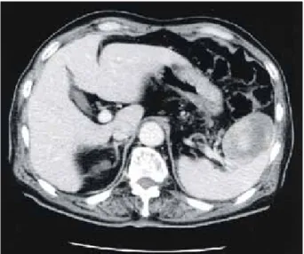
Fig. 1. Abdomen and pelvis CT scan showed about 6-cm sized, ovoid-shaped, hypodense mass in the spleen, suggestive of splenic neoplasm.
Fig. 2. Pathologic specimen showed a well-circumscribed, ovoid, bulging-out soft mass, measuring 6.7×5×5 cm in the spleen.
G
C
Fig. 3. Microscopic examination showed chronic granulomatous inflammation (G) with Langhans' giant cells (black arrow) and caseous necrosis (C), consistent with splenic tuber- culosis (Hematoxylin and eosin stain, ×100).
hypoechoic lesion in the spleen. Abdomen and pelvis CT scan showed about 6-cm sized, ovoid-shaped, hypodense mass in the spleen, suggestive of splenic neoplasm such as hemangioma or lymphoma (Fig. 1).
The patient underwent splenectomy under the preoperative impression of a splenic neoplasm, and no other lesions were observed in the operative findings. On gross examination, there was a well- circumscribed, ovoid, bulging-out soft mass, measur- ing 6.7×5×5 cm (Fig. 2). Microscopic examination showed chronic granulomatous inflammation with Langhans' giant cells and caseous necrosis, consistent
with splenic tuberculosis, although the smear and culture from the resected specimen were negative (Fig. 3). Postoperative sputum smear was also nega- tive. Combination anti-tuberculous therapy was adm- inistered and the patient was discharged without complications.
DISCUSSION
Abdominal tuberculosis continues to be endemic in the developing world and has shown a resurgence in the west due to extensive use of immunosup- pressive drugs and increasing incidence of HIV infection.(1-3) Tuberculosis of the spleen is rarely seen in isolation and is more frequently a part of multi-focal or disseminated disease. Although the majority of the patients described in the literature were immunosuppressed patients, splenic tuberculosis may be seen in otherwise healthy, immunocompetent patients.(5-9) However, there are no specific sym- ptoms suggestive of splenic tuberculosis.(7)
CT evaluation is singularly informative in patients with abdominal tuberculosis as it demonstrates involvement of the bowel, peritoneum, lymph nodes, and solid organs in a single examination.(1) Almost all cases of splenic tuberculosis present as multiple hypoechoic foci (<2 cm) on US and multiple focal hypodense lesions on contrast enhanced CT scan.
(1,10) However, these radiological features are not
188 대한외과학회지:제 69 권 제 2 호 2005
pathognomonic, but can be suggestive when consid- ered along with the clinical presentation, immune status, and demographic background of the patient.
Splenic tuberculosis is rare and delay in diagnosis is common especially in atypical patients with no evidence of pulmonary involvement because of non- specific symptomatology and non-pathognomonic radiological features. As in any other forms of vis- ceral tuberculosis, evidence of simultaneous or pre- vious involvement of other sites can be obtained in most patients with or without HIV infection.(11) However, our case had no history of either pulmo- nary or intestinal tuberculosis. US and CT findings were considered primarily as a splenic neoplasm. A splenectomy was performed, and no other lesions were observed in the operative findings. Microscopic examination of the specimens revealed tuberculosis.
This therefore seemed to be an unusual course for Mycobacterium tuberculosis infection, and the route of infection was not clear. In the current literature, splenectomy for isolated splenic tuberculosis is not recommended unless there is an abscess formation or the biopsy is nondiagnostic.(12) Patients usually have a favorable response to anti-tuberculous treatment and splenectomy is rarely necessary.(7) However, in our case, we performed splenectomy under the preo- perative impression of a splenic neoplasm.
We report an extremely rare case of splenic tuber- culosis presenting as a large solitary splenic mass on US and contrast enhanced CT scan in an old-aged man and suggest that regardless of the immune status, splenic tuberculosis should be considered as a diagnostic possibility when dealing with the solitary
nodules of the spleen.
REFERENCES
1) Gulati MS, Sarma D, Paul SB. CT appearances in abdominal tuberculosis. A pictorial essay. Clin Imaging 1999;23:51-9.
2) Özgüroglu M, ÇCelik AF, Demir G, Aki H, Demirelli F, Mandel N, et al. Primary splenic tuberculosis in a patient with nasal angiocentric lymphoma: mimicking metastatic tumor on abdominal CT. J Clin Gastroenterol 1999;29:96-8.
3) MacGregor BR. Tuberculosis: from history to current man- agement. Semin Roentgenol 1993;28:101-8.
4) Horne NW. Problems of tuberculosis in decline. BMJ 1984;
288:1249-52.
5) Yoon HJ, Song YG, Park WI, Choi JP, Chang KH, Kim JM.
Clinical manifestations and diagnosis of extrapulmonary tuberculosis. Yonsei Med J 2004;45:453-61.
6) Lee JO, Lee BK, Lee SD, Seo JK, Park YH. Intraabdominal tuberculosis. J Korean Surg Soc 1989;36:323-31.
7) Gonzalez-Lopez A, Dronda F, Alonso-Sanz M, Chaves F, Fernandez-Martin I, Lopez-Cubero L. Clinical significance of splenic tuberculosis in patients infected with human immunodeficiency virus. Clin Infect Dis 1997;24:1248-51.
8) Reichel C, Theisen A, Rockstroh JK, Muller-Miny H, Speng- ler U, Sauerbruch T. Splenic abscesses and abdominal tuber- culosis in patients with AIDS. Z Gastroenterol 1996;34:494-6.
9) Adil A, Chikhaouni N, Ousehal A, Kadiri R. Splenic tuber- culosis. A propos of 12 cases. Ann Radiol 1995;38:403-7.
10) Chandra S, Srivastava DN, Gandhi D. Splenic tuberculosis:
an unusual sonographic presentation. Int J Clin Pract 1999;
53:318-9.
11) Bankier AA, Fleischmann D, Wiesmayr MN, Putz D, Kontrus M, Hubsch P, et al. Update: abdominal tuberculosis-unusual findings on CT. Clin Radiol 1995;50:223-8.
12) Nayyar V, Ramakrishna B, Mathew G, Williams RR, Khan- duri P. Response to antituberculous chemotherapy after splenectomy. J Intern Med 1993;223:81-3.