RISK FACTORS OF LYMPHOCELE AFTER RAH WITH PELVIC LYMPH NODE DISSECTION FOR WOMEN WITH CERVICAL CANCER
Ji Yen Lee, MD, Jaeman Bae, MD, Sun-Joo Lee, MD, Ji Yeon Kim, MD, Soo Nyung Kim, MD
Department of Obstetrics and Gynecology, Konkuk University School of Medicine, Seoul, Korea
Objective
To identify the risk factors of lymphocele formation in patients with cervical cancer who underwent radical abdominal hysterectomy and pelvic lymph node dissection.
Methods
We conducted a retrospective study of 62 patients diagnosed with cervical cancer who underwent radical abdominal hysterectomy, including pelvic lymph node dissection between April 2005 and June 2010. Lymphocele was confirmed by imaging studies such as computed tomography or ultrasonography. Included risk factors were patient’s age, body mass index, surgeon’s experience, tumor histology, stage of cancer, previous surgery, number of retrieved lymph nodes, and radiation therapy. Multiple logistic regression analysis was performed to evaluate the risk factors of lymphocele formation.
Results
During median follow-up of 34.5 months (range, 12−69 months), 20 patients (32%) out of 62 had developed lymphocele. Eight patients developed symptomatic lymphocle. Univariate analysis showed the radiation therapy, and the number of retrieved lymph nodes as significant risk factors of lymphocele formation. When applying mutivariate analysis using logistic regression, radiation therapy (odds ratio=5.19, P=0.010) and the number of retrieved lymph node (odds ratio=4.80, P=0.021) were independent risk factors of lymphocele formation.
Conclusion
Radiation therapy and the number of retrieved lymph node were significant risk factors of lymphocele formation in patients with cervical cancer who underwent radical abdominal hysterectomy and pelvic lymph node dissection.
Keywords: Lymphocele; Cervical cancer; Risk factors
Received: 2012.4.27. Revised: 2012.9.10. Accepted: 2012.11.28.
Corresponding author: Soo Nyung Kim, MD
Department of Obstetrics and Gynecology, Konkuk University Medical Center, Konkuk University School of Medicine, 120 Neungdong-ro, Gwangjin-gu, Seoul 143-729, Korea Tel: +82-2-2030-7641 Fax: +82-2-2030-7748 E-mail: [email protected]
This is an Open Access article distributed under the terms of the Creative Commons Attribution Non-Commercial License (http://creativecommons.org/licenses/
by-nc/3.0/) which permits unrestricted non-commercial use, distribution, and reproduction in any medium, provided the original work is properly cited.
Copyright © 2012. Korean Society of Obstetrics and Gynecology 자궁경부암은 전세계적으로 두 번째로 흔한 여성암이며 우리나라의
경우 다섯 번째로 많이 발생하는 암으로 부인암 중에서 가장 높은 빈 도를 차지한다[1]. 자궁경부암 발병의 평균 나이는 40세이며 최근 젊은 여성에서의 발병률이 증가하는 추세로 다른 암에 비하여 평균 여명이 길어 치료 후 환자의 삶의 질에 대한 관점이 중요시 되고 있다[2]. 자궁 경부암의 치료는 림프혈관강 침범이 없는 병기 Ia1에서는 제I형 자궁절 제술을 시행하며, 림프혈관강 침범이 있는 병기 Ia1 및 자궁방결합조 직 침범이 없는 병기 IIA 이하에서는 근치적 자궁절제술 및 골반림프절 절제술을 시행하는 것이 원칙이고 자궁방결합조직을 침범하는 병기 IIB 이상에서는 항암방사선동시요법이 원칙이다. 수술적 치료 시 시행하게 되는 골반림프절절제술의 가장 흔한 합병증이 림프낭종이다[3,4].
http://dx.doi.org/10.5468/KJOG.2012.55.12.907 pISSN 2233-5188 · eISSN 2233-5196
림프낭종이란 후복막강에 림프액이 저류되는 상태를 의미하며, 발 생원인은 정확히 알려져 있지는 않다. 주로 장골동맥 주위에 잘 생기 는 것으로 알려져 있다. 대부분의 경우 무증상으로 별다른 치료가 필요 치 않은 경우가 많으나 골반통, 하지부종 및 동통, 신기능 저하를 동반 한 수신증, 심부정맥 혈전증, 폐색전증, 이차적 감염, 패혈증 등의 증상 을 유발하여 치료를 요하는 경우도 있다[3-7]. 하지부종, 통증과 같은 증상이 있는 경우 경피적 흡인 또는 배액관을 삽입하여 감압을 통해 증 상을 완화하고 얻어진 검체로 미생물학적 검사를 시행해 볼 수 있으며 배액량이 지속적으로 많은 경우 경화요법 또는 수술적 접근을 시행해 볼 수 있다. 또한 감염이 의심되는 경우 배액과 더불어 항생제를 사용 해야 한다[8-13]. 초기에 적극적으로 치료하지 않으면 이차적 감염으 로 패혈증이 나타날 수 있으며 골반 혈관의 압박에 의해 심부정맥 혈전 증 및 폐색전증이 발생하는 경우 생명을 위협하는 심각한 문제로 이어 질 수 있기에 림프낭종이 의심되는 증상이 발생했을 때 의료진을 방문 하여 초기에 적극적인 치료가 이루어질 수 있도록 사전 교육이 필요하 다[14,15].
외국 연구문헌에서 자궁경부암 환자의 근치적 자궁절제술 및 림프절 절제술 후 림프낭종의 발생률은 2%-9.1% 정도로 다양하게 보고되고 있으며[5], 림프절절제술이 광범위할수록 림프낭종의 발생빈도가 증가 하는 것으로 알려져 있다[16-18]. 본 연구에서는 근치적 자궁절제술 및 골반림프절절제술을 시행받은 자궁경부암 환자에서의 림프낭종에 대한 발생 위험인자에 대해 분석해 보았다.
연구대상 및 방법
이번 연구는 2005년 4월부터 2010년 6월까지 단일기관에서 자궁경 부암으로 진단받고 근치적 자궁절제술 및 골반 림프절절제술을 시행받 은 병력이 있는 62명의 환자들을 대상으로 하여 림프낭종 발생여부를 조사하였다.
연구대상의 의무기록을 통해 환자군의 나이, 산과력, 병기, 과거 복 부수술력, 병리결과(조직형, 종양의 크기, 획득한 림프절의 개수, 림 프관 및 미세혈관 침범여부 등), 수술 전 편평세포암(squamous cell carcinoma, SCC) 값, 수술명, 집도의 및 체질량지수(body mass index, BMI) 등에 대해 조사하였다. BMI는 체중(kg)을 키(m)의 제곱으로 나누 어 계산하였다.
모든 수술은 개복수술로 진행하였고, 병기 Ia1 환자의 경우 림프혈관 강 침범이 있는 고위험군에 한해 근치적 자궁절제술 및 골반림프절절 제술을 시행하였다. 집도의 간 수술방법의 차이, 원칙의 차이, 수술범위 의 차이는 없었다. 골반림프절절제술의 범위는 외측으로는 음부대퇴신 경(genitofemoral nerve), 내측으로는 방광벽을 경계로 하였다. 또한 폐 쇄신경(obturator nerve) 주위와 폐쇄동맥 앞쪽의 모든 림프조직을 제 거하였다. 위쪽 경계로는 총장골동맥(common iliac artery)가 분지되는 지점, 아래로는 클로림프절(Cloquet node)까지이다. 획득한 림프조직 은 외장골 동맥부근, 내장골동맥부근, 폐쇄오목(obturator fossa) 부근으
로 분리하여 병리과로 보냈다. 수술 시 모든 환자들에게 양쪽 후복막강 에 21프렌치의 배액관을 삽입하고, 후복막을 봉합하여 닫았다. 배액관 은 30 mL/day 이하로 배액됐을 때 제거하였다. 모든 환자들에게 예방 적 항생제를 사용하였으며, 혈전증 예방을 위해 입원 전일부터 수술 후 7일간 4,000 U의 저분자량의 헤파린(low molecular weight heparin)을 사용하였다. 병기 IIB 이상에서는 항암방사선동시요법을 시행하였으며, 방사선치료 진행 시 모든 환자가 수술 후 보조적 항암방사선동시요법 치료를 하였던 환자로 치료 시작 시기는 별다른 차이가 없었다.
수술 후 시행한 초음파 혹은 골반 및 복부 전산화단층촬영을 조사 하여 림프낭종이 있는지 여부를 확인하였다. 또한 림프낭종이 발견 된 경우 동반된 증상으로 발열(6시간 이상 간격으로 2회 이상 체온이 38.0oC 이상인 경우), 골반통, 하지부종 및 하지동통, 수신증, 심부정맥 혈전증, 폐색전증, 감염 혹은 패혈증 등을 조사하였다.
연관성이 있을 수 있는 가능성이 있는 독립변수들에 대해 림프낭종 과의 연관성에 대해 이변량 분석 및 다변량 분석을 통해 분석해 보았 다. 위험인자와 림프낭종의 빈도의 연관성 분석은 이변량 분석으로 카 이제곱 검정 분석을 이용하였고, 다변량 분석은 로지스틱 회귀 분석을 이용하였다. 통계 분석은 dBSTAT ver. 5 (DBSTAT Co., Chuncheon, Korea) 프로그램을 사용하였다. P-value가 0.05 미만인 경우 통계학적 으로 유의하다고 판정하였다.
결 과
연구대상자는 총 62명으로, 추적관찰 기간의 중앙값은 34.5개월(범 위, 12-69개월)이었다. 전체 환자군에 대한 특징들에 대하여 Table 1 과 같다. 연령분포는 30세부터 70세까지로 평균 연령은 49.5세였고, 수술한 의사는 부인종양 전문의로서 20년 이상의 경력을 가진 2명의 의사(1, 2)와 20년 미만의 경력을 가진 의사(3, 4)로 분류하였다. 조직 형은 편평세포암과 비편평세포암으로 분류하였고, 편평세포암이 51명 (82%)로 다수를 차지하였다. 수술 후 병리 결과에 따라 보조적 방사선 치료를 받았던 환자가 32명(52%)으로 절반을 약간 넘었다.
림프낭종의 유병률은 62명 중 20명으로 32%였고, 그 중 증상이 있는 경우가 8명, 증상을 보이지 않았던 경우가 12명이었다. 가장 흔한 증상 은 감염에 의한 동통으로 8명의 환자 중 5명이었고, 발열 증상을 보인 경우가 4명, 그 외 하지 부종 1명, 복부팽만과 하지저림 증상이 1명이었 다. 발생시기의 중앙값은 수술 후 5.5개월(범위, 1-35개월)이었다.
림프낭종의 위험인자로 나이는 50세 이상과 미만으로 분류하였고, BMI는 25 kg/m2을 기준으로, 획득한 림프절의 개수는 35개 이상과 미 만으로, 종양의 크기는 4 cm 이상과 미만을 기준으로 분류하였다.
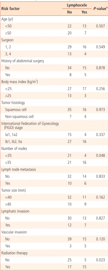
각 위험인자에 대한 이변량 분석 결과 방사선치료를 시행한 군, 획득 한 림프절의 개수 35개 이상인 군에서 림프낭종의 발생률이 유의하게 높게 나타났다(Table 2).
이변량 분석에서 통계적으로 유의하였던 방사선치료 유무 및 획득 한 림프절의 개수를 변수로 하고 로지스틱 회귀 분석을 이용하여 교차
비를 분석한 결과 방사선치료를 시행한 군이 임파낭종 발생에 대한 교 차비가 4.8배로 통계학적으로 유의하게 높았고, 획득한 림프절의 개수 35개 이상인 군에서도 림프낭종 발생에 대한 교차비가 5.2배로 통계학 적으로 유의하였다(Table 3).
고 찰
골반림프절절제술은 부인암 치료에 있어서 림프절 전이가 의심되는 경우 시행되며 림프절 전이가 없는 경우에도 생존율을 향상시키는 이점 을 가지고 있다. 골반림프절절제술 후 나타나는 가장 흔한 합병증은 림 프낭종이며[3,4], 후복막강에 림프액이 저류되어 발생하며 림프절절제 Table 1. Characteristics of patients
Characteristic No. of patients
(%)
Mean age (range) 49.5 (30−74)
Surgeon
1, 2 45 (72.6)
3, 4 17 (27.4)
Body mass index (kg/m2)
<25 44 (70.1)
≥25 16 (25.8)
Tumor histology
Squamous cell carcinoma 51 (82.3)
Non‐squamous cell carcinoma 11 (17.7)
In ternational Federation of Gynecology (FIGO) stage
Ia1 16 (25.8)
Ia2 3 (4.8)
Ib1 31 (50.0)
Ib2 9 (14.5)
IIA 3 (4.8)
Radiation therapy
No 30 (48.4)
Yes 32 (51.6)
Mean number of nodes (range) 38.7 (4−71) Lymph node metastasis
LC (−) 46 (74.2)
LC (+) 16 (25.8)
Lymphocele 20 (32.3)
Symptom (+) 8 (12.6)
Symptom (−) 12 (19.4)
Median size of lymphocele (range, mm) 37 (10−90)
Table 2. Bivariate analyses for risk factors of lymphocele
Risk factor Lymphocele
P‐valuea
No Yes
Age (yr)
<50 22 13 0.507
≥50 20 7
Surgeon
1, 2 29 16 0.549
3, 4 13 4
History of abdominal surgery
No 34 15 0.878
Yes 8 5
Body mass index (kg/m2)
<25 27 17 0.256
≥25 13 3
Tumor histology
Squamous cell 35 16 0.973
Non‐squamous cell 7 4
In ternational Federation of Gynecology (FIGO) stage
Ia1, 1a2 15 4 0.337
Ib1, Ib2, IIa 27 16
Number of nodes
<35 21 4 0.048
≥35 21 16
Lymph node metastasis
No 32 14 0.833
Yes 10 6
Tumor size (mm)
<40 32 11 0.162
≥40 10 9
Lymphatic invasion
No 30 13 0.827
Yes 12 7
Vascular invasion
No 39 15 0.120
Yes 3 5
Radiation therapy
No 25 5 0.023
Yes 17 15
aP‐value by chi‐square test.
술이 광범위할수록 발생 빈도가 증가하는 것으로 알려져 있다[16-18].
국내에서는 림프절절제술과 림프낭종에 대한 연구가 거의 이루어지 지 않고 있으며, 해외 연구문헌만이 일부 있는 데 비하여 림프절절제술 과 림프부종에 대한 연구는 국내외적으로 많은 편이다.
림프부종은 림프순환 장애로 인하여 조직액이 피부와 피하조직에 축 적되어 발생하는 질환으로 부인암 치료 후 발생하는 이차성 림프부종 에 대한 국내 연구에서는 연령, 항암화학요법, 고혈압, 당뇨, 부인암 수 술방법은 림프부종 발생에 대해 통계학적으로 유의한 연관성이 없고, 25 이상의 신체비만지수, 방사선 치료의 유무, 림프절절제 유무가 통계 학적으로 유의한 것으로 나타났다[15]. 외국 연구문헌에 따르면 Ryan 등[19]은 25 이상의 신체비만지수와 광범위한 림프절절제가 하지 림프 부종의 발생률을 높인다고 보고하였으며, Abu-Rustum 등[20]은 자궁 체부암에서 광범위한 림프절절제가 하지 림프부종 발생에 영향을 미치 지 못한다고 보고하였다. Werngren-Elgström과 Lidman [21], Füller 등 [22]은 자궁경부암에서 방사선치료가 하지 림프부종 발생과 유의한 상 관관계가 있다고 보고하였다.
골반림프절절제술과 림프낭종과의 상관관계는 부인과보다 비뇨기과 고환암 수술에서 연구가 활발히 이루어져 왔다. 근치적 고환절제술 및 골반림프절절제술을 시행받은 고환암 환자를 대상으로 림프낭종의 발 생과 위험인자에 대한 연구 논문에서는 림프낭종의 발생에 연령, 수술 한 집도의, 수술 전후로 저분자량의 헤파린 사용유무, 병기, 전보조호르 몬요법 유무, American Society of Anesthesiologists score, Gleason score, 전립선특이항원(prostate-specific antigen) value는 통계학적으 로 유의한 상관관계가 없으며[5,18], 제한된 림프절절제술보다는 광범 위한 림프절절제술, 절제된 림프절의 개수가 통계학적으로 유의한 것 으로 나타났다[5,16-18].
일부 연구문헌에 따르면 근치적 자궁절제술 및 골반림프절절제술을 시행 받은 부인암환자에서 광범위한 림프절절제술, 절제된 림프절의 개수, 림프절 전이, 방사선 치료, 예방적 항응고제 사용이 림프낭종 발 생과 연관이 있다고 보고하였다[5,18,23,24]. Franchi 등[25]은 수술 후 배액관 삽입이 림프낭종의 발생률에 영향을 미치지 않는다고 하였으 며, Benedetti-Panici 등[26]은 배액관 삽입이 이물질로 작용하여 림프 부종의 발생률을 높인다고 하였으며, Srisomboon 등[27]과 Morice 등
[28]은 배액관 삽입이 림프부종의 발생률을 낮추고 수술 후 며칠간 배 액관을 유지하는 것이 도움이 된다고 하였다.
림프낭종의 발생을 최소화하기 위해서는 엄격한 수술 적응증의 적용 및 세심한 수술적 접근이 요구되며, 감시림프절 개념의 도입[29], 림프 관 손상을 최소화하기 위한 복강경, 양극성 소작기, harmonic shears, Ligaclip 등의 사용, TachoSil과 같은 surgical patch의 사용, 복막피복술 생략, 대망이식술, 대망고정술, 배액관 삽입 생략, 헤파린 사용 제한 등 이 림프낭종 예방에 도움이 된다고 알려져 있다[23,24,26,30,31].
본 연구에서는 근치적 자궁절제술 및 골반림프절절제술을 시행받은 자궁경부암 환자에서 림프낭종의 발생에 연령, 신체비만지수, 수술한 집도의, 병기, 림프절 전이여부, 림프관 및 미세혈관 침범여부는 통계 학적으로 유의한 연관성이 없으며, 방사선치료 유무와 절제된 림프절 의 개수가 통계학적으로 유의한 것으로 나타났다. 저분자량의 헤파린 사용이 림프낭종 생성의 위험인자로 알려져 있으나[5,18] 본 연구에서 는 심부정맥 혈전증 예방을 위해 모든 환자에게 수술 전후로 저분자량 의 헤파린을 사용하였으며, 모든 환자에게 배액관 삽입을 시행하고 30 mL/day 이하로 배액되었을 때 제거하였다.
본 연구의 한계점으로 대상군의 수가 일반화하기에 충분하지 못하 며, 후향적 연구를 시행하였기에 배액관 삽입 여부와 저분자량의 헤파 린 사용 여부가 림프낭종에 어떤 영향을 미치는지 알 수 없었다. 이에 대하여 대규모의 전향적 대조연구가 필요할 것으로 생각되며, 현재까 지 림프낭종의 발생원인 및 생성 기전이 정확히 알려져 있지 않아 이에 대한 지속적인 연구가 필요할 것으로 생각된다.
감사의 글
이 논문은 2010년도 건국대학교병원 임상연구비 지원을 받았음.
References
1. Ohba Y, Todo Y, Kobayashi N, Kaneuchi M, Watari H, Takeda M, et al. Risk factors for lower-limb lymphedema after surgery for cervical cancer. Int J Clin Oncol 2011;16:238-43.
2. Matsuda T, Marugame T, Kamo K, Katanoda K, Ajiki W, Sobue T, et al. Cancer incidence and incidence rates in Japan in 2003:
based on data from 13 population-based cancer registries in the Monitoring of Cancer Incidence in Japan (MCIJ) Project.
Jpn J Clin Oncol 2009;39:850-8.
3. Musch M, Klevecka V, Roggenbuck U, Kroepfl D. Complica- tions of pelvic lymphadenectomy in 1,380 patients undergo- ing radical retropubic prostatectomy between 1993 and 2006.
J Urol 2008;179:923-8.
4. Briganti A, Chun FK, Salonia A, Suardi N, Gallina A, Da Pozzo Table 3. Multivariate analysis for risk factors of lymphocele
Risk factor
Multivariate analysis Odds ratio
(confidence interval) P‐valuea
No. of nodes 0.021
<35 1
≥35 4.80 (1.26-18.28)
Radiation therapy 0.010
No 1
Yes 5.19 (1.47-18.27)
aP‐value by logistic regression analysis.
LF, et al. Complications and other surgical outcomes associ- ated with extended pelvic lymphadenectomy in men with localized prostate cancer. Eur Urol 2006;50:1006-13.
5. Naselli A, Andreatta R, Introini C, Fontana V, Puppo P. Predic- tors of symptomatic lymphocele after lymph node excision and radical prostatectomy. Urology 2010;75:630-5.
6. Augustin H, Hammerer P, Graefen M, Palisaar J, Noldus J, Fer- nandez S, et al. Intraoperative and perioperative morbidity of contemporary radical retropubic prostatectomy in a consecu- tive series of 1243 patients: results of a single center between 1999 and 2002. Eur Urol 2003;43:113-8.
7. Suzuki M, Ohwada M, Sato I. Pelvic lymphocysts following retroperitoneal lymphadenectomy: retroperitoneal partial “no- closure” for ovarian and endometrial cancers. J Surg Oncol 1998;68:149-52.
8. Hakenberg OW. The incidence and treatment of lymphoceles after radical retropubic prostatectomy. BJU Int 2005;96:1422.
9. Kim JK, Jeong YY, Kim YH, Kim YC, Kang HK, Choi HS. Postop- erative pelvic lymphocele: treatment with simple percutaneous catheter drainage. Radiology 1999;212:390-4.
10. Fallick ML, Long JP. Laparoscopic marsupialization of lym- phocele after laparoscopic lymph node dissection. J Endourol 1996;10:533-4.
11. Gilliland JD, Spies JB, Brown SB, Yrizarry JM, Greenwood LH.
Lymphoceles: percutaneous treatment with povidone-iodine sclerosis. Radiology 1989;171:227-9.
12. Sawhney R, D’Agostino HB, Zinck S, Rose SC, Kinney TB, Og- levie SB, et al. Treatment of postoperative lymphoceles with percutaneous drainage and alcohol sclerotherapy. J Vasc Interv Radiol 1996;7:241-5.
13. Zanetta G, Trio D, Lissoni A, Dalla Valle C, Rangoni G, Pittelli M, et al. Early and short-term complications after US-guided puncture of gynecologic lesions: evaluation after 1,000 con- secutive cases. Radiology 1993;189:161-4.
14. Yamamoto R, Saitoh T, Kusaka T, Todo Y, Takeda M, Okamoto K, et al. Prevention of lymphocyst formation following systematic lymphadenectomy. Jpn J Clin Oncol 2000;30:397-400.
15. Kang SH, Hwang KH, Sim YJ, Jeong HJ, Lee TH, Kim SH. The prevalence and risk factors of lower limb lymphedema in the patients with gynecologic neoplasms. Korean J Obstet Gynecol 2009;52:815-20.
16. Heidenreich A, Varga Z, Von Knobloch R. Extended pelvic lymphadenectomy in patients undergoing radical prosta- tectomy: high incidence of lymph node metastasis. J Urol 2002;167:1681-6.
17. Clark T, Parekh DJ, Cookson MS, Chang SS, Smith ER Jr, Wells N, et al. Randomized prospective evaluation of extended versus limited lymph node dissection in patients with clinically local- ized prostate cancer. J Urol 2003;169:145-7.
18. Khoder WY, Trottmann M, Buchner A, Stuber A, Hoffmann S, Stief CG, et al. Risk factors for pelvic lymphoceles post-radical prostatectomy. Int J Urol 2011;18:638-43.
19. Ryan M, Stainton MC, Jaconelli C, Watts S, MacKenzie P, Man- sberg T. The experience of lower limb lymphedema for women after treatment for gynecologic cancer. Oncol Nurs Forum 2003;30:417-23.
20. Abu-Rustum NR, Alektiar K, Iasonos A, Lev G, Sonoda Y, Agha- janian C, et al. The incidence of symptomatic lower-extremity lymphedema following treatment of uterine corpus malignan- cies: a 12-year experience at Memorial Sloan-Kettering Cancer Center. Gynecol Oncol 2006;103:714-8.
21. Werngren-Elgström M, Lidman D. Lymphoedema of the lower extremities after surgery and radiotherapy for cancer of the cervix. Scand J Plast Reconstr Surg Hand Surg 1994;28:289-93.
22. Füller J, Guderian D, Köhler C, Schneider A, Wendt TG. Lymph edema of the lower extremities after lymphadenectomy and radiotherapy for cervical cancer. Strahlenther Onkol 2008;184:206-11.
23. Gallotta V, Fanfani F, Rossitto C, Vizzielli G, Testa A, Scambia G, et al. A randomized study comparing the use of the Ligaclip with bipolar energy to prevent lymphocele during laparoscopic pelvic lymphadenectomy for gynecologic cancer. Am J Obstet Gynecol 2010;203:483.e1-6.
24. Kim HY, Kim JW, Kim SH, Kim YT, Kim JH. An analysis of the risk factors and management of lymphocele after pelvic lymphadenectomy in patients with gynecologic malignancies.
Cancer Res Treat 2004;36:377-83.
25. Franchi M, Trimbos JB, Zanaboni F, v d Velden J, Reed N, Coens C, et al. Randomised trial of drains versus no drains follow- ing radical hysterectomy and pelvic lymph node dissection: a European Organisation for Research and Treatment of Cancer- Gynaecological Cancer Group (EORTC-GCG) study in 234 patients. Eur J Cancer 2007;43:1265-8.
26. Benedetti-Panici P, Maneschi F, Cutillo G, D’Andrea G, di Pa- lumbo VS, Conte M, et al. A randomized study comparing retro- peritoneal drainage with no drainage after lymphadenectomy in gynecologic malignancies. Gynecol Oncol 1997;65:478-82.
27. Srisomboon J, Phongnarisorn C, Suprasert P, Cheewakriangkrai C, Siriaree S, Charoenkwan K. A prospective randomized study comparing retroperitoneal drainage with no drainage and
no peritonization following radical hysterectomy and pelvic lymphadenectomy for invasive cervical cancer. J Obstet Gynae- col Res 2002;28:149-53.
28. Morice P, Lassau N, Pautier P, Haie-Meder C, Lhomme C, Castaigne D. Retroperitoneal drainage after complete Para- aortic lymphadenectomy for gynecologic cancer: a randomized trial. Obstet Gynecol 2001;97:243-7.
29. Abu-Rustum NR, Barakat RR. Observations on the role of circumflex iliac node resection and the etiology of lower ex- tremity lymphedema following pelvic lymphadenectomy for
gynecologic malignancy. Gynecol Oncol 2007;106:4-5.
30. Park NY, Seong WJ, Chong GO, Hong DG, Cho YL, Park IS, et al. The effect of nonperitonization and laparoscopic lymphade- nectomy for minimizing the incidence of lymphocyst formation after radical hysterectomy for cervical cancer. Int J Gynecol Cancer 2010;20:443-8.
31. Tinelli A, Giorda G, Manca C, Pellegrino M, Prudenzano R, Guido M, et al. Prevention of lymphocele in female pelvic lymphadenectomy by a collagen patch coated with the human coagulation factors: a pilot study. J Surg Oncol 2012;105:835-40.
자궁경부암 환자에서 근치적 자궁절제술 및 골반림프절절제술 시행 후 발생하는 림프낭종의 위험인자
건국대학교 의과대학 산부인과학교실 이지연, 배재만, 이선주, 김지연, 김수녕
목적
자궁경부암 환자에서 근치적 자궁절제술 및 골반림프절절제술 시행 후 발생하는 림프낭종의 위험인자에 대해 분석해 보았다.
연구방법
2005년 4월부터 2010년 6월까지 단일기관에서 자궁경부암으로 진단받고 근치적 자궁절제술 및 골반림프절절제술을 시행받은 병력이 있는 62명의 환자의 의무기록을 후향적으로 분석하였다. 림프낭종의 발생 여부는 초음파 혹은 골반 및 복부 전산화단층촬영을 통하여 확 인하였다. 환자군의 나이, 체질량지수, 수술한 의사, 과거 수술력, 병기, 종양의 조직형, 획득한 림프절의 개수, 방사선치료 여부 등에 대하 여 조사하였다. 이변량 분석 및 다변량 분석을 통하여 림프낭종에 대한 위험인자를 평가하였다.
결과
평균 추적관찰 기간 34.5개월(범위, 12-69개월) 동안 62명의 환자 중 20명(32%)에서 림프낭종이 있었고, 그 중 증상이 있는 경우가 8명 이었다. 단변량 분석 결과 방사선치료 병력과 획득한 림프절의 개수가 많을수록 림프낭종의 발생률이 유의하게 높았다. 로지스틱 회귀 분 석을 통해 방사선치료 병력과 획득한 림프절의 개수가 림프낭종에 대한 독립적인 위험인자임을 보였다.
결론
방사선치료 병력과 획득한 림프절의 개수는 근치적 자궁절제술 및 골반림프절절제술을 시행받은 자궁경부암 환자에서의 림프낭종의 발 생과 유의한 연관성을 보였다.
중심단어: 림프낭종, 자궁경부암, 위험인자