224
Print ISSN 1738-5520 / On-line ISSN 1738-5555 Copyright © 2011 The Korean Society of Cardiology IMAGES IN CARDIOVASCULAR MEDICINE
DOI 10.4070/kcj.2011.41.4.224
Open Access
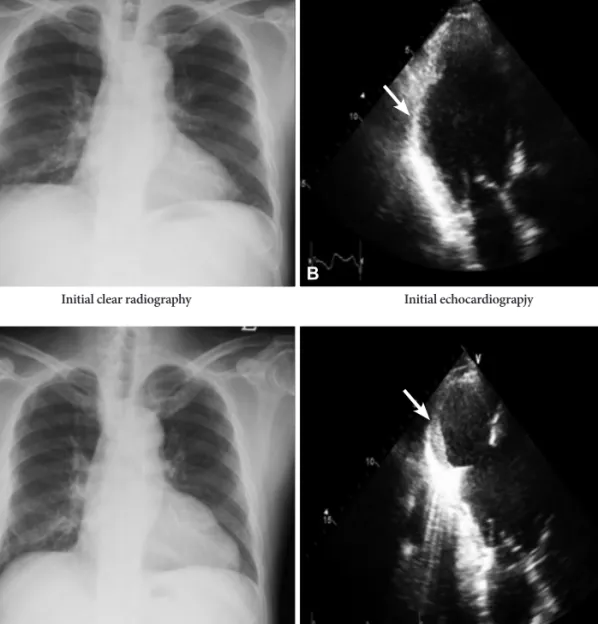
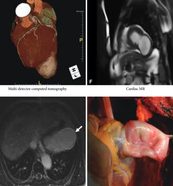
Asymptomatic Left Ventricular Pseudoaneurysm Evaluated With Multimodality Cardiac Imaging
Young-Ran Kang, MD
1, Young-Hoon Jeong, MD
1, Jin-Sin Koh, MD
1, Ho-Cheol Choi, MD
2, Jong-Woo Kim, MD
3, So-Ra Park, MD
1, Jeong Rang Park, MD
1, Yongwhi Park, MD
1, Seok-Jae Hwang, MD
1, Choong Hwan Kwak, MD
1, and Jin-Yong Hwang, MD
11
Division of Cardiology, Departments of Internal Medicine,
2Radiology and
3Thoracic and Cardiaovascular Surgery, Gyeongsang National University College of Medicine, Jinju, Korea
Received: June 26, 2010 / Revision Received: August 20, 2010 / Accepted: August 31, 2010
Correspondence: Young-Hoon Jeong, MD, Division of Cardiology, Department of Internal Medicine, Gyeongsang National University College of Medicine, 90 Chiram-dong, Jinju 660-702, Korea
Tel: 82-55-750-8873, Fax: 82-55-758-9122, E-mail: [email protected]
• The authors have no financial conflicts of interest.
cc