© 2019 The Korean Ophthalmological Society
This is an Open Access article distributed under the terms of the Creative Commons Attribution Non-Commercial License (http://creativecommons.org/licenses /by-nc/3.0/) which permits unrestricted non-commercial use, distribution, and reproduction in any medium, provided the original work is properly cited.
Original Article
Myopia, especially high myopia, is associated with vari- ous vision-threatening diseases such as myopic macular degeneration [1,2], retinal breaks and detachment [3], and
glaucoma [4,5]. Since myopia is growing in prevalence, particularly among younger people [6-10], ocular morbidity related to myopia may be more of problem in future elder- ly populations, thereby constituting an important clinical and public health problem [11].
It has long been recognized that myopia increases the risk of glaucoma [4,5]. Several hypotheses have been pro- posed for the association between myopia and glaucoma.
Cahane and Bartov [12] suggested that myopic eyes are
Received: July 23, 2018 Accepted: August 7, 2018
Corresponding Author: Tae-Woo Kim, MD. Department of Ophthalmol- ogy, Seoul National University Bundang Hospital, 82 Gumi-ro, 173beon- gil, Bundang-gu, Seongnam 13620, Korea. Tel: 82-31-787-7374, Fax: 82-31- 787-4057, E-mail: [email protected]
Development of Optic Disc Torsion in Children
Ji-Ah Kim, Tae-Woo Kim, Eun Ji Lee, Jeong-Min Hwang
Department of Ophthalmology, Seoul National University Bundang Hospital, Seongnam, Korea
Purpose: To document the development of disc torsion.
Methods: Consecutive disc photographs obtained at an interval of at least 1 year were reviewed retrospectively in 173 eyes of 173 Korean children. The angle of the vertical disc axis (AVDA) was measured in each fundus photograph with the fovea-disc center axis set at 0°. The associated change in the morphology of the optic disc was assessed by measuring the ratio of the horizontal to vertical disc diameters and the ratio of the max- imum parapapillary atrophy width to vertical disc diameter. Eyes were divided into two groups with respect to the development of disc torsion: torsion and non-torsion group. Progressive torsion was defined as a change in AVDA between baseline and follow-up photographs beyond the coefficient of intraobserver repeatab ility.
Factors associated with optic disc torsion were evaluated using logistic regression analysis.
Results: Mean subject age and refractive error at the time of baseline fundus examination were 6.8 ± 1.7 (range, 2 to 11) years and 0.2 ± 2.6 (range, -6.0 to +5.5) diopters, respectively. Mean follow-up period was 44.8 ± 21.1 (range, 12 to 103) months. Forty-two eyes (24%) were classified as torsion group who showed changes in AVDA that were greater than the intraobserver measurement variability (4.5°) during the follow-up period. The development of optic disc torsion was associated with greater myopic shift, a decrease in horizontal to vertical disc diameters, and an increase in parapapillary atrophy width to vertical disc diameter.
Conclusions: Progressive optic disc torsion was a common phenomenon in the children included in this study.
Torsion occurred as the result of optic disc tilt in an oblique axis in most cases. The findings provide a frame- work for understanding torsion-related glaucomatous optic nerve damage.
Key Words: Disc tilt, Disc torsion, Optic disk
subject to greater stress due to their axial elongation. La- place’s law states that the wall tension of a sphere is pro- portional to its radius, such that wall tension in the lamina cribrosa and/or parapapillary sclera may be increased in myopic eyes. Quigley [13] suggested that myopic eyes are anatomically weaker due to scleral stretching. In line with this, Ren et al. [14] reported that the lamina cribrosa and peripapillary sclera were thinner in eyes with longer axial length. However, the definitive pathologic relationship be- tween myopia and glaucomatous optic neuropathy (GON) remains to be determined.
Optic disc torsion has recently become a focus of interest in relation to glaucoma in myopic eyes. Park et al. [15] re- ported that the direction of optic disc torsion was related to the location of visual field defects. Lee et al. [16] demon- strated that the prevalence and degree of optic disc torsion were significantly greater in the affected eyes of young myopic patients with a unilateral visual field defect than in contralateral normal eyes. These data together suggest that optic disc torsion significantly influences the development of GON in myopic eyes. However, the precise mechanism underlying how optic disc torsion is associated with glau- coma remains unclear. Elucidating this association first re- quires clarification of what torsion is and how it develops.
Our group proposed that the horizontally oval optic disc (i.e., disc torsion) may represent horizontal disc tilt rather than true rotation of the optic disc based on the lamina cribrosa configuration [17]. However, the true nature of op- tic disc torsion can be confirmed only by observing the de- velopment of a torted disc in a longitudinal study. The purpose of the present study was to document the develop- ment of disc torsion in children.
Materials and Methods
This study performed a retrospective analysis of serial optic disc photographs obtained from subjects aged younger than 18 years who were consecutively enrolled from a data- base of patients first examined for suspected glaucomatous optic disc between October 2004 and October 2014 at Seoul National University Bundang Hospital. This retrospective, observational study was approved by the institutional re- view board of Seoul National University Bundang Hospital (B-1705/396-110). The requirement for informed consent was waived owing to the retrospective nature of the study.
The study followed the tenets of the Declaration of Helsinki.
Each subject underwent ophthalmic examination that in- cluded best-corrected visual acuity, cycloplegic refraction, slit-lamp biomicroscopy, and disc photography. Subjects were eligible for inclusion when their medical records in- cluded serial color- or red-free fundus photographs with an interval between the first and last photographs of at least 1 year. Eyes with a history of intraocular surgery, a history or evidence of glaucoma, other optic neuropathies or reti- nal disease, an elevated intraocular pressure (IOP), or a spherical equivalent (SE) of less than -6 diopters were ex- cluded. The medical records of selected subjects were re- viewed, and data on sex, age, laterality, and cycloplegic re- fractive error at the initial and final examinations were collected. When both eyes were eligible for inclusion, one eye was randomly selected.
Fundus photography
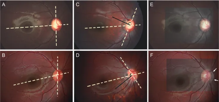
Color- or red-free fundus photographs were acquired us- ing a digital fundus camera (EOS D60, Canon, Utsunomi- yashi, Japan or Kowa VX-10a, Kowa, Tokyo, Japan). Serial photographs were assessed on an LCD monitor by an ob- server (JAK) who was masked to the clinical information of the subjects. The angle of the vertical optic disc axis (AVDA) in each fundus photograph was measured with the fovea-disc enter axis set at 0° using ImageJ ver. 1.52 (devel- oped by Wayne Rasband, National Institutes of Health, Bethesda, MD, USA) (Fig. 1A, 1B). Progressive torsion was defined in this study as a change in AVDA between the baseline and follow-up photographs beyond the coefficient of intraobserver repeatability. “Considerable torsion” was deemed to be present when the AVDA change exceeded 15°
based on criteria often used in this field [18,19].
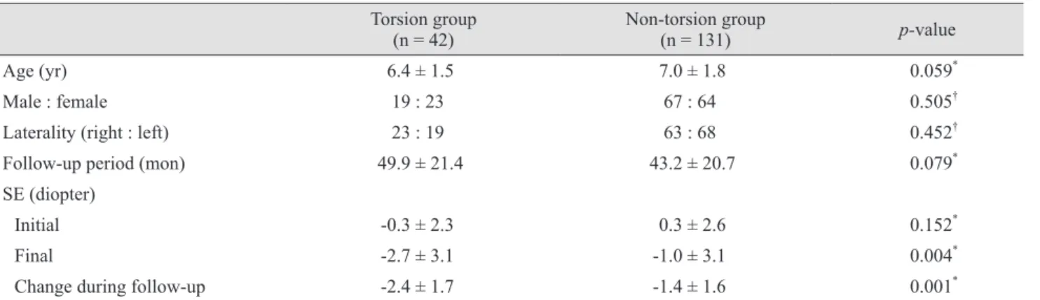
Changes associated with torsion were evaluated by mea- suring the ratio of horizontal to vertical disc diameters (HVDR) and the ratio of maximum parapapillary atrophy (PPA) width to vertical disc diameter (PVDR) in each pho- tograph. To determine whether torsion can be fully ex- plained by disc tilt, the baseline (Fig. 2A, 2B) and fol- low-up (Fig. 2C, 2D) photographs were superimposed with blood vessels aligned. Optic disc margin was expected to overlap at least partially when torsion was the result of the optic disc tilt (Fig. 2E). However, when torsion represented the true rotation of the optic disc axis, not even a small seg- ment of the disc margin was expected to overlap (Fig. 2F).
This analysis was performed only in eyes with consider- able torsion in order to maximize the clarity of the analysis.
Data analysis
To evaluate the intraobserver and interobserver repro- ducibilities of measurements of the optic disc axis and PPA
parameters, 30 randomly selected disc photographs were evaluated by two independent observers (JAK and TWK).
The coefficient of intraobserver repeatability was defined as 1.96 times the intermeasurement standard deviation.
Based on the coefficient of intraobserver repeatability, eyes were classified into two groups: eyes with torsion (tor- sion group) and eyes without torsion (non-torsion group).
Fig. 1. Measuring the vertical disc axis. (A,B) Baseline and follow-up red-free fundus photograph and disc photograph (inset). The angle of the vertical disc axis (solid line) was measured from the reference line connecting the fovea and the disc center (dashed line).
A
Fig. 2. Representative cases showing the development of optic-disc torsion. (A) Baseline fundus photograph of a 4-year-old girl. (B) Base- line fundus photograph of a 3-year-old girl. (C) Superior optic-disc torsion was observed 6 years later. (D) Inferior optic-disc torsion was observed 3 years later. (E) When the baseline and follow-up photographs were superimposed using the blood vessels as references, the na- sal disc margin of the two photographs overlapped, indicating that the apparent torsion resulted from disc tilt on an oblique axis. (F) The disc margin of the two photographs did not overlap at any region when the two photographs were superimposed. The current disc margin is vaguely seen (arrows). (C,D) Note that the vertical disc axis (dashed line) and direction of the longest parapapillary atrophy width (black line) are approximately perpendicular.
A
B
C
D
E
f B
Comparisons between these two groups were performed with the chi-square and Student’s t tests. Logistic regres- sion analysis was performed to identify factors associated with optic disc torsion. All statistical tests were performed using IBM SPSS Statistics ver. 20.0 (IBM Corp., Chicago, IL, USA). A probability value of p < 0.05 was considered to be indicative of statistical significance.
Results
This study initially included the 398 eyes of 199 subjects who had more than 1 year of follow-up using serial disc photography. Of these, 65 eyes were excluded because of a history or evidence of congenital glaucoma (n = 16), juve- nile glaucoma (n = 21), or other optic neuropathy (n = 10), or SE <-6 diopters (n = 18), leaving a final sample of 333
eyes of 173 subjects. When both eyes were eligible for in- clusion, one eye was randomly selected.
Subject age and refractive error at the time of initial fun- dus examination were 6.8 ± 1.7 years (range, 2 to 11 years) and 0.2 ± 2.6 diopters (range, -6.0 to +5.5 diopters), respec- tively. The follow-up period was 44.8 ± 21.1 months (range, 12 to 103 months). The coefficient of the intraobserver re- peatability for measuring AVDA was 4.5°. Based on this value, progressive torsion was defined as an AVDA change exceeding 5°.
Of the 173 finally included eyes, 42 (24.3%) had progres- sive torsion and were classified as the torsion group; the re- maining 131 eyes were classified as the non-torsion group.
Of the 42 eyes in the torsion group, 15 and 27 had superior and inferior torsion, respectively, as defined by clockwise and counterclockwise rotations, respectively, of the verti- cal diameter axis at a right-eye orientation (Fig. 3). Consid- erable torsion was found in 21 eyes (12.1%).
Table 1 compares the baseline characteristics between the torsion and non-torsion groups. No significant differ- ence was found between groups with regard to age, sex, laterality, or follow-up period. The final SE was more my- opic in the torsion group than in the non-torsio n group (p
= 0.004), and the degree of myopic shift was also signifi- cantly greater in the torsion group than in the non-torsion group (-2.4 ± 1.7 vs. -1.4 ± 1.6 diopters, p = 0.001).
Table 2 compares changes in the optic disc and parapap- illary region between the two groups. The final HVDR was smaller in the torsion group than in the non-torsion group (p = 0.005). In addition, the change in HVDR was signifi- cantly larger in the torsion group (p < 0.001). PVDR did not
Torsion (°)
No. of eyes
-60 -55 -50 -45 -40 -35 -30 -25 -20 -15 -10 -5 0 5 10 15 20 25 30 35 40 45 90
80 70 60 50 40 30 20 10 0
Fig. 3. Frequency distribution of the degree of torsion. Note that inferior torsion was more common than superior torsion.
Table 1. Demographic and clinical characteristics
Torsion group
(n = 42) Non-torsion group
(n = 131) p-value
Age (yr) 6.4 ± 1.5 7.0 ± 1.8 0.059*
Male : female 19 : 23 67 : 64 0.505†
Laterality (right : left) 23 : 19 63 : 68 0.452†
Follow-up period (mon) 49.9 ± 21.4 43.2 ± 20.7 0.079*
SE (diopter)
Initial -0.3 ± 2.3 0.3 ± 2.6 0.152*
Final -2.7 ± 3.1 -1.0 ± 3.1 0.004*
Change during follow-up -2.4 ± 1.7 -1.4 ± 1.6 0.001*
Values are presented as mean ± standard deviation or number.
SE = spherical equivalent.
*Analyzed with the Student t-test; †Analyzed with the chi-square test.
differ significantly between the two groups on initial exam- ination (p = 0.183), but the subsequent change in PVDR was significantly larger in the torsion group (p < 0.001).
The torsion in 19 of the 21 eyes with considerable disc torsion was fully explained by the tilt of the optic disc on an oblique axis. In the remaining eyes, torsion could not be explained solely by tilt, and additional rotation was consid- ered to be present in those eyes (Fig. 2D-2F).
In univariate analysis, torsion was significantly associat- ed with the last measured value of SE (p = 0.004), changes in SEs (myopic shift, p = 0.001), and changes in HVDR
and PVDR during follow-up (p < 0.001 and p < 0.001, re- spectively). Since changes in HVDR and PVDR represent different aspects of optic disc changes associated with my- opic shift [20], multivariate analysis was performed in two ways to avoid multicollinearity between them. Changes in HVDR and PVDR during follow-up remained statistically significant in multivariate analysis (p < 0.001) (Table 3).
Table 2. Comparison of the changes in the optic disc and parapapillary region between the two groups Torsion group
(n = 42) Non-torsion group
(n = 131) p-value*
HVDR ratio
Initial 0.85 ± 0.08 0.84 ± 0.10 0.361
Final 0.78 ± 0.08 0.83 ± 0.11 0.005
Change during follow-up -0.07 ± 0.06 -0.01 ± 0.05 <0.001
PVDR ratio
Initial 0.06 ± 0.07 0.04 ± 0.09 0.183
Final 0.19 ± 0.11 0.07 ± 0.11 <0.001
Change during follow-up 0.13 ± 0.09 0.03 ± 0.06 <0.001
Values are presented as mean ± standard deviation.
HVDR = ratio of horizontal disc diameter to vertical disc diameter; PVDR = ratio of maximum parapapillary atrophy width to vertical disc diameter.
*Analyzed with the Student’s t-test.
Table 3. Factors associated with optic nerve head torsion
Variables Univariate analysis Multivariate analysis 1 Multivariate analysis 2
OR 95% CI p-value OR 95% CI p-value OR 95% CI p-value
Age (yr) 0.831 0.674–1.023 0.081 0.943 0.687–1.293 0.713 0.880 0.625–1.241 0.467
Sex (male) 0.789 0.393–1.585 0.506 - - - - - -
Laterality (right) 1.307 0.650–2.625 0.452 - - - - - -
Follow-up periods (mon) 1.015 0.999–1.032 0.073 1.003 0.978–1.028 0.840 0.997 0.971–1.024 0.825
Initial SE (D) 0.909 0.790–1.045 0.181 - - - - - -
Last SE (D) 0.843 0.749–0.948 0.004 0.938 0.790–1.113 0.461 0.969 0.807–1.163 0.732 Change of SE
during follow-up (D) 0.683 0.547–0.853 0.001 0.895 0.644–1.243 0.507 1.037 0.727–1.480 0.840 HVDR difference
(per 0.01) 0.835 0.776–0.899 <0.001 0.860 0.797–0.929 <0.001 - - -
PVDR difference
(per 0.01) 1.165 1.106–1.227 <0.001 - - - 1.166 1.096–1.241 <0.001
OR = odds ratio; CI = confidence interval; SE = spherical equivalent; D = diopters; HVDR = ratio of horizontal disc diameter to vertical disc diameter; PVDR = ratio of maximum parapapillary atrophy width to vertical disc.
Discussion
We identified 42 cases in which progressive optic disc torsion occurred during the study period. Torsion was as- sociated with disc tilt and the development/enlargement of PPA in all eyes. Additional rotation of the optic disc axis was observed in two patients. To the best of our knowl- edge, this study is the first to document the development of disc torsion using serial fundus photographs.
We previously demonstrated the presence of optic disc tilt in children with incipient myopia [20]. Tilt was accom- panied by the development/enlargement of PPA and was associated with myopic shift, suggesting that disc tilt de- rives from scleral stretching secondary to axial elongation.
In the present study, torsion was universally associated with a decrease in HVDR (i.e., disc tilt), and also with the development/enlargement of PPA and myopic shift. These findings suggest that torsion represents tilt on an oblique axis. Axial elongation can occur in various directions [21], and the direction of axial elongation can influence the di- rection of disc tilt. For instance, when the posterior center of the axis of elongation is located superotemporally, scler- al stretching would occur in the superotemporal direction, resulting in PPA in the superotemporal region, with oblique disc tilt being perpendicular to the direction of scleral stretching. The obliquely tilted disc would have an oblique disc axis, and be identified as torsion.
The changes in almost all cases in the torsion group were explainable by oblique disc tilt. However, in two eyes torsion could not be explained by oblique tilt because the nasal disc margin was deviated from the expected disc margin after oblique disc tilt. This finding suggests that true rotation of the disc axis also occurs in some eyes. Al- though we do not have a clear explanation for such rota- tion, we speculate that it is attributable to axial elongation in multiple directions. Such axial elongation could result in complex changes in the optic disc, resulting in apparent rotation of the optic disc axis.
Torsion is a common feature in myopic eyes [15], and it has recently become a focus of interest in relation to how it influences the development of GON. In young patients with unilateral myopic glaucoma, greater torsion was found in the affected eye. In particular, it is intriguing that the direction of torsion was related to the location of reti- nal nerve fiber layer (RNFL) defect: this defect was seen in the superotemporal sector in eyes with superior torsion,
and in the inferotemporal sector in eyes with inferior tor- sion [15]. Our findings may provide a plausible explanation for the relationship between optic disc torsion and the lo- cation of glaucomatous damage. The superotemporal and inferotemporal sectors are preferentially involved in glau- coma due to the presence of less support for the axons from connective tissue [22]. In eyes with optic disc tilt, tensile and/or shearing stresses arising from scleral stretch- ing may induce axonal damage. In this process the direc- tion of scleral stretching may result in stress varying by region of the optic nerve head. For instance, in eyes with superior torsion, the change would be largest near the su- perior disc margin and thereby increase the possibility of axonal damage in the superior optic nerve (and vice versa for eyes with inferior torsion).
An RNFL defect was not detected on follow-up photo- graphs in any of the eyes in the torsion group in this study (data not presented). However, especially in myopic pa- tients aged in their 20s or early 30s, RNFL defect is not rarely seen despite low IOP, particularly among those with disc torsion [23,24]. We speculate that the tensile stress de- rived from torsion does not immediately lead to develop- ment of axonal damage. However, eyes with torsion will be subject to sustained tensile stress, which may eventually damage the axons. An analogy to this finding may be found in the relationship between ocular hypertension and glaucoma. Eyes with high IOP do not necessarily exhibit optic nerve damage (i.e., ocular hypertension) at initial presentation, but some develop glaucomatous damage years later [25].
AVDA was larger than 15° at baseline in 10 of the 173 included eyes. These eyes all had PPA and a tilted disc ap- pearance. We speculate that those discs were torted before the baseline photograph was obtained. Progressive consid- erable torsion was not observed during the study period in these 10 eyes, and so the incidence of considerable disc torsion may have been underestimated in this study.
The relationship between myopia and glaucoma remains unclear. It is generally acknowledged that myopia is a risk factor for glaucoma. In contrast, recent studies have demonstrated that glaucoma progression does not occur more rapidly in myopic eyes [26,27]. Our data may provide some insight into this puzzle. The tensile stress associated with torsion may increase the susceptibility of axons to glaucomatous damage. Thus, it is possible that eyes subject to torsion will develop glaucoma despite the relatively low
IOP-induced stress not being sufficient to induce glauco- matous damage in eyes without torsion. Meanwhile, the tensile stress would remain focused near the superior or inferior pole of the optic disc, depending on the direction of torsion. Thus, once glaucomatous damage occurs in the respective polar area in these eyes, the remaining axons may remain healthy if the glaucomatous insult (i.e., IOP-induced stress) is not sufficient to induce glaucoma by itself. In this situation the rate of disease progression would decrease. This may be a factor contributing to the slow rate of disease progression in myopic patients.
This study was subject to several limitations. First, we did not measure axial length, and myopic shift was used as a surrogate of axial elongation. However, myopic shift is attributed to axial elongation that occurs in childhood [28].
Second, we could not measure the true length or distance values in fundus photographs because we could not correct for the magnification effect due to the actual axial length being unknown. However, all measurements in the study were of angles and ratio parameters that would not be af- fected by magnification errors. Third, the subjects were glaucoma suspect patients, and so the findings of this study might not be applicable to a general population. Fourth, the subjects were not observed until the end of eye growth. It is possible that the non-torsion group could show changes in the future. Lastly, disc changes were evaluated based on two-dimensional analyses. This was because three-dimen- sional measurements of the optic nerve head were not fea- sible in most of the children, who were aged 6.8 ± 1.7 years at the time of baseline examination. Three-dimensional analysis may provide greater insight into optic disc tilt.
Nonetheless, our data clearly indicate that optic disc tor- sion can be an acquired feature arising from scleral stretching.
In conclusion, we have demonstrated the development of optic disc torsion in children. Torsion was mostly explained by optic disc tilt on the oblique axis. The findings provide a framework for understanding torsion-related GON.
Conflict of Interest
No potential conflict of interest relevant to this article was reported.
References
1. Ito-Ohara M, Seko Y, Morita H, et al. Clinical course of newly developed or progressive patchy chorioretinal atro- phy in pathological myopia. Ophthalmologica 1998;212:23- 9.
2. Avila MP, Weiter JJ, Jalkh AE, et al. Natural history of choroidal neovascularization in degenerative myopia. Oph- thalmology 1984;91:1573-81.
3. Pierro L, Camesasca FI, Mischi M, Brancato R. Peripheral retinal changes and axial myopia. Retina 1992;12:12-7.
4. Suzuki Y, Iwase A, Araie M, et al. Risk factors for open-angle glaucoma in a Japanese population: the Tajimi Study. Ophthalmology 2006;113:1613-7.
5. Mitchell P, Hourihan F, Sandbach J, Wang JJ. The relation- ship between glaucoma and myopia: the Blue Mountains Eye Study. Ophthalmology 1999;106:2010-5.
6. Kempen JH, Mitchell P, Lee KE, et al. The prevalence of refractive errors among adults in the United States, West- ern Europe, and Australia. Arch Ophthalmol 2004;122:495- 505.
7. Guo K, Yang DY, Wang Y, et al. Prevalence of myopia in schoolchildren in Ejina: the Gobi Desert Children Eye Study. Invest Ophthalmol Vis Sci 2015;56:1769-74.
8. Williams KM, Verhoeven VJ, Cumberland P, et al.
Prevalence of refractive error in Europe: the European Eye Epidemiology (E(3)) Consortium. Eur J Epidemiol 2015;30:305-15.
9. Saxena R, Vashist P, Tandon R, et al. Prevalence of myo- pia and its risk factors in urban school children in Delhi:
the North India Myopia Study (NIM Study). PLoS One 2015;10:e0117349.
10. Foster PJ, Jiang Y. Epidemiology of myopia. Eye (Lond) 2014;28:202-8.
11. Seet B, Wong TY, Tan DT, et al. Myopia in Singapore: tak- ing a public health approach. Br J Ophthalmol 2001;85:521-6.
12. Cahane M, Bartov E. Axial length and scleral thickness effect on susceptibility to glaucomatous damage: a theo- retical model implementing Laplace’s law. Ophthalmic Res 1992;24:280-4.
13. Quigley HA. Reappraisal of the mechanisms of glaucoma- tous optic nerve damage. Eye (Lond) 1987;1:318-22.
14. Ren R, Wang N, Li B, et al. Lamina cribrosa and peripap- illary sclera histomorphometry in normal and advanced glaucomatous Chinese eyes with various axial length. In- vest Ophthalmol Vis Sci 2009;50:2175-84.
15. Park HY, Lee K, Park CK. Optic disc torsion direction pre- dicts the location of glaucomatous damage in normal-ten- sion glaucoma patients with myopia. Ophthalmology 2012;119:1844-51.
16. Lee KS, Lee JR, Kook MS. Optic disc torsion presenting as unilateral glaucomatous-appearing visual field defect in young myopic Korean eyes. Ophthalmology 2014;121:1013- 9.
17. Lee KM, Lee EJ, Kim TW. Lamina cribrosa configuration in tilted optic discs with different tilt axes: a new hypothe- sis regarding optic disc tilt and torsion. Invest Ophthalmol Vis Sci 2015;56:2958-67.
18. Witmer MT, Margo CE, Drucker M. Tilted optic disks.
Surv Ophthalmol 2010;55:403-28.
19. How AC, Tan GS, Chan YH, et al. Population prevalence of tilted and torted optic discs among an adult Chinese population in Singapore: the Tanjong Pagar Study. Arch Ophthalmol 2009;127:894-9.
20. Kim TW, Kim M, Weinreb RN, et al. Optic disc change with incipient myopia of childhood. Ophthalmology 2012;119:21-6.
21. Moriyama M, Ohno-Matsui K, Hayashi K, et al. Topo- graphic analyses of shape of eyes with pathologic myopia by high-resolution three-dimensional magnetic resonance imaging. Ophthalmology 2011;118:1626-37.
22. Dandona L, Quigley HA, Brown AE, Enger C. Quantita- tive regional structure of the normal human lamina cribro- sa. A racial comparison. Arch Ophthalmol 1990;108:393-8.
23. Doshi A, Kreidl KO, Lombardi L, et al. Nonprogressive glaucomatous cupping and visual field abnormalities in young Chinese males. Ophthalmology 2007;114:472-9.
24. Park HY, Lee KI, Lee K, et al. Torsion of the optic nerve head is a prominent feature of normal-tension glaucoma.
Invest Ophthalmol Vis Sci 2014;56:156-63.
25. Kass MA, Heuer DK, Higginbotham EJ, et al. The Ocular Hypertension Treatment Study: a randomized trial deter- mines that topical ocular hypotensive medication delays or prevents the onset of primary open-angle glaucoma. Arch Ophthalmol 2002;120:701-13.
26. Araie M, Shirato S, Yamazaki Y, et al. Risk factors for progression of normal-tension glaucoma under β-blocker monotherapy. Acta Ophthalmol 2012;90:e337-43.
27. Lee JY, Sung KR, Han S, Na JH. Effect of myopia on the progression of primary open-angle glaucoma. Invest Oph- thalmol Vis Sci 2015;56:1775-81.
28. Hyman L, Gwiazda J, Hussein M, et al. Relationship of age, sex, and ethnicity with myopia progression and axial elongation in the correction of myopia evaluation trial.
Arch Ophthalmol 2005;123:977-87.