393 REVIEW
DOI 10.4070 / kcj.2009.39.10.393
Print ISSN 1738-5520 / On-line ISSN 1738-5555 Copyright ⓒ 2009 The Korean Society of Cardiology
Open Access
The Inflammatory Response and Cardiac Repair After Myocardial Infarction
Deuk-Young Nah, MD and Moo-Yong Rhee, MD
Division of Cardiology, Department of Internal Medicine, College of Medicine, Dongguk University, Gyeongju, Korea ABSTRACT
One of the most important therapeutic targets of current cardiology practice is to determine optimal strategies for the minimization of myocardial necrosis and optimization of cardiac repair following an acute myocardial infarc- tion. Myocardial necrosis after acute myocardial infarction induces complement activation and free radical gene- ration, triggering a cytokine cascade initiated by tumor necrosis factor-alpha (TNF-α) release. When reperfusion of the infarcted area is initiated, intense inflammation follows. Chemokines, cytokines and the complement system play an important role in recruiting neutrophils in the ischemic and reperfused myocardium. Cytokines promote adhesive interactions between leukocytes and endothelial cells, resulting in transmigration of inflammatory cells into the site of injury. The recruited neutrophils have potent cytotoxic effects through the release of proteolytic enzymes, and they interact with adhesion molecules on cardiomyocytes. In spite of the potential injury, reperfusion enhances cardiac repair; this may be related to the inflammatory response. Monocyte chemoattractant protein (MCP)-1 is upregulated in reperfused myocardium and can induce monocyte recruitment in the infarcted area.
Monocyte subsets play a role in phagocytosis of dead cardiomyocytes and in granulation tissue formation. In ad- dition, the transforming growth factor (TGF)-β plays a crucial role in cardiac repair by suppressing inflam- mation. Resolution of inflammatory infiltration, containment of inflammation and the reparative response af- fecting the infarcted area are essential for optimal infarct healing. Here, we review the current literature on the inflammatory response and cardiac repair after myocardial infarction. (Korean Circ J 2009;39:393-398)
KEY WORDS:Myocardial infarction; Inflammation; Left ventricle remodeling; Cytokines.
Introduction
Acute myocardial infarction is a potentially fatal event and a common causes of death in adults. Sudden coro- nary artery occlusion results in ischemic related death of cardiomyocytes.1) The human heart has low regenerative ability. The inflammatory response and cytokine release from the myocardium are essential components of the host response to acute myocardial infarction, and play a crucial role in cardiac repair. Tumor necrosis factor-alpha (TNF-α) and interleukin-6 (IL-6) are increased after an
acute myocardial infarction and can regulate myocyte survival and induce additional cellular inflammatory re- sponses.2)3) Chemokines stimulate the recruitment of in- flammatory leukocytes to the infarct related myocar- dium.4) Monocyte chemoattractant protein (MCP-I)/
CCL2 has a potent effect on macrophage recruitment and myofibroblast accumulation in healing myocardium and also plays an important role in postinfarct left ven- tricular healing.5) Transforming growth factor (TGF)-β also plays a crucial role in cardiac repair by the suppres- sion of inflammation.6) Timely resolution of the inflam- mation and inhibition of the inflammatory response and recovery from tissue injury of the infarcted area are essential for optimal healing of the myocardium. Here we review recent researches and evolving concepts of the inflammatory responses and cardiac repair after acute myocardial infarction.
Inflammatory Response
The complement cascade, reactive oxygen species
Correspondence: Deuk-Young Nah, MD, Division of Cardiology, Department of Internal Medicine, College of Medicine, Dongguk University, 1090-1 Seockjang-dong, Gyeongju 780-350, Korea
Tel: 82-54-770-8561, Fax: 82-54-770-8378 E-mail: [email protected]
○cc This is an Open Access article distributed under the terms of the Creative Commons Attribution Non-Commercial License (http://creativecommons.
org/licenses/by-nc/3.0) which permits unrestricted non-commercial use, distribution, and reproduction in any medium, provided the original work is properly cited.
394·Inflammatory Response and Cardiac Repair After MI
(ROS) and cytokine cascade mediated pathway play an important role in the post infarction inflammatory re- sponse.
Complement cascade activation
Hill and Ward were the first to report that ischemic myocardial injury can induce activation of the comple- ment cascade in a rat infarct model.7) Pinckard et al.8) also showed that ischemic myocardial necrosis was as- sociated with the release of subcellular membrane cons- tituents that are triggered by the early acting components of the complement cascade (C1, C4, C2 and C3) in pa- tients with an acute myocardial infarction. Yasojima et al.9) demonstrated that complement gene expression was upregulated by ischemia and reperfusion in the rabbit heart. Complement activation was reported to induce neutrophil and monocyte recruitment in the ischemic myocardium.10) Weisman et al.11) reported that infusion of soluble human complement receptor type 1 (sCR1) significantly suppressed post-infarct inflammation and necrosis in a rat model of myocardial ischemia and reperfusion. In spite of promising experimental results, recent clinical trials testing the effects of complement inhibition, in patients with acute myocardial infarction, showed disappointing results. Administration of the human anti-C5 monoclonal antibody pexelizumab, in patients with an acute myocardial infarction, had no effect on infarct size and clinical outcome.12-14)
Reactive oxygen species
Meldrum et al.15) demonstrated that H2O2 alone in- duced myocardial TNF-α mediated cardiac injury by a p38 mitogen-activated protein kinase (MAPK)-depen- dent mechanism. Reactive oxygen intermediates may generate a leukotatic stimulus that includes, comple- ment activation, induction of hemorrhagic shock-in- duced P-selectin expression, chemokine upregulation and an increase in the endothelial intercellular adhe- sion molecule (ICAM)-1 ability to bind neutrophils.16-19) The use of the antioxidant enzymes superoxide dismutase and catalase reduced the infarct size in dogs with myo- cardial ischemia and reperfusion.20) However, there have been some failed studies of antioxidant treatment used to prevent myocardial ischemic injury.21)22) Two clinical studies using recombinant human superoxide dismutase in patients with an acute myocardial infarction undergo- ing percutaneous coronary intervention or thrombolysis
showed no significant improvement of left ventricular function.23)24)
Cytokine amplication
Cytokines can self-amplify through a positive feed- back loop targeting the nuclear factor (NF)-κB. Upre- gulation of TNF-α in the infarct myocardium can upregulate the levels of TNF-α in the neighboring nor- mal myocardium, leading to amplified cytokine effects.3) TNF-α stimulates expression of proinflammatory cy- tokines, chemokines and adhesion molecules by leuko- cytes and endothelial cells and regulates extracellular matrix metabolism by reducing collagen synthesis and by enhancing matrix metalloprotease (MMP) activity in cardiac fibroblasts;25) other adhesive cytokines such as monocyte chemoattractant protein (MCP)-1 is also in- duced in the ischemic and reperfused canine myocardi- um. Kumar et al.26) suggested a significant role for MCP- 1 in monocyte trafficking in reperfused myocardium.
Cytokine and chemokine upregulation
Chemokine upregulation is a noted feature of the post- infarction inflammatory response (Table 1).27) Recent investigators have demonstrated strong induction of sev- eral chemokines in the ischemic myocardium supporting their role in leukocyte recruitment.4) MCP-1 upregula- tion has been demonstrated in a mouse model.28) Fran- gogiannis reported that a MCP-1 -/- infarct mouse mo- del had decreased mesenger ribonucleic acid (mRNA) expression of the cytokine TNF-α, IL-1β, TGF-β and IL-10 and showed defective macrophage differentia- tion.27) Induction and release of cytokines such as TNF- α and IL-6 are rapidly released in the central zone dur- ing a myocardial infarction; however, they are usually maximal in the border zone.3)29) This robust upregulation may return to baseline levels if the infarction is small, and if the infarction is large and the inflammatory re- sponse is excessive, there can be sustained cytokine up- regulation, corresponding to a chronic remodeling phase.
Cellular inflammatory response to myocardial infarction
The neutrophils
Neutrophils are recruited during the initial stage of cardiac ischemic injury. Neutrophil transmigration in the infarcted myocardium requires adhesive interactions with
Table 1. Upregulated chemokines and their role after myocardial ischemia and reperfusion
CXCL8/Interleukin (IL)-8 Induce neutrophil infiltration
CCL2/Monocyte Chemoattractant Protein (MCP)-1 Regulate monocyte and lymphocyte recruitment CCL3/Macrophage Inflallatroy Protein (MIP)-1α Regulate monocyte and lymphocyte recruitment CCL4/Macrophage Inflallatroy Protein (MIP)-1β Regulate monocyte and lymphocyte recruitment
CXCL10/Interferon-10 Angiostatic factor with antifibrotic properties
Deuk-Young Nah, et al.·395
activated vascular endothelial cells. Neutrophils may se- crete oxidants and proteases and possibly express media- tors capable of amplifying cell recruitment.30) Neutrophil depletion in animals undergoing reperfused myocardial infarction has been reported to significantly decrease the infarct size suggesting that a significant amount of myo- cardial injury may be induced by neutrophil dependent mechanisms.31)32)
However, the mechanisms associated with neutrophil- induced myocardial ischemic injury have not been iden- tified. Jaeschke et al.33) suggested that neutrophils may di- rectly injure parenchymal cells through release of specific toxic products. The selectin family consists of L-selectin, P-selectin and E-selectin. P-selectin expression occurs rapidly in endothelial cells during cardiac ischemic in- jury. Experimental study has suggested that monoclonal antibodies against P-selectin reduced myocardial necrosis, preserving coronary endothelial function and attenuat- ing neutrophil infiltration in ischemic and reperfused myocardium.34) However, there have been inconsistent results of selectin-related interventions in experimental models of myocardial ischemia.35)36)
The mononuclear cells
As previously mentioned, MCP-1/CCL2 plays an im- portant role in monocyte recruitment to the infarcted myocardium.5) Cytokines, such as TGF-β, free radical oxygen, complement, and the CC chemokine may also play a role in monocyte infiltration. Infiltration of mo- nocytes into the infarcted myocardium is followed by maturation and differentiation of these blood-derived cells into macrophages.
Cardiac Repair After Myocardial Infaction
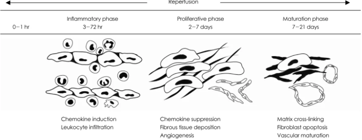
Healing of an infarction can be divided into three overlapping phases: the inflammatory phase, the proli- ferative phase and the maturation phase (Fig. 1).37) The acute repair process is mediated by cytokines and inflam-
matory cells in the infarcted myocardium. The induction of pro-inflammatory mediators and leukocyte infiltra- tion play a crucial role in phagocytosis and removal of necrotic cells and matrix debris from infarcted myocar- dium. Moreover, they promote tissue repair and scar for- mation.38) The strategy for optimal infarct healing re- quires inhibition of cytokines and chemokine synthesis after myocardial infarction.
Transforming growth factor-β as a key regulator in cardiac repair
TGF-β is a multifunctional cytokine that controls proliferation and cellular differentiation in most cells.
TGF-β has been shown to be significantly upregulated in an experimental rat model of myocardial infarction;
in addition, TGF-β mRNA and protein was signifi- cantly increased at the infarct border zone.39)40) During infarct healing, TGF-β may play a role in the suppres- sion of chemokine and cytokine synthesis and is thought to be a key mediator of the transition from inflamma- tion to fibrosis.41) Lefer et al.42) reported that TGF-β in- jections reduced myocardial ischemic injury mediated by proinflammatory cytokines such as TNF-α during the inflammatory phase of myocardial healing. Anti- TGF-β treatment before or after coronary artery liga- tion increased mortality and worsened the left ventricular remodeling in mice with non-reperfused myocardial in- farction.43) The inhibition of TGF-β signaling by in- jection of a TGF-β II receptor resulted in reduction of left ventricular remodeling by modulation of cardiac fibrosis; early TGF-β inhibition increased mortality and left ventricular dilatation.44)45) Youn et al.46) reported that the angiotensin converting enzyme inhibitor and angio- tensin receptor blockade resulted in decreased TGF-β mRNA expression after nontransmural infarction in the rat.
However, the exact role of TGF-β signaling in the in- farcted and remodeled heart is poorly understood con- sidering the complex and unusual biology of TGF-β (Table 2).47)
Fig. 1. The phases of healing in murine myocardial infarction.
Chemokine induction Leukocyte infiltration
Chemokine suppression Fibrous tissue deposition Angiogenesis
Matrix cross-linking Fibroblast apoptosis Vascular maturation Maturation phase
7-21 days Proliferative phase
2-7 days Inflammatory phase
3-72 hr 0-1 hr
Reperfusion
396·Inflammatory Response and Cardiac Repair After MI
The role of other cytokines in cardiac repair There are three IL-1 molecules consisting of IL-1α, IL-β and IL-1 Ra that are specific receptor antago- nists.48) Bujak et al.49) demonstrated that IL-1 signaling is essential for activation of inflammatory and fibrogenic pathways in the healing infarct and plays an important role in the pathogenesis of remodeling after infarction.
IL-10 exerts potent anti-inflammatory effects and modu- lates MMP expression.50-52) However, Zymek et al.53) and our group reported that IL-10 signaling plays a non- critical role in the suppression of inflammatory media- tors, resolution of the inflammatory response and fibrous tissue deposition following myocardial infarction in the mouse. This result may be due to the involvement of multiple overlapping regulatory mechanisms controll- ing various proinflammatory pathways activated in the infarcted myocardium.
The role of proteins in cardiac repair
CD44 is a cell surface glycoprotein involved in cell- cell interaction and cell adhesion and migration. CD44- hyaluronan interactions play a role in leukocyte extrav- asation at the inflammatory site and serves as a key factor in the resolution of inflammation through removal of matrix breakdown products and clearance of apoptotic neutrophils.54-56) Huebener et al.57) tested the role of CD44 in infarct healing and demonstrated that CD44 mRNA levels were significantly induced in the infarcted heart; CD44 null mice showed enhanced and prolong- ed inflammation in the infarcted heart followed by decreased myofibroblast infiltration, reduced collagen deposition and diminished proliferative activity. Hue- bener et al.57) concluded that CD44 is critically involved in infarct healing by regulating the inflammatory and fibrotic response. Thrombospondin (TSP)-1 is an adhe- sive glycoprotein involved in cell to cell and cell to ma- trix interaction with potent angiostatic properties; it is a TGF-β activator.58) TSP-1 showed strikingly selective localization in the infarct border zone, and TSP-1 knock- out animals had markedly increased macrophage and myofibroblast density in the infarct and in remodeling of noninfarcted myocardial areas, and was more exten- sive in the postinfarction remodeling than in the wild-
type mice. Frangogiannis et al.59) concluded that the selective endogenous expression of TSP-1 at the infarct border zone may serve as a “barrier,” limiting expansion of granulation tissue and protecting the noninfarcted myocardium from fibrotic remodeling. Smad is an es- sential protein component of the TGF-β pathway.60) Hao et al.61) showed that TGF-β mRNA was signifi- cantly increased in the infarct scar compared to viable myocardium, and that Cardiac Smad 2, 3 and 4 pro- teins were significantly increased in the border and scar tissues when compared to viable myocardium. These re- sults indicate that TGF-beta/Smad signaling may be involved in the remodeling of the infarct scar. Optimal cardiac repair requires containment of the inflamma- tion in the infarcted area. Extension of the inflamma- tion into the non-infarcted area could result in expan- sion of the neutrophil infiltration and worsening of the remodeling. Currently, investigators are testing potential treatment strategies for prevention of the expansion of the inflammatory response into viable myocardium.
Conclusions
The inflammatory response and cytokines such as TNF-α and IL-6 are integral components of the host response to tissue injury and play an important role after acute myocardial infarction. Furthermore, TGF-β plays an important role in cardiac repair after myocardial infarction. Many experimental studies have shown a dramatic reduction in infarct size with the use of anti- inflammatory treatment and inhibition of cytokine signaling. However, treatment with specific cytokine in- hibitors has been unsuccessful in clinical practice; ex- planations include: the animal models have fundamen- tal differences compared to the human disease process and the inflammatory cascade is a complex network of multiple overlapping regulatory mechanisms that controls various pro-inflammatory pathways. The properties of cytokines in the network include redundancy, pleiotropy, synergistic activity, and the antagonistic effects on each other. We are now only beginning to elucidate the roles and significance of various cytokines and growth factors in healing of a myocardial infarction.
If we get the knowledge of inflammatory response to ischemic myocardial injury and role of cytokines after myocardial infarction, the effective inflammation-relat- ed interventions may allow us to promote improved heal- ing and cardiac remodeling after myocardial infarction.
REFERENCES
1) Jennings RB, Murry CE, Steenbergen C Jr, Reimer KA. Devel- opment of cell injury in sustained acute ischemia. Circulation 1990;82(3 Suppl):II2-12.
2) Deten A, Volz HC, Briest W, Zimmer HG. Cardiac cytokine ex- pression is upregulated in the acute phase after myocardial in- Table 2. The complex role of TGF-β signaling in myocardial
infarction
Cadiomyocyte hypertrophy Angiogenic or angiostatic effects Reduced adhesion molecule expression Macrophage deactivation
Chemokine and cytokine repression Myofibroblast differenciation Fibroblast proliferation
Extracellular matrix protein synthesis
Deuk-Young Nah, et al.·397
farction: experimental studies in rats. Cardiovasc Res 2002;55:
329-40.
3) Irwin M, Mak S, Mann DL, et al. Tissue expression and im- munolocalization of tumour necrosis factor-alpha in post in- farction-dysfunctional myocardium. Circulation 1999;99:1492-8.
4) Birdsall HH, Green DM, Trial J, et al. Complement C5a, TGF- beta 1, and MCP-1, in sequence, induce migration of monocytes into ischemic canine myocardium within the first one to five hours after reperfusion. Circulation 1997;95:684-92.
5) Dewald O, Zymek P, Winkelmann K, et al. CCL2/monocyte che- moattractant protein-1 regulates inflammatory responses critical to healing myocardial infarcts. Circ Res 2005;96:881-9.
6) Bujak M, Frangogiannis NG. The role of TGF-beta signaling in myocardial infarction and cardiac remodeling. Cardiovasc Res 2007;74:184-95.
7) Hill JH, Ward PA. The phlogistic role of C3 leukotactic fragments in myocardial infarcts of rats. J Exp Med 1971;133:885-900.
8) Pinckard RN, Olson MS, Giclas PC, Terry R, Boyer JT, O’Rourke RA. Consumption of classical complement components by heart subcellular membranes in vitro and in patients after acute myo- cardial infarction. J Clin Invest 1975;56:740-50.
9) Yasojima K, Schwab C, McGeer EG, McGeer PL. Human heart generates complement proteins that are upregulated and activated after myocardial infarction. Circ Res 1998;83:860-9.
10) Dreyer WJ, Michael LH, Nguyen T, et al. Kinetics of C5a release in cardiac lymph of dogs experiencing coronary artery ischemia- reperfusion injury. Circ Res 1992;71:1518-24.
11) Weisman HF, Bartow T, Leppo MK, et al. Soluble human com- plement receptor type 1: in vivo inhibitor of complement sup- pressing post-ischemic myocardial inflammation and necrosis.
Science 1990;249:146-51.
12) Granger CB, Mahaffey KW, Weaver WD, et al. Pexelizumab, an anti-C5 complement antibody, as adjunctive therapy to primary percutaneous coronary intervention in acute myocardial infarc- tion: the complement inhibition inmyocardial infarction treated with angioplasty (COMMA) trial. Circulation 2003;108:1184-90.
13) Mahaffey KW, Granger CB, Nicolau JC, et al. Effect of pexeli- zumab, an anti-C5 complement antibody, as adjunctive therapy to fibrinolysis in acute myocardial infarction: the COMPlement inhibition in myocardial infarction treated with thromboLYtics (COMPLY) trial. Circulation 2003;108:1176-83.
14) Armstrong PW, Granger CB, Adams PX, et al. Pexelizumab for acute ST-elevation myocardial infarction in patients undergoing primary percutaneous coronary intervention: a randomized con- trolled trial. JAMA 2007;297:43-51.
15) Meldrum DR, Dinarello CA, Cleveland JC Jr, et al. Hydrogen peroxide induces tumor necrosis factor alphamediated cardiac injury by a p38 mitogen activated protein kinasedependent me- chanisms. Surgery 1998;124:291-6; discussion 297.
16) Shingu M, Nobunaga M. Chemotactic activity generated in human serum from the fifth component of complement by hydrogen per- oxide. Am J Pathol 1984;117:201-6.
17) Akgur FM, Brown MF, Zibari GB, et al. Role of superoxide in hemorrhagic shock-induced P-selectin expression. Am J Physiol Heart Circ Physiol 2000;279:H791-7.
18) Lakshminarayanan V, Beno DW, Costa RH, Roebuck KA. Dif- ferential regulation of interleukin-8 and intercellular adhesion molecule-1 by H2O2 and tumor necrosis factor-alpha in endo- thelial and epithelial cells. J Biol Chem 1997;272:32910-8.
19) Sellak H, Franzini E, Hakim J, Pasquier C. Reactive oxygen spe- cies rapidly increase endothelial ICAM-1 ability to bind neutro- phils without detectable upregulation. Blood 1994;83:2669-77.
20) Jolly SR, Kane WJ, Bailie MB, Abrams GD, Lucchesi BR. Canine myocardial reperfusion injury: its reduction by the combined ad-
ministration of superoxide dismutase and catalase. Circ Res 1984;
54:277-85.
21) Uraizee A, Reimer KA, Murry CE, Jennings RB. Failure of su- peroxide dismutase to limit size of myocardial infarction after 40 minutes of ischemia and 4 days of reperfusion in dogs. Circulation 1987;75:1237-48.
22) Gallagher KP, Buda AJ, Pace D, Gerren RA, Shlafer M. Failure of superoxide dismutase and catalase to alter size of infarction in conscious dogs after 3 hours of occlusion followed by reperfusion.
Circulation 1986;73:1065-76.
23) Murohara Y, Yui Y, Hattori R, Kawai C. Effects of superoxide dismutase on reperfusion arrhythmias and left ventricular func- tion in patients undergoing thrombolysis for anterior wall acute myocardial infarction. Am J Cardiol 1991;67:765-7.
24) Flaherty JT, Pitt B, Gruber JW, et al. Recombinant human su- peroxide dismutase (h-SOD) fails to improve recovery of ventri- cular function in patients undergoing coronary angioplasty for acute myocardial infarction. Circulation 1994;89:1982-91.
25) Siwik DA, Chang DL, Coluci WS. Interleukin-1 beta and tumor necrosis factor-alpha decrease collagen synthesis and increase matrix metalloproteinase activity in cardiac fibroblasts in vitro.
Circ Res 2000;86:1259-65.
26) Kumar AG, Ballantyne CM, Michael LH, et al. Induction of monocyte chemoattractant protein-1 in the small veins of the is- chemic and reperfused canine myocardium. Circulation 1997;
95:693-700.
27) Frangogiannis NG. Chemokines in ischemia and reperfusion.
Thromb Haemost 2007;97:738-47.
28) Tarzami ST, Cheng R, Miao W, Kitsis RN, Berman JW. Chemo- kine expression in myocardial ischemia: MIP-2 dependent MCP- 1 expression protects cardiomyocytes from cell death. J Mol Cell Cardiol 2002;34:209-21.
29) Gwechenberger M, Mendoza LH, Youker KA, et al. Cardiac myocytes produce interleukin-6 in culture and in viable border zone of reperfused infarctions. Circulation 1999;99:546-51.
30) Frangogiannis NG, Youker KA, Entman ML. The role of the neu- trophil in myocardial ischemia and reperfusion. EXS 1996;76:
263-84.
31) Romson JL, Hook BG, Kunkel SL, Abrams GD, Schork MA, Lucchesi BR. Reduction of the extent of ischemic myocardial injury by neutrophil depletion in the dog. Circulation 1983;67:
1016-23.
32) Jordan JE, Zhao ZQ, Vinten-Johansen J. The role of neutrophils in myocardial ischemia-reperfusion injury. Cardiovasc Res 1999;
43:860-78.
33) Jaeschke H, Smith CW. Mechanisms of neutrophil-induced pa- renchymal cell injury. J Leukoc Biol 1997;61:647-53.
34) Palazzo AJ, Jones SP, Anderson DC, Granger DN, Lefer DJ. Cor- onary endothelial P-selectin in pathogenesis of myocardial is- chemia-reperfusion injury. Am J Physiol 1998;275:H1865-72.
35) Jones SP, Girod WG, Granger DN, Palazzo AJ, Lefer DJ. Reper- fusion injury is not affected by blockade of P-selectin in the dia- betic mouse heart. Am J Physiol 1999;277:H763-9.
36) Birnbaum Y, Patterson M, Kloner RA. The effect of CY1503, a sialyl Lewisx analog blocker of the selectin adhesion molecules, on infarct size and “noreflow” in the rabbit model of acute myo- cardial infarction/reperfusion. J Mol Cell Cardiol 1997;29:2013-25.
37) Frangogiannis NG. The mechanistic basis of infarct healing. An- tioxid Redox Signal 2006;8:1907-39.
38) Nathan C. Points of control in inflammation. Nature 2002;420:
846-52.
39) Thompson NL, Bazoberry F, Speir EH, et al. Transforming growth factor beta-1 in acute myocardial infarction in rats. Growth Fac- tors 1988;1:91-9.
398·Inflammatory Response and Cardiac Repair After MI
40) Dean RG, Balding LC, Candido R, et al. Connective tissue growth factor and cardiac fibrosis after myocardial infarction. J Histo- chem Cytochem 2005;53:1245-56.
41) Bassols A, Massague J. Transforming growth factor beta regulates the expression and structure of extracellular matrix chondroitin/
dermatan sulfate proteoglycans. J Biol Chem 1988;263:3039-45.
42) Lefer AM, Tsao P, Aoki N, Palladino MA Jr. Mediation of car- dioprotection by transforming growth factor-beta. Science 1990;
249:61-4.
43) Frantz S, Hu K, Adammek A, et al. Transforming growth factor beta inhibition increases mortality and left ventricular dilatation after myocardial infarction. Basic Res Cardiol 2008;103:485-92.
44) Ikeuchi M, Tsutsui H, Shiomi T, et al. Inhibition of TGF-beta signaling exacerbates early cardiac dysfunction but prevents late remodeling after infarction. Cardiovasc Res 2004;64:526-35.
45) Okada H, Takemura G, Kosai K, et al. Postinfarction gene ther- apy against transforming growth factor-beta signal modulates infarct tissue dynamics and attenuates left ventricular remodeling and heart failure. Circulation 2005;111:2430-7.
46) Youn TJ, Kim HS, Oh BH. Ventricular remodeling and trans- forming growth factor-beta 1 mRNA expression after nontrans- mural myocardial infarction in rats: effects of angiotensin con- verting enzyme inhibition and angiotensin II type 1 receptor blockade. Basic Res Cardiol 1999;94:246-53.
47) Frangogiannis NG. The immune system and cardiac repair. Ph- armacol Res 2008;58:88-111.
48) Dinarello CA . Biologic basis for interleukin-1 in disease. Blood 1996;87:2095-147.
49) Bujak M, Dobaczewski M, Chatila K, et al. Interleukin-1 recep- tor type I signaling critically regulates infarct healing and cardiac remodeling. Am J Pathol 2008;173:57-67.
50) de Waal Malefyt R, Abrams J, Bennett B, Figdor CG, de Vries JE. Interleukin 10 (IL-10) inhibits cytokine synthesis by human monocytes: an autoregulatory role of IL-10 produced by mono-
cytes. J Exp Med 1991;174:1209-20.
51) Moore KW, deWaal Malefyt R, Coffman RL, O’Garra A. In- terleukin-10 and the interleukin-10 receptor. Annu Rev Immunol 2001;19:683-765.
52) Lacraz S, Nicod LP, Chicheportiche R, Welgus HG, Dayer JM.
IL-10 inhibits metalloproteinase and stimulates TIMP-1 produc- tion in human mononuclear phagocytes. J Clin Invest 1995;96:
2304-10.
53) Zymek P, Nah DY, Bujak M, et al. Interleukin-10 is not a critical regulator of infarct healing and left ventricular remodeling. Car- diovasc Res 2007;74:313-22.
54) Mikecz K, Brennan FR, Kim JH, Glant TT. Anti-CD44 treatment abrogates tissue edema and leukocyte infiltration in murine ar- thritis. Nat Med 1995;1:558-63.
55) DeGrendele HC, Estess P, Siegelman MH. Requirement for CD44 in activated T cell extravasation into an inflammatory site. Science 1997;278:672-5.
56) Teder P, Vandivier RW, Jiang D, et al. Resolution of lung inflam- mation by CD44. Science 2002;296:155-8.
57) Huebener P, Abou-Khamis T, Zymek P, et al. CD44 is critically involved in infarct healing by regulating the inflammatory and fi- brotic response. J Immunol 2008;180:2625-33.
58) Lawler J. Thrombospondin-1 as an endogenous inhibitor of an- giogenesis and tumor growth. J Cell Mol Med 2002;6:1-12.
59) Frangogiannis NG, Ren G, Dewald O, et al. The critical role of endogenous thrombospondin (TSP)-1 in preventing expansion of healing myocardial infarcts. Circulation 2005;111:2935-42.
60) Shi Y, Massague J. Mechanisms of TGF-beta signaling from cell membrane to the nucleus. Cell 2003;113:685-700.
61) Hao J, Ju H, Zhao S, Junail A, Scammell-La Fleur, Dixon IM.
Elevation of expression of Smads 2, 3, and 4, decorin and TGF- beta in the chronic phase of myocardial infarct scar healing. J Mol Cell Cardiol 1999;31:667-78.