599
Safety and Efficacy of Peroxisome Proliferator-Activated Receptor-αAgonist for Treating Cardiovascular Disease
Young-Ran Kang, MD, Choong-Hwan Kwak, MD and Jin-Yong Hwang, MD
Department of Internal Medicine, College of Medicine, Gyeongsang National University, Jinju, Korea ABSTRACT
Peroxisome proliferator-activated receptor (PPAR) -α belongs to the nuclear family of ligand-activated transcriptional factors. The main role of PPAR-α is to activate the expression of the genes that are involved in fatty acid oxidation to achieve energy homeostasis. Fibrates are a known class of PPAR-α agonists, and they been used clinically for their effects of lowering triglycerides and elevating high-density lipoprotein-cholesterol (HDL-C). Further, recent experimental studies have demonstrated the anti-inflammatory and anti-atherosclerotic actions of PPAR-α ago- nists directly on the vascular wall. PPAR agonists are currently emerging as a promising therapeutic option to control systemic and vascular atherogenic factors. Regardless of their strong anti-atherosclerotic properties, large clinical studies have demonstrated inconsistent results for the cardioprotective effect of PPAR-α agonists; moreover, it has been observed that they did not decrease the total mortality, which stands in contrast to the statin trials.
This review summarizes the current knowledge regarding the PPAR biology and the mechanisms of the effects of PPAR-α on lipid metabolism, the vessel wall and the cardiac metabolism. We also describe the results and lessons learned from the important clinical trials of PPAR-α agonists and we discuss these drugs’ efficacy and safety.
(Korean Circ J 2007;37:599-608)
KEY WORDS: PPAR-α; Fibrates; Safety.
Introduction
It is well known that atherosclerosis has a complex pathogenesis that involves local factors in the vessel wall such as vascular inflammation and also systemic factors such as dyslipidemia and insulin resistance.1) Peroxisome proliferator-activated receptor (PPAR) ago- nists have been regarded as one of the promising anti- atherogenic agents to control both local and systemic atherogenic factors.2) Experimental animal and in vitro studies and some clinical studies too have demon- strated that PPAR agonists limit vascular inflammation and improve insulin resistance and the lipid profiles.3-5) Fibric acid derivatives are known as PPAR-α agonists, and they are already being clinically used for lowering lipid levels. Previous clinical trials of fibrates have de- monstrated their beneficial effects on cardiovascular clinical outcomes, and particularly among patients suf- fering with metabolic syndrome and diabetes.6-10) How- ever, the recent large randomized clinical studies11)12) that
have evaluated the more potent PPAR-α agonist have questioned the beneficial effects of PPAR-α agonists, and these studies have presented a variety of debatable issues to clinicians who have prescribed PPAR-α ago- nists for such diseases.
This review 1) describes the PPAR biology and the mechanism of action of PPAR-α agonists and 2) it summarizes the results of clinical trials of PPAR ago- nists on atherosclerotic diseases, with a special focus on their safety and efficacy.
Brief Peroxisome Proliferator- Activated Receptor Biology
Issemann and Green13) discovered that clofibrate ac- tivated an orphan nuclear receptor, which they named the PPAR.
This name was based on the observation that these agents induce the proliferation of peroxisomes, a cell or- ganelle, in rodents. The PPAR family consists of 3 members, namely, PPAR-α, PPAR-γ and PPAR-β/δ, which all share approximately 60-80% homology in their ligand-binding domains (LBD) and DNA-binding domains (DBD). Each subtype of PPAR has a distinct tissue distribution, target gene, individual encoding gene
Correspondence: Jin-Yong Hwang, MD,Department of Internal Medicine, College of Medicine, Gyeongsang National University, 90 Chiram-dong, Jinju 660-702, Korea
Tel: 82-55-750-8064, Fax: 82-55-758-9122 E-mail: [email protected]
and physiological action (Table 1).
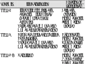
PPARs have 5 domains14) (Fig. 1A): 1) the LBD, to which the specific PPAR agonist binds; 2) the N-ter- minal AF2 domain, which can be activated by phos- phorylation through the mitogen-activated protein (MAP) kinase sites via ligand-independent activation; 3) the DBD, which interacts with specific PPAR response ele- ments (PPRE) in the promoter region of the PPAR-acti- vated target genes; 4) the C-terminal AF2 domain, which, in response to ligand binding, undergoes a permissive conformational change that’s required for transcriptional activation; 5) the hinge domain, whose function is not well understood, but whose structure is very flexible and it may be crucial for efficient binding of the DBD.
PPAR activation via the binding of LBD to specific ligands leads to its heterodimerization with the retinoid X receptor (RXR), and it undergoes a conformational
change in the AF2 domain, which facilitates the release of co-repressors and the recruitment of coactivators (Fig. 1B).15) In contrast to the positive transcriptional regulation of PPAR as described above, PPAR activation can also repress the transcription of the target genes by unknown mechanisms.16) This repressive action of PPARs is the major mechanism that underlies its anti-infla- mmatory action.
As described above, transcriptional regulation by PP- AR agonists requires multiple levels of control as fol- lows: PPAR ligand with synthetic ligands and natural ligands, corepressors and coactivators, homology of the LBD between each subtype of PPAR, and many known and unknown target genes in multiple organs. In fact, synthetic agonists can activate only a specific subtype of PPAR. For example, fibrates bind PPAR-α, which controls lipid metabolism, and thiazolinediones bind PPAR-γ, which controls hyperglycemia. However the ef- fects of synthetic PPAR agonists in vivo differ from those of PPAR itself because the synthetic agonists may be greatly influenced by natural ligands in vivo, and these natural ligands are as yet unknown.5) Corepressors and coactivators form a large, diverse family with members such as nuclear corespressor,17) PPAR-binding protein,18) PPAR-γ coactivator19) and cAMP response element- binding protein.20) PPAR activation by synthetic ligands may modulate a large number of genes, and some of which produce unknown effects. Particularly, the dual or broad PPAR agonists may be more dangerous.21) PPAR activation may be beneficial in one organ, but harmful in another organ. The clinical application of PPAR agonists for therapeutic targets may require a more comprehensive understanding about PPAR bio- logy and this must be carefully approached.
Table 1. Tissue distribution of PPARs and the target genes Subfamily Tissue distribution Function of
target genes PPAR-α Tissues with high fatty acid
catabolism (liver, kidney, skeletal muscle, heart, brown fat)
Vessel cells and immune cells Human atherosclerotic lesions
Fatty acid oxidation Lipid metabolism Inflammation/
vascular
PPAR-γ Brown and white adipose tissue Large intestine
Vascular and immune cells Human atherosclerotic lesions
Adipogenesis Lipid metabolism Glucose control Inflammation/
vascular PPAR-β/δ Ubiquitous Lipid metabolism
Wound healing Brain metabolism PPAR: peroxisome proliferator-activated receptor
Fig. 1.A: schematic structure of PPAR. B: PPAR mechanism of transcriptional regulation by ligand binding transactivation. In response to PPAR ligand, a conformational change in the AF2 domain induces the release of the corepressor and this recruits the coactivator.
PPAR binds with the Retinoid X receptor (RXR) to form a heterodimeric complex to regulate transcription. This complex interacts with the specific PPAR response element (PPRE) in the promotor region, which activates target gene transcription. PPAR activation also can repress the expression of target genes via unknown mechanism. AF: activation function, PPAR: peroxisome proliferators-activated receptor.
Hinge
DNA-Binding (D) Ligand-Binding (L)
AF1 AF2
NH2- -COOH
L AF2
D
L AF2
D
DNA: promotor region PPRE
PPAR RXR
Target gene activation
PPAR ligand RXR ligand
Co-repressor
Co-activator
B A
Effects of Peroxisome Proliferator- Activated Receptor-αon Lipid Metabolism, the Vessel Wall and
the Heart
PPAR-α is metabolically active in the liver, heart, kidney, skeletal muscle and brown fat.22)23) It is also present in all vascular cells, including endothelial cells, smooth muscle cells and monocytes/macrophages.24-26) The effects of PPAR-α include hypolipidemic action, an anti-inflammatory effect on the vascular wall and metabolic effects on the myocardium.
Hypolipidemic action
PPAR-α agonists, i.e., the fibrates (clofibrate, gemfi- brozil, fenofibrate, benzafibrate, and ciprofibrate), have been used as lipid-lowering agents for over 40 years, and this is mainly due to their action of lowering trigl- yceride (TG) levels and raising high density lipoprotein cholesterol (HDL-C) levels.
The lowering of TG levels in plasma after PPAR-α activation is attributed to the following mechanisms:
1) The increased diversion of fatty acids into β-oxi- dation, thereby limiting their availability for TG and very low-density lipoprotein (VLDL) synthesis,27) 2) the inhibition of apo CIII, which is an inhibitor of lipo- protein lipase (LPL)28) and this increases LPL activity,29) which enhances the hydrolysis of TG-rich particles and improves the uptake of their remnants.
The increased HDL-cholesterol levels in plasma after PPAR-α activation may be explained by the following factors: 1) increased production of apo AI and apo AII, which are the major HDL components,30)31) 2) increased transfer of the other surface components of triglyceride- rich particles to HDL by enhancing the LPL activity,32) and 3) an enhanced APT-binding cassette transporter A1 (ABCA1) expression as a result of PPAR-α acti- vation.33)
It has recently been recognized that fibrates decrease the levels of small, dense low density lipoprotein choles- terol (LDL-C) particles.34)35)
Effects on the vascular wall
The anti-inflammatory action of PPAR-α on all vascular cells has been studied and reported on, and most notably by both in vitro and in vivo studies.4) PPAR-α activators inhibit the production of inflamma- tory response markers such as endothelin-1, vascular adhesion molecule-1 (VCAM-1), interleukin (IL)-6 and tissue factors in endothelial cells, smooth muscle cells and macrophages.36-45) In patients with dyslipidemia, PPAR-α agonists reduce the levels of inflammatory markers such as IL-6, fibronogen, C-reactive protein, serum amyloid A, plasminogen, α2-macroglobin, inter- feron-γ, IL-2, tumor necrosis factor-α and IL-1β.42)46-51)
These effects of PPAR-α agonists on the vessel wall may explain their cardiovascular protective effects that extend beyond their lipid lowering effect.10)
Effects on myocardium
The cardiac metabolic effects of PPAR-α activation are less well defined. Although the fetal heart obtains most of its energy from glucose and lactate, the adult heart obtains its energy from PPAR-α -dependent fatty acid oxidation, as well as from glucose and lactate, in order to meet its energy demands under varying die- tary and physiological conditions.27)52) In a murine model of pressure induced cardiac hypertrophy, PPAR- α was observed to be down-regulated,52)53) which indi- cates that the cardiac metabolism shifted from fatty acid oxidation to glucose utilization. PPAR-α activation in this model resulted in severe left ventricular dysfunc- tion. It is suggested that PPAR-α downregulation in a hypertrophic heart may be an adaptive process that is essential for maintaining normal heart function.54) The significance of PPAR-α activation on human cardiac hypertrophy has not yet been established.
In the hearts of patients with uncontrolled diabetes, impaired glucose utilization results in almost exclusive use of fatty acid oxidation to provide for the ATP needs of the myocardium.55) In that case, PPAR-α activation may theoretically include conflicting effects on the myocardial energy metabolism. The beneficial effect is that PPAR-α agonists cause a hypolipemic state that may reduce the amount of fatty acids, which is a subs- trate of fatty acid oxidation, delivered to the myocardium.
However, another potentially harmful effect is that PPAR-α activation triggers the shift from glucose utili- zation to fatty acid oxidation as an energy source. In- creased fatty acid oxidation may lead to an increased oxygen demand, and a high uptake of fatty acid by the myocardium may lead to lipid accumulation in the myocardium, which predisposes it to systolic dysfunction and heart failure. Therefore, questions have been rais- ed regarding the net effect of PPAR-α activation on the diabetic myocardium. In a diabetic murine model, PPAR-α activation revealed that the reduction of the delivered substrate was more important than the energy switch.56) The treatment with PPAR-α agonist nor- malized the free fatty acid, TG and glucose levels; more- over, it also reduced myocardial fatty acid oxidation by 50% and increased glucose utilization. In contrast, the cardiac-specific overexpression of PPAR-α in non-di- abetic mice revealed increased fatty acid oxidation and decreased glucose utilization in the myocardium, and this induced a diabetic-type cardiomyopathy in otherwise normal mice. In diabetic patients, the effects of PPAR- α activation on myocardial metabolism are still under debate and this requires further study.
In the ischemic heart, glucose utilization requires less
oxygen than fatty acid oxidation; further, it does not worsen acidosis in the ischemic myocardium. There- fore, inhibitors of fatty acid oxidation such as trimeta- zidine are being used in clinical practice.57) Theoretically, PPAR-α agonist may compromise this anti-anginal ef- fect of fatty acid oxidation inhibitors. However, since severe ischemia itself can turn off fatty acid oxidation, the role of PPAR-α in the ischemic human myocar- dium needs to be clearly defined. The pretreatment of an infarction and an ischemia-reperfusion model with fibrates demonstrated a reduction in the size of infarc- tion and improved postischemic contractile function because PPAR-α activation had an anti-inflamma- tory effect.58)59) These results suggest that pre-ischemic treatment with PPAR-α agonist may limit the ische- mic damage; however, post-ischemic treatment might be theoretically harmful if it shifts the cardiac meta- bolism from glucose utilization to fatty acid oxidation.
Summary of the Important Clinical Trials That Used Peroxisome
Proliferator-Activated Receptor-αAgonists
Several large clinical studies have investigated the potential cardioprotective effects of PPAR-α agonists, and particularly the fibrate derivatives. These studies have exhibited a wide range of results (Table 2):
• No beneficial results or harmful results: the World Health Organization (WHO) cooperative trial, the Coro- nary Drug Project (CDP) trial, the Lower Extremity Arterial Disease Event Reduction Study (LEADER), and the Bezafibrate Infarction Prevention Trial (BIP)
• Beneficial results: the Helsinki Heart Study (HHS) and the Veteran’s Affairs-HDL Intervention Trial (VA- HIT)
• Mixed results: the Fenofibrate Intervention and Event Lowering in Diabetes (FIELD) study
World health organization cooperative trial
Male patients (n=15,745) without coronary heart disease (CHD) were enrolled and followed up for a mean period of 5.3 years.60) Of the major CHD events, only the incidence of non-fatal myocardial infarction (MI) was significantly reduced in the fibrate group (relative risk reduction: 20%). There was no difference in the number of deaths due to cardiac causes between the groups. The overall mortality was higher in the clofibrate group; this was attributed to diseases of the liver, in- testines and gall bladder.61)
Coronary drug project trial
Male patients (n=8, 341) with one or more MI attacks were randomized to 1 of 6 treatment groups and they were followed up for 5-8.5 years.62) Follow up was discon- tinued early in 3 of those 6 groups due to the increased incidence of cardiac events. The remaining 3 groups were comprised of patients who were treated with clofi- brate, niacin and placebo. Overall, there was no statis- tically significant difference in the total mortality, and the incidence of nonfatal MI and cardiac deaths. Fur- thermore, this trial showed a statistically nonsignificant increase in thromboembolism, angina, intermittent clau- dication, cardiac arrhythmia and gall stones, along with an increase of the nonfatal cardiovascular events.
Lower extremity arterial disease event reduction trial The treatment of 1,568 men with either bezafibrate or placebo demonstrated that the incidence of CHD and stroke was not reduced in the bezafibrate treated group.63) The beneficial effects on non-fatal cardiac events were the greatest in men aged <65 years at study entry;
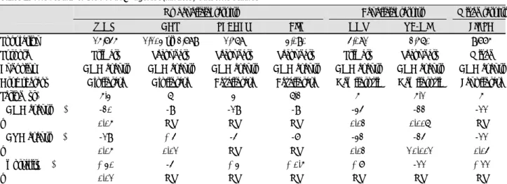
Table 2. The results of the PPAR-α agonist (fibrates) outcome studies
No beneficial results Beneficial results Mixed results
WHO CDP LEADER BIP HHS VA-HIT FIELD
Population 15,745 1,103 vs. 2,789 1,568 3,090 4,081 2,560 9,775
Purpose Primary Secondary Secondary Secondary Primary Secondary Mixed
Endpoint CHD events CHD events CHD events CHD events CHD events CHD events CHD events Drug therapy Clofibrate Clofibrate Bezafibrate Bezafibrate Gemfibrozil Gemfibrozil Fenofibrate
Period (yr) 5.3 6 3 6.2 5 5.1 5
ΔCHD events (%) -20 -9 -19 -9 -34 -22 -11
p 0.05 NS NS NS 0.02 0.006 NS
ΔCVD events (%) -19 +4 -4 -7 -32 -24 -11
p 0.05 0.01 NS NS 0.02 <0.001 0.04
Δmortality (%) +30 -4 +3 +0.5 +7 -11 +11
p 0.01 NS NS NS NS NS NS
PPAR: peroxisome proliferator-activated receptor, WHO: world health organization, CDP: coronary drug project, LEADER: lower extremity arterial disease event reduction, BIP: bezafibrate infarction prevention, HHS: helsinki heart study, VA-HIT: veteran’s administration-HDL intervention trial, FIELD: fenofibrate intervention and event lowering in diabetes, CHD: coronary heart disease, CVD: cardiovascular disease, NS: not significant
a beneficial effect on all coronary events was also ob- served in this group of patients. There were no signifi- cant effects in men aged ≥65 years. There was no dif- ference between the 2 groups with respect to all causes of deaths. Bezafibrate only reduced the severity of inter- mittent claudication for up to 3 years.
Bezafibrate infarction prevention trial
In this trial,7) 3,090 patients with CHD and dyslipi- demia (total cholesterol: 180-250 mg/dL, HDL-C≤45 mg/dL, TG≤300 mg/dL and LDL-C≤180 mg/dL) were randomized to receive either bezafibrate or a pla- cebo. After follow up (mean duration: 6.2 years), there was no difference in the incidence of fatal and non- fatal MI or sudden death between the 2 groups. The total mortality and noncardiac mortality were also si- milar, and the incidences of adverse events and cancer were equally distributed. Only the subgroup with a high baseline TG level (≥200 mg/dL) enjoyed the re- duction in the cumulative probability of myocardial infarction or sudden death by bezafibrate administration.
Helsinki heart study
This primary prevention trial6) with using gemfibrozil was performed for 5 years on 4,081 asymptomatic mid- dle-aged men (age range: 40-55 years) with primary dyslipidemia (non-HDL cholesterol≤200 mg/dL). This trial demonstrated a 34% reduction in the incidence of overall cardiac events, including fatal and nonfatal MI and cardiac death. This effect is more marked in patients suffering with diabetic and metabolic synd- rome.64) No difference was noted in the mortality due to all-causes; however, an increase was noted in the de- aths due to noncoronary causes.
A substudy of the HHS,65) which employed the males excluded from the primary prevention cohort due to a history of MI, angina or prior ECG changes, also show- ed no difference between the gemfirozil group and the placebo group
Veteran’s administration-HDL intervention trial In this study,8) 2,531 men with CHD, low HDL (≤
40 mg/dL), and moderately elevated LDL-C (≤140 mg/dL) were randomized and treated with either gem- fibrozil or placebo for 5.1 years. Although the LDL-C levels did not significantly differ between the groups, the gemfibrozil group showed a significant reduction in the risk of nonfatal MI and cardiac death (relative risk reduction: 22%). Further, a 24% reduction was noted in the combined outcome of death from CHD, nonfatal MI and stroke. There were no significant differences in the rates of coronary revascularization, hospitalization for unstable angina, death from any cause and cancer.
The experts concluded that the beneficial effect of gem- fibrozil on cardiovascular events may have been driven
largely by the characteristics of the enrolled group in the VA-HIT as the subjects were mostly patients with diabetes and/or metabolic syndrome, along with the lipid inclusion criteria.9)10)
Fenofibrate intervention and event lowering in diabetes
This study11) was comprised of a mixed population of 9,795 low-risk “primary prevention” and medium-risk
“secondary prevention” diabetic patients. Fenofibrate treatment showed a significant 24% reduction in the incidence of non-fatal MI and a non-significant increase in CHD mortality. The total cardiovascular disease events were significantly reduced, and this was mainly due to a reduction in coronary revascularization and nonfatal MI. The total mortality was similar in both the groups.
No benefit was observed in the “secondary prevention”
subgroup. Unexpectedly, fenofibrate treatment was as- sociated with a lower incidence of microvascular com- plications such as albuminuria progression and retinop- athy that required laser treatment. However, except for a slight increase in the incidence of pancreatitis and pulmonary embolism, no other significant adverse ef- fects were noted. The authors concluded that the higher use of statins in the placebo group might have masked a moderately larger treatment benefit.
More potent and selective peroxisome proliferator-activated receptor-αagonist, LY518674, trial
The novel selective PPAR-α agonist, LY518674, is approximately 10,000 times more potent than fenofi- brate. This study12) aimed at examining the safety and efficacy of LY518674 among 2 populations of patients with atherogenic dyslipidemia and hypercholesterolemia.
In a trial of 309 patients with atherogenic dyslipidemia, LY518674 and fenofibrate both individually outper- formed the placebo, with no significant differences being noted between the 2 PPAR-α agonists. LY518674 also raised the LDL level in a dose-dependent fashion and it did so much more than fenofibrate. In the other trial that was comprised 304 hypercholesterolemic and statin-naive patients, LY518674 and atorvastatin each significantly reduced the TG and LDL levels, and they both increased the HDL levels. For the atorvastatin recipients, the addition of LY518674 further increased the HDL levels (by 1-12%) and reduced the TG levels;
however, it had little effect on the LDL levels. LY518674 and fenofibrate demonstrated evidence of increasing the serum creatinine levels; this effect was substantial in some cases.
The novel PPAR-α agonist was not better than fe- nofibrate or statin monotherapy in achieving the in- tended improvement of the lipid profile, and it ap- peared to worsen renal function.
Efficacy from the Clinical Trials of Peroxisome Proliferator-Activated
Receptor-αAgonist
In the LOCAT66) (Lopid Coronary Angiography Trial), the DAIS67) (Diabetes Atherosclerosis Intervention Study) and other clinical studies,68) fibrates have demonstrated a reduction in the rates of progression of coronary ath- eromas as measured by quantitative angiography. How- ever, the cardioprotective effects of PPAR-α agonists were not consistent in the individual clinical trials.
When comparing the results of 2 initial large-scale trials of clofibrate, i.e., the CDP trial62) and the WHO cooperative trial,60) the benefit for reducing CHD events was not significant in the former, but it was significant in the latter (relative risk reduction: 20%, p<0.05). How- ever, both studies showed a significant increase in the total mortality (p<0.05). In the BIP trial,7) bezafibrate treatment showed an 11% relative reduction in the risk of CHD events, but this was not statistically significant (p=0.26). However, post hoc analysis revealed a signi- ficant reduction in the risk of CHD events (relative risk reduction: 22%, p=0.02) in those patients with higher baseline TG levels (≥200 mg/dL). The LEADER trial63) showed no beneficial effect of bezafibrate treatment on cardiovascular disease except for a reduction in the risk of nonfatal MI in young men. All the patients of the LEADER trial were also treated with statins, and the out- come results were compounded by the effects of statins.
In contrast to the disappointing results of the clofibrate and bezafibrate trials, the gemfibrozil trials (the HHS and VA-HIT) demonstrated a statistically significant reduc- tion in the incidence of cardiovascular events. The VA- HIT results helped to stimulate considerable anticipation for the results of the FIELD study11) that used fenofi- brate for treating first or recurrent cardiovascular events in patients with type 2 diabetes. However, the results of the FIELD study showed a statistically nonsignificant difference between the treatment and placebo groups with respect to reducing the primary endpoints, in- cluding CHD death and non-fatal MI, and there was a slightly increase of the total and cardiovascular mortality.
In the “primary prevention” group, fenofibrate treatment resulted in a cardioprotective effect only against the se- condary endpoints such as nonfatal MI and revasulari- zation procedures. This major discrepancy between the results of the VA-HIT and FIELD has led to several debates and it has raised many questions.
First, the cardioprotective effects of gemfibrozil in the HHS and VA-HIT were greater than those of other fibrates in the WHO cooperative trail, the BIP trial and the FIELD study. Some researchers suggest that gem- fibrozil has less severe adverse effects than do the other fibrates. Gemfibrozil does not increase the levels of homocysteine and creatinine to the same extent that has
been observed with using the other fibrates.69)70) Indeed, in the VA-HIT study, the benefit of gemfibrozil due to the increase in HDL-C could account for only 20% of the CHD reduction in terms of the lipid changes.71) This result suggests that gemfibrozil itself has other beneficial effects that extend beyond its lipid-modulating effect.
Second, the cohort of the FIELD study had different characteristics from those subjects of the VA-HIT. The baseline HDL-C levels of the FIELD cohort were rela- tively high (mean HDL-C: 42 mg/dL); this might have masked the cardioprotective effects of fenofibrate. The VA-HIT was designed to evaluate the effect of gemfi- brozil in patients who were not using statin. However, the control group of the FIELD study had an asymmet- rical higher statin drop-in due to the LDL-C-lowering effect of fenofibrate, which might have also masked the beneficial effects of fenofibrate. In contrast, some might argue that the beneficial effect in the FIELD study might be attributed to the modest LDL-C low-ering effect of fenofibrate.72)
Third, some suggest that the potency of PPAR-α agonist might have had an influence on the cardiac out- come results. PPAR is an extremely critical transcrip- tional regulator, and a number of factors can affect it. In view of the mechanism of action of PPAR agonists, it may be very difficult to get clinically beneficial effects by using only a synthetic agent, which may result in bene- ficial and/or unwanted effects. Theoretically, the final net effect of both the beneficial and unwanted effects must be considered to be more important. Gemfibrozil is less potent than fenofibrate. The result of the VA- HIT suggests that more potent PPAR activation may not be essential for achieving clinical benefits. Furthermore, LY518674, which is a potent PPAR-α agonist, showed no better results in achieving an improved lipid profile than did fenofibrate or statin monotherapy.12)
Fourth, we do not have a clear answer as to whether combination therapy with statin and fibrate may lead to a beneficial cardiac outcome. Combination therapy may achieve an optimal improvement in the lipid profile and offer greater cardiovascular risk reduction; how- ever, it might increase the risk of rhabdomyolysis.73) When the Action to Control Cardiometabolic Risk in Diabetes (ACCORD) study is completed, good evidence regarding combination therapy will become available.
In the ACCORD study design, fenofibrate is added to baseline 20-40 mg simvastatin administration in 5,800 patients in a 2×2 design trial with ultratight glycemic control versus the usual glycemic control.
Safety Issues from the Clinical Trials of Peroxisome Proliferator-Activated
Receptor-αAgonist
The safety issues of PPAR agonists have recently
entered the spotlight since Nissen et al.74) argued about the potentially cardiotoxic effects of rosiglitazone, a PPAR-γ agonist that’s widely used as a hypoglycemic agent. Fortunately, none of the failed PPAR agonists that showed severe side effects were pure PPAR-α or PPAR-α-preferential dual agonists when they tested on the human PPAR isoforms.75) Indeed, for most of the failed PPAR agonists, their apparent affinity for PPAR-γ is higher than their affinity for PPAR-α. Furthermore, because the PPAR-α agonists, i.e., fibrates, have been used for 40 years, they are generally considered as safe drugs with only a few side effects. However, the safety issues of PPAR-α agonists include increased total mor- tality, muscle toxicity such as myopathy or rhabdomyo- pathy, increased levels of plasma creatinine and homo- cysteine, and lithogenicity. We will briefly discuss here the unwanted effects of PPAR-α agonists based on the results of clinical trials.
Increased total mortality
Although clinical trials have demonstrated that treat- ment with fibrates reduces nonfatal MI, all the clinical trials of fibrates, other than the VA-HIT, showed either no increase or a slight increase in the total mortality due to noncardiovascular causes in the treatment group.
More recently, the FIELD study11) showed a nonsignifi- cant increase in the cardiovascular, noncardiovascular, coronary and total mortality associated with fenofi- brate treatment. However, the relationship between total mortality and fibrate treatment was not statisti- cally powerful, and the increased mortality was not at- tributable to any specific causes of death (such as invasive cancer).76) It is not known whether the increased or unchanged rates of mortality are a chance finding or if they are side effects of this drug. However, concern could be raised that the unwanted effects of PPAR-α agonists might be related to the increased mortality and this should be resolved in the future.
Rhadomyolysis, myopathy and myalgia
Monotherpy as well as combined therapy with fibrates may be associated with cases of myopathy, myalgia, and in extremely rare cases, with rhabdomyolysis.77) The mechanism of fibrate-induced muscle toxicity is un- known; however, it may be explained on the basis of PPAR-α-dependent and compound-dependent actions.
Severe myopathy in mice correlates with an increased expression of the LPL-target protein of PPAR-α in skeletal muscles.78) Some authors suggest that the ener- gy imbalance due to increased fatty acid oxidation and decreased TG in skeletal muscle may cause the degra- dation of muscle protein.79)80) Muscle toxicity has been noted with both gemfibrozil and fenofibrate admini- stration, but it is more frequent with the former (rha- domyolysis incidence: 59.6/million and 5.5/million, re-
spectively).81)
In clinical practice, combination therapy with both fibrate and statin definitely increases the risk of rha- domyolysis and myopathy. Furthermore, combined the- rapy with gemfibrozil plus statin is considered more risky than that with fenofibrate and statin. Recent reviews of the Food and Drug Administration’s Adverse Event Reporting System database reported that the rate of myopathy with combined gemfibrozil and statin thera- py was 33 times more than that with combined fenofi- brate and statin therapy.73)81) This difference is attributed to the ability of gemfibrozil to increase the plasma statin level to some extent because it uses enzymes from the same family of glucuronidation enzymes as do the statins;
however, fenofibrate uses enzymes from a different en- zyme family.73)
Increased plasma creatinine
A modest and reversible elevation of the plasma cre- atinine level has been reported in clinical trials that have used bezafibrate82)83) and fenofibrate82)84)85) and less commonly, in trials that have used gemfibrozil.84) Some authors have suggested that creatininemia may be re- lated to a PPAR-α activation-induced inhibition of the cyclooxygenase (COX-2) gene expression in the kidney86) and the resultant decrease in the synthesis of the va- sodilator prostaglandin.87) It’s interesting that the use of tesaglitazar, a dual PPAR-α/γ agonist, was discon- tinued because of severe renal toxicity.88) However, recent reports suggest that fibrate-induced creatininemia was not associated with the glomerular filtration rate (GFR), which is assessed by using the insulin or creatinine clear- ance,89) and this was attributed to increased creatinine production and particularly from muscle tissue. Al- though the harmful renal effect of PPAR-α agonist is uncertain, using these drugs clinically in patients with renal insufficiency should be carefully considered.
Increased plasma homocysteine
Fenofibrate, ciprofibrate and bezafibrate all increase the plasma homocysteine concentration of dyslipide- mic and diabetic patients,90-93) whereas gemfibrozil does so to a lesser extent. Although the exact mechanism for this is not known, fibrate treatment did not cause homocysteinemia in PPAR-α knockout mice; however, it led to homocysteinemia in wild mice.94) This finding suggests that fibrate-induced homocysteinemia may be related to PPAR-α activation. The clinical significance of hyperhomocysteinemia remains a matter of discussion.
The DAIS67) showed that fenofibrate-induced homo- cysteinemia did not attenuate its beneficial effect for inhibiting the progression of atherosclerosis.
Increased gallstones
There was a marked association between clofibrate
and gallstones in the WHO cooperative trial60)61) and the CDP trial,62) and the latter trial showed a 2-3 times higher rate of cholecystectomy in the treatment group as compared with the placebo group. A possible me- chanism of lithogenicity could be that PPAR-α agonists reduce the expression of cholesterol 7α-hydroxylase and sterol 27-hydroxylase, and this results in an increased cholesterol saturation index.95)
It is unknown whether gemfibrozil or fenofibrate can be a causative factor for gallstones because the rates of cholecystectomy in the clinical trials, including the HHS, VA-HIT and FIELD study, were not reported.
Conclusion
Considering the benefits and safety concerns, the PPAR-α agonists (fibrates) that are currently in use may definitely be beneficial for decreasing the rate of clinical cardiovascular events in a specific population, and particularly among those patients suffering with metabolic syndrome and atherogenic dyslipidemia.
Large-scale long-term clinical trials of PPAR-α agonists have shown that these drugs very rarely have side effects;
therefore, clinicians consider fibrates to be safe drugs.
However, it should be noted that long-term treatment with most fibrates, unlike that with statins, has not been shown to decrease total mortality; in fact, it has been demonstrated to increase the total mortality in some instances. Because PPAR is a very critical and important gene regulator in the cell and its natural controlling factors are yet unknown, the actions of PPAR-α activation may include not only the known beneficial effects, such as the hypolipidemic and anti- inflammatory actions, but also the unknown harmful effects. Therefore, clinicians should bear in mind the risk/benefit ratio of a PPAR-α agonist in each parti- cular case before prescribing it in clinical practice. In the future, intensive research on this interesting thera- peutic target, PPAR, will lead to developing safer and more effective PPAR-α agonists.
REFERENCES
1) Libby P. Inflammation in atherosclerosis. Nature 2002;420:868-74.
2) Plutzky J. Medicine: PPARs as therapeutic targets: reverse car- diology? Science 2003;302:406-7.
3) Kho JS, Park SJ, Im SI, Choi BR, Kwak CH, Hwang JY. Pero- xisome proliferator-activated receptor gamma (PPAR-γ) agonist improves endothelial function in diabetic patients with mMeta- bolic syndrome: pivotal role of NOx and inflammation. Korean Circ J 2007;37:221-9.
4) Libby P, Plutzky J. Inflammation in diabetes mellitus: role of pe- roxisome proliferator-activated receptor-alpha and peroxisome proliferator-activated receptor-gamma agonists. Am J Cardiol 2007;99:27B-40B.
5) Brown JD, Plutzky J. Peroxisome proliferator-activated recep- tors as transcriptional nodal points and therapeutic targets. Cir- culation 2007;115:518-33.
6) Frick MH, Elo O, Haapa K, et al. Helsinki Heart Study: primary prevention trial with gemfibrozil in middle-aged men with dysli- pidemia: safety of treatment, changes in risk factors, and inci- dence of coronary heart disease. N Engl J Med 1987;317:1237-45.
7) The BIP Study Group. Secondary prevention by raising HDL cholesterol and reducing triglycerides in patients with coronary artery disease. Circulation 2000;102:21-7.
8) Rubins HB, Robins SJ, Collins D, et al. Gemfibrozil for the secondary prevention of coronary heart disease in men with low levels of high-density lipoprotein cholesterol. N Engl J Med 1999;341:410-8.
9) Rubins HB, Robins SJ, Collins D, et al. Diabetes, plasma insulin, and cardiovascular disease: subgroup analysis from the Depart- ment of Veterans Affairs High-Density Lipoprotein Intervention Trial (VA-HIT). Arch Intern Med 2002;162:2597-604.
10) Robins SJ, Rubins HB, Faas FH, et al. Insulin resistance and cardiovascular events with low HDL cholesterol. Diabetes Care 2003;26:1513-7.
11) Keech A, Simes RJ, Barter P, et al. Effects of long-term feno- fibrate therapy on cardiovascular events in 9795 people with type 2 diabetes mellitus (the FIELD study): randomised controlled trial.
Lancet 2005;366:1849-61.
12) Nissen SE, Nicholls SJ, Wolski K, et al. Effects of a potent and selective PPAR-alpha agonist in patients with atherogenic dysli- pidemia or hypercholesterolemia: two randomized controlled tri- als. JAMA 2007;297:1362-73.
13) Issemann I, Green S. Activation of a member of the steroid hor- mone receptor superfamily by peroxisome proliferators. Nature 1990;347:645-50.
14) Glass CK, Ogawa S. Combinatorial roles of nuclear receptors in inflammation and immunity. Nat Rev Immunol 2006;6:44-55.
15) Shulman AI, Mangelsdorf DJ. Retinoid X receptor heterodimers in the metabolic syndrome. N Engl J Med 2005;353:604-15.
16) Pascual G, Fong AL, Ogawa S, et al. A SUMOylation-dependent pathway mediates transrepression of inflammatory response genes by PPAR-gamma. Nature 2005;437:759-63.
17) Mizukami J, Taniguchi T. The antidiabetic agent thiazolidine- dione stimulates the interaction between PPAR gamma and CBP.
Biochem Biophys Res Commun 1997;240:61-4.
18) Puigserver P, Wu Z, Park CW, Graves R, Wright M, Spiegelman BM. A cold-inducible coactivator of nuclear receptors linked to adaptive thermogenesis. Cell 1998;92:829-39.
19) Zhu Y, Kan L, Qi C, et al. Isolation and characterization of pero- xisome proliferator-activated receptor (PPAR) interacting pro- tein (PRIP) as a coactivator for PPAR. J Biol Chem 2000;275:
13510-6.
20) Jia Y, Guo GL, Surapureddi S, et al. Transcription coactivator peroxisome proliferator-activated receptor-binding protein/medi- ator 1 deficiency abrogates acetaminophen hepatotoxicity. Proc Natl Acad Sci U S A 2005;102:12531-6.
21) Nissen SE, Wolski K, Topol EJ. Effect of muraglitazar on death and major adverse cardiovascular events in patients with type 2 diabetes mellitus. JAMA 2005;294:2581-6.
22) Auboeuf D, Rieusset J, Fajas L, et al. Tissue distribution and quantification of the expression of PPARs and LXR in humans:
no alterations in adipose tissue of obese and NIDDM patients.
Diabetes 1997;46:1319-27.
23) Braissant O, Foufelle F, Scotto C, Dauca M, Wahli W. Differen- tial expression of peroxisome proliferator-activated receptors (PPARs): tissue distribution of PPAR -α,-β, and -γ in the adult rat. Endocrinology 1996;137:354-66.
24) Chinetti G, Griglio S, Antonucci M, et al. Activation of proli- ferator-activated receptors alpha and gamma induces apoptosis of human monocyte-derived macrophages. J Biol Chem 1998;
273:25573-80.
25) Inoue I, Shino K, Noji S, Awata T, Katayama S. Expression of peroxisome proliferator-activated receptor alpha (PPAR alpha) in primary cultures of human vascular endothelial cells. Bio- chem Biophys Res Commun 1998;246:370-4.
26) Staels B, Koenig W, Habib A, et al. Activation of human aortic smooth-muscle cells is inhibited by PPARalpha but not by PPAR gamma activators. Nature 1998;393:790-3.
27) Vosper H, Khoudoli GA, Graham TL, Palmer CN. Peroxisome proliferator-activated receptor agonists, hyperlipidaemia, and at- herosclerosis. Pharmacol Ther 2002;95:47-62.
28) Haubenwallner S, Essenburg AD, Barnett BC, et al. Hypoli- pidemic activity of select fibrates correlates to changes in hepatic apolipoprotein C-III expression: a potential physiologic basis for their mode of action. J Lipid Res 1995;36:2541-51.
29) Schoonjans K, Peinado-Onsurbe J, Lefebvre AM, et al. PPAR-α and PPAR-γ activators direct a tissue-specific transcriptional response via a PPRE in the lipoprotein lipase gene. EMBO J 1996;15:5336-48.
30) Duez H, Lefebvre B, Poulain P, et al. Regulation of human apoA-I by gemfibrozil and fenofibrate through selective peroxi- some proliferator-activated receptor alpha modulation. Arterio- scler Thromb Vasc Biol 2005;25:585-91.
31) Schultze AE, Alborn WE, Newton RK, Konrad RJ. Admini- stration of a PPARalpha agonist increases serum apolipoprotein A-V levels and the apolipoprotein A-VI apolipoprotein C-III ratio.
J Lipid Res 2005;46:1591-5.
32) Havel RJ, Kane JP. Structure and metabolism of plasma lipopro- teins. In: The Metabolic and Molecular Bases of Inherited Di- sease. 7th ed. New York: McGraw Hill; 1995. p.1841-52.
33) Chawla A, Boisvert WA, Lee CH, et al. A PPAR gamma-LXR- ABCA1 pathway in macrophages is involved in cholesterol efflux and atherogenesis. Mol Cell 2001;7:161-71.
34) Feher MD, Caslake M, Foxton J, Cox A, Packard CJ. Atheroge- nic lipoprotein phenotype in type 2 diabetes: reversal with mic- ronised fenofibrate. Diabetes Metab Res Rev 1999;15:395-9.
35) Staels B, Dallongeville J, Auwerx J, Schoonjans K, Leitersdorf E, Fruchart JC. Mechanism of action of fibrates on lipid and lipo- protein metabolism. Circulation 1998;98:2088-93.
36) Lee H, Shi W, Tontonoz P, et al. Role for peroxisome prolife- rator-activated receptor alpha in oxidized phospholipid-induced synthesis of monocyte chemotactic protein-1 and interleukin-8 by endothelial cells. Circ Res 2000;87:516-21.
37) Marx N, Sukhova GK, Collins T, Libby P, Plutzky J. PPAR-α activators inhibit cytokine-induced vascular cell adhesion mole- cule-1 expression in human endothelial cells. Circulation 1999;
99:3125-31.
38) Jackson SM, Parhami F, Xi XP, et al. Peroxisome proliferator- activated receptor activators target human endothelial cells to inhibit leukocyte-endothelial cell interaction. Arterioscler Thro- mb Vasc Biol 1999;19:2094-104.
39) Xu X, Otsuki M, Saito H, et al. PPARalpha and GR differentially down-regulate the expression of nuclear factor-kappaB-respon- sive genes in vascular endothelial cells. Endocrinology 2001;
142:3332-9.
40) Delerive P, Martin-Nizard F, Chinetti G, et al. Peroxisome pro- liferator-activated receptor activators inhibit thrombin-induced endothelin-1 production in human vascular endothelial cells by inhibiting the activator protein-1 signaling pathway. Circ Res 1999;85:394-402.
41) Satoh H, Tsukamoto K, Hashimoto Y, et al. Thiazolidinediones suppress endothelin-1 secretion from bovine vascular endothelial cells: a new possible role of PPARgamma on vascular endothe- lial function. Biochem Biophys Res Commun 1999;254:757-63.
42) Staels B, Koenig W, Habib A, et al. Activation of human aortic smooth-muscle cells is inhibited by PPARalpha but not by PPAR gamma activators. Nature 1998;393:790-3.
43) Delerive P, Gervois P, Fruchart JC, Staels B. Induction of Ika- ppaBalpha expression as a mechanism contributing to the anti- inflammatory activities of peroxisome proliferator-activated re- ceptor-alpha activators. J Biol Chem 2000;275:36703-7.
44) Marx N, Mackman N, SchOnbeck U, et al. PPARalpha activa- tors inhibit tissue factor expression and activity in human mono- cytes. Circulation 2001;103:213-9.
45) Neve BP, Corseaux D, Chinetti G, et al. PPARalpha agonists inhibit tissue factor expression in human monocytes and macro- phages. Circulation 2001;103:207-12.
46) Frost RJ, Otto C, Geiss HC, Schwandt P, Parhofer KG. Effects of atorvastatin versus fenofibrate on lipoprotein profiles, low-den- sity lipoprotein subfraction distribution, and hemorheologic pa- rameters in type 2 diabetes mellitus with mixed hyperlipoprotein- emia. Am J Cardiol 2001;87:44-8.
47) Despres JP, Lemieux I, Pascot A, et al. Gemfibrozil reduces plasma C-reactive protein levels in abdominally obese men with the atherogenic dyslipidemia of the metabolic syndrome. Arterio- scler Thromb Vasc Biol 2003;23:702-3.
48) Gervois P, Kleemann R, Pilon A, et al. Global suppression of IL- 6-induced acute phase response gene expression after chronic in vivo treatment with the peroxisome proliferator-activated recep- tor-alpha activator fenofibrate. J Biol Chem 2004;279:16154-60.
49) Okopien B, Krysiak R, Kowalski J, et al. The effect of statins and fibrates on interferon-γ and interleukin-2 release in patients with primary type II dyslipidemia. Atherosclerosis 2004;176: 327-35.
50) Sebestjen M, Keber I, Zegura B, et al. Statin and fibrate treat- ment of combined hyperlipidemia: the effects on some novel risk factors. Thromb haemost 2004;92:1129-35.
51) Okopien B, Krysiak R, Kowalski J, et al. Monocyte release of tumor necrosis factor-α and interleukin-1β in primary type IIa and IIb dyslipidemic patients treated with statins or fibrates.
J Cardiovasc Pharmacol 2005;46:377-86.
52) Sack MN, Rader TA, Park S, Bastin J, McCune SA, Kelly DP.
Fatty acid oxidation enzyme gene expression is downregulated in the failing heart. Circulation 1996;94:2837-42.
53) Barger PM, Brandt JM, Leone TC, Weinheimer CJ, Kelly DP.
Deactivation of peroxisome proliferator-activated receptor-du- ring cardiac hypertrophic growth. J Clin Invest 2000;105:1723-30.
54) Young ME, Laws FA, Goodwin GW, Taegtmeyer H. Reacti- vation of peroxisome proliferator-activated receptor alpha is as- sociated with contractile dysfunction in hypertrophied rat heart.
J Biol Chem 2001;276:44390-5.
55) Stanley WC, Lopaschuk GD, McCormack JG. Regulation of en- ergy substrate metabolism in the diabetic heart. Cardiovasc Res 1997;34:25-33.
56) Aasum E, Belke DD, Severson DL, et al. Cardiac function and metabolism in Type 2 diabetic mice after treatment with BM 17.0744, a novel PPAR-α activator. Am J Physiol Heart Circ Physiol 2002;283:H949-57.
57) Lopaschuk GD. Optimizing cardiac energy metabolism: how can fatty acid and carbohydrate metabolism be manipulated? Coron Artery Dis 2001;12(Suppl 1):S8-11.
58) Tabernero A, Schoonjans K, Jesel L, Carpusca I, Auwerx J, Andriantsitohaina R. Activation of the peroxisome proliferator- activated receptor alpha protects against myocardial ischaemic injury and improves endothelial vasodilatation. BMC Pharmacol 2002;2:10.
59) Wayman NS, Hattori Y, McDonald MC, et al. Ligands of the peroxisome proliferator-activated receptors (PPAR-gamma and PPAR-alpha) reduce myocardial infarct size. FASEB J 2002;16:
1027-40.
60) Committee of Principal Investigators. A co-operative trial in the primary prevention of ischaemic heart disease using clofibrate.
Br Heart J 1978;40:1069-118.
61) Committee of Principal Investigators. WHO cooperative trial on primary prevention of ischaemic heart disease with clofibrate to lower serum cholesterol: final mortality follow-up. Lancet 1984;
2:600-4.
62) Coronary Drug Project Research Group. Clofibrate and niacin in coronary heart disease. JAMA 1975;231:360-81.
63) Meade T, Zuhrie R, Cook C, Cooper J. Bezafibrate in men with lower extremity arterial disease: randomised controlled trial. BMJ 2002;325:1139.
64) Tenkanen L, Manttari M, Manninen V. Some coronary risk fac- tors related to the insulin resistance syndromes and treatment with gemfibrozil. Circulation 1995;92:1779-85.
65) Frick MH, Heinonen OP, Huttunen JK, Koskinen P, Manttari M, Manninen V. Efficacy of gemfibrozil in dyslipidemic subjects with suspected heart disease. Ann Med 1993;25:41-5.
66) Frick MH, Syvanne M, Nieminen MS, et al. Prevention of the angiographic progression of coronary and vein-graft atheroscle- rosis by gemfibrozil after coronary bypass surgery in men with low levels of HDL cholesterol. Circulation 1997;96:2137-43.
67) Diabetes Atherosclerosis Intervention Investigators. Effect of fenofibrate on progression of coronary-artery disease in type 2 diabetes: the Diabetes Atherosclerosis Intervention Study, a ran- domised study. Lancet 2001;357:905-10.
68) Ericsson CG, Hamsten A, Nilsson J, Grip L, Svane B, de Faire U. Angiographic assessment of effects of bezafibrate on pro- gression of coronary artery disease in young male postinfarction patients. Lancet 1996;347:849-53.
69) Westphal S, Dierkes J, Luley C. Effects of fenofibrate and gem- fibrozil on plasma homocysteine. Lancet 2001;358:39-40.
70) Elisaf M. Effects of fibrates on serum metabolic parameters.
Curr Med Res Opin 2002;18:269-76.
71) Barter PJ, Rye KA. Cardioprotective properties of fibrates: which fibrate, which patients, what mechanism? Circulation 2006;113:
1553-5.
72) Colhoun H. After FIELD: should fibrates be used to prevent cardiovascular disease in diabetes? Lancet 2005;366:1829-31.
73) Jones PH, Davidson MH. Reporting rate of rhabdomyolysis with fenofibrate+statin versus gemfibrozil+any statin. Am J Cardiol 2005;95:120-2.
74) Nissen SE, Wolski K. Effect of rosiglitazone on the risk of myo- cardial infarction and death from cardiovascular causes. N Engl J Med 2007;356:2457-71.
75) Rubenstrunk A, Hanf R, Hum DW, Fruchart JC, Staels B. Safety issues and prospects for future generations of PPAR modulators.
Biochim Biophys Acta 2007;1771:1065-81.
76) Davidson MH, Armani A, McKenney JM, Jacobson TA. Safety considerations with fibrate therapy. Am J Cardiol 2007;99:3C- 18C.
77) Graham DJ, Staffa JA, Shatin D, et al. Incidence of hospitalized rhabdomyolysis in patients treated with lipid-lowering drug.
JAMA 2004;292:2585-90.
78) Levak-Frank S, Radner H, Walsh A, et al. Muscle-specific ove-
rexpression of lipoprotein lipase causes a severe myopathy cha- racterized by proliferation of mitochondria and peroxisomes in transgenic mice. J Clin Invest 1995;96:976-86.
79) Motojima K. A metabolic switching hypothesis for the first step in the hypolipidemic effects of fibrates. Biol Pharm Bull 2002;
25:1509-11.
80) Motojima K, Seto K. Fibrates and statins rapidly and syner- gistically induce pyruvate dehydrogenase kinase 4 mRNA in the liver and muscles of mice. Biol Pharm Bull 2003;26:954-8.
81) Alsheikh-Ali AA, Kuvin JT, Karas RH. Risk of adverse events with fibrates. Am J Cardiol 2004;94:935-8.
82) Decaudin B, Beraud G, Lannoy D, et al. Fibrate-induced in- crease in serum creatinine levels: two cases. Therapie 2005;60:
601-2.
83) Lipkin GW, Tomson CR. Severe reversible renal failure with bezafibrate. Lancet 1993;341:371.
84) Broeders N, Knoop C, Antoine M, Tielemans C, Abramowicz D.
Fibrate-induced increase in blood urea and creatinine: is gemfi- brozil the only innocuous agent? Nephrol Dial Transplant 2000;
15:1993-9.
85) Ritter JL, Nabulsi S. Fenofibrate-induced elevation in serum cre- atinine. Pharmacotherapy 2001;21:1145-9.
86) Ledwith BJ, Pauley CJ , Wagner LK, Rokos CL, Alberts DW, Manam S. Induction of cyclooxygenase-2 expression by pero- xisome proliferators and non-tetradecanoylphorbol 12, 13-myri- state-type tumor promoters in immortalized mouse liver cells. J Biol Chem 1997;272:3707-14.
87) Devuyst O, Goffin E, Pirson Y, van Ypersele de Strihou C. Cre- atinine rise after fibrate therapy in renal graft recipients. Lancet 1993;341:840.
88) Goldstein BJ, Rosenstock J, Anzalone D, Tou C, Ohman KP.
Effect of tesaglitazar, a dual PPAR alpha/gamma agonist, on glu- cose and lipid abnormalities in patients with type 2 diabetes: a 12- week dose-ranging trial. Curr Med Res Opin 2006;22:2575-90.
89) Hottelart C, el Esper N, Achard JM, Pruna A, Fournier A.
Fenofibrate increases blood creatinine, but does not change the glomerular filtration rate in patients with mild renal insufficiency.
Nephrologie 1999;20:41-4.
90) Dierkes J, Westphal S, Luley C. Serum homocysteine increases after therapy with fenofibrate or bezafibrate. Lancet 1999;354:
219-20.
91) Westphal S, Dierkes J, Luley C. Effects of fenofibrate and gem- fibrozil on plasma homocystein. Lancet 2001;358:39-40.
92) de Lorgeril M, Salen P, Paillard F, Lacan P, Richard G. Lipid- lowering drugs and homocysteine. Lancet 1999;353:209-10.
93) Harats D, Yodfat O, Doolman R, et al. Homocysteine elevation with fibrates: is it a class effect? Isr Med Assoc J 2001;3:243-6.
94) Franken DG, Boers GH, Blom HJ, Trijbels FJ, Kloppenborg PW.
Treatment of mild hyperhomocysteinemia in vascular disease pa- tients, Arterioscler Thromb 1994;14:465-70.
95) Post SM, Duez H, Gervois PP, Staels B, Kuipers F, Princen HM.
Fibrates suppress bile acid synthesis via peroxisome prolifera- tor-activated receptor-alpha-mediated downregulation of choles- terol 7alpha-hydroxylase and sterol 27-hydroxylase expression.
Arterioscler Thromb Vasc Biol 2001;21:1840-5.