D I A B E T E S & M E T A B O L I S M J O U R N A L
This is an Open Access article distributed under the terms of the Creative Commons At- tribution Non-Commercial License (http://creativecommons.org/licenses/by-nc/3.0/) which permits unrestricted non-commercial use, distribution, and reproduction in any medium, provided the original work is properly cited.
Effects of Sulfonylureas on Peroxisome Proliferator- Activated Receptor γ Activity and on Glucose Uptake by Thiazolidinediones
Kyeong Won Lee, Yun Hyi Ku, Min Kim, Byung Yong Ahn, Sung Soo Chung, Kyong Soo Park
Department of Internal Medicine, Seoul National University College of Medicine, Seoul, Korea
Background: Sulfonylurea primarily stimulates insulin secretion by binding to its receptor on the pancreatic β-cells. Recent studies have suggested that sulfonylureas induce insulin sensitivity through peroxisome proliferator-activated receptor γ (PPARγ), one of the nuclear receptors. In this study, we investigated the effects of sulfonylurea on PPARγ transcriptional activity and on the glucose uptake via PPARγ.
Methods: Transcription reporter assays using Cos7 cells were performed to determine if specific sulfonylureas stimulate PPARγ transactivation. Glimepiride, gliquidone, and glipizide (1 to 500 µM) were used as treatment, and rosiglitazone at 1 and 10 µM was used as a control. The effects of sulfonylurea and rosiglitazone treatments on the transcriptional activity of endogenous PPARγ were observed. In addition, 3T3-L1 adipocytes were treated with rosiglitazone (10 µM), glimepiride (100 µM) or both to verify the effect of glimepiride on rosiglitazone-induced glucose uptake.
Results: Sulfonylureas, including glimepiride, gliquidone and glipizide, increased PPARγ transcriptional activity, gliquidone be- ing the most potent PPARγ agonist. However, no additive effects were observed in the presence of rosiglitazone. When rosigli- tazone was co-treated with glimepiride, PPARγ transcriptional activity and glucose uptake were reduced compared to those after treatment with rosiglitazone alone. This competitive effect of glimepiride was observed only at high concentrations that are not achieved with clinical doses.
Conclusion: Sulfonylureas like glimepiride, gliquidone and glipizide increased the transcriptional activity of PPARγ. Also, glimepiride was able to reduce the effect of rosiglitazone on PPARγ agonistic activity and glucose uptake. However, the competi- tive effect does not seem to occur at clinically feasible concentrations.
Keywords: Diabetes mellitus, type 2; Peroxisome proliferator-activated receptors; PPAR gamma; Sulfonylurea compounds; Thi- azolidinediones
Corresponding author: Kyong Soo Park
Department of Internal Medicine & WCU Department of Molecular Medicine and Biopharmaceutical Sciences, Seoul National University College of Medicine, 28 Yeongeon-dong, Jongno-gu, Seoul 110-744, Korea
INTRODUCTION
Insulin resistance is a characteristic feature in the pathogenesis of type 2 diabetes mellitus [1-3]. Thiazolidinediones (TZDs) are a class of antidiabetic agents that improve peripheral insu- lin resistance and lead to lower fasting and postprandial glu- cose levels, as well as circulating insulin levels [3]. TZDs bind to peroxisome proliferator-activated receptor γ (PPARγ) with-
in the cell nucleus and activate the transcription of several specific genes which result in glycemic control, increase in high density lipoprotein, reduction of triglyceride, free fatty acids and small, dense low density lipoprotein, inhibition of tumoral angiogenesis, anti-inflammatory effects and adipose cell differentiation [4,5]. PPARγ is a member of the nuclear re- ceptor superfamily and functions as a heterodimer with reti- noid X receptors (RXRs) [4,6]. The C-terminal region of pISSN 2233-6079 · eISSN 2233-6087
PPARγ, called ligand binding domain, is dimerized with RXR and contains the major transcriptional activation domain, termed the activation-function 2 (AF2) domain. This region is important for the docking of coactivators and forms a ligand- binding pocket through conformational change [4]. Also, the N-terminal of PPARγ has transcriptional activity and leads to diverse biological actions [4,7].
Various substances have been suggested to be natural ligands for PPARγ, such as fatty acids and eicosanoids [8], components of oxidized low-density lipoproteins [9] and nitrolinoleic acid [10,11]. Additionally, some synthetic compounds, including TZDs, some non-steroidal anti-inflammatory drugs [12] and telmisartan, an angiotensin II receptor antagonist [13,14], showed a partial agonistic effect to PPARγ. Recently, investiga- tors demonstrated that some antidiabetic sulfonylureas, whose main mechanism in glycemic control is to stimulate insulin secretion by binding to the sulfonylurea receptor of pancreatic β-cells, played roles as PPAR agonists [15-18].
In the present work, we compared PPARγ activating prop- erties of sulfonylureas to those of rosiglitazone and also inves- tigated their effects on PPARγ activity when combined with rosiglitazone.
METHODS
Materials
Rosiglitazone was purchased from Cayman Chemical (Ann Arbor, MI, USA), gliquidone from Apin Chemicals (Abing- don, UK), and glipizide from Sigma-Aldrich (Louis, MO, USA).
Glimepiride was a kind gift from the Handok Co. (Seoul, Ko- rea). Agents were dissolved in dimethyl sulfoxide (Sigma, St.
Louis, MO, USA).
Cell culture
COS7 cells were maintained in Dulbecco’s modified Eagle’s medium (DMEM) with 10% fetal bovine serum (FBS). 3T3- L1 preadipocytes were cultured in DMEM with 10% calf se- rum. Two days after 3T3-L1 cells had reached confluence, dif- ferentiation was induced by treating the cells with 10% FBS- supplemented DMEM containing 0.5 nM 3-isobutyl-1-meth- ylxanthine (IBMX), 0.25 µM dexamethasone and 5 µg/mL in- sulin for 48 hours. Cells were refed in DMEM with 10% FBS and 1 µg/mL insulin for the following two days and were then maintained in DMEM with 10% FBS for the following four days.
Construction of plasmids
The Gal4 DNA binding domain fused expression vectors en- coding the deletion mutant of mouse PPARγ2 were prepared by subcloning the corresponding cDNAs into a pM vector.
The cDNAs encoding the full-length DNA (WT, amino acid 1-505), the activation function-1 domain (AF1, amino acid 1-138), the ligand binding domain-deleted construct (∆LBD, amino acid 1-311) and only the ligand binding domain (LBD, amino acid 203-505) of the mouse PPARγ type 2 were gener- ated using polymerase chain reaction with oligonucleotide primers. Primer sets were as follows: WT, AF1, and ∆LBD sense primer, 5′-agt cga ctg ggt gaa act ctg gga gat tc-3′; LBD sense primer, 5′- atg tcg acg gat gtc tca caa tgc cat cag g-3′; WT and LBD antisense primer, 5′-ggt cta gac ggg tgg gac ttt cct gc- 3′; AF1 antisense primer, 5′-cct cta gac tca atg gcc atg agg-3′;
∆LBD antisense primer, 5′-att cta gat tga aaa att cgg atg gcc ac- 3′. The amplicons were ligated into the pM vector containing the Gal4 DNA binding domain at the N-terminal using the SalI and XbaI sites. Gal4-responsive tk-Luc reporters (Gal4 tk-Luc) were used to evaluate the transcriptional activity of PPARγ, and β-galactosidase (pCMV-β-gal) was used to normalize the transient transfection efficiency.
Transient transfection, treatment and reporter assay COS7 cells in 12-well plates were transfected with Gal4 tk-Luc (0.1 µg), pCMV-β-gal (0.1 µg) and pM-PPARγ constructs (0.03 µg) using LipofectAMIN Plus (Invitrogen, Carlsbad, CA, USA). Cells were incubated in DMEM supplemented with 10%
FBS and were treated with glimepiride, gliquidone, glipizide and rosiglitazone at the indicated doses for 24 hours. In order to identify the concentration-dependent activation of PPARγ by sulfonylureas, 1, 10, and 100 µM of glimepiride and 1, 10, and 30 µM of gliquidone were used. Luciferase activity was determined using the Luciferase Assay System Kit (Promega, Madison, WI, USA) and Lumat LB9507 (Berthold, Bad Wild- bad, Germany). Luciferase activity was normalized according to β-galactosidase activity.
Glucose uptake
3T3-L1 adipocytes were treated with rosiglitazone (10 µM), glimepiride (100 µM) or both for 48 hours. After the addition of insulin (100 nM) to the medium for 30 minutes, cells were washed with salt-HEPES buffer (4.7 mM KCl, 130 mM NaCl, 1.25 mM CaCl2, 2.5 mM NaH2PO4, 1.2 mM MgSO4, and 10 mM HEPES). Cells were incubated in salt-HEPES buffer con-
taining 0.2 µCi of [3H]deoxyglucose for 15 minutes. Uptake was terminated via five rapid washes with cold phosphate-buffered saline (PBS). Cells were lysed by 0.5N NaOH and neutralized by HCl. The radioactivity was determined using liquid scintil- lation counting in a β-counter and normalized according to total protein level.
Statistics
SPSS version 12.0 (SPSS Inc., Chicago, IL, USA) was used for statistical analysis. The data are expressed as mean±standard error. The differences between the means were calculated us- ing the Mann-Whitney U-test. A P value less than 0.05 denot- ed the presence of a statistically significant difference.
RESULTS
PPARγ transcriptional activity by sulfonylureas
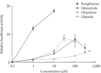
Transcription reporter assays were used to determine the ef- fects of sulfonylureas including glimepiride, gliquidone, and glipizide on the transcriptional activity of PPARγ. Each sulfo- nylurea was tested at the following concentrations: glimepiride, 1, 10, 100, and 300 µM; gliquidone, 1, 10, 30, and 100 µM; glipi- zide, 1, 10, 100, 300, and 500 µM and rosiglitazone, 1 and 10 µM. We obtained the peak concentration at which glimepiride and gliquidone induced the highest transcriptional activity;
gliquidone, 30 µM and glimepiride, 100 µM. Glipizide reached saturation at concentrations greater than 500 µM, therefore, tests were performed at lower concentrations, although we did not obtain the peak value. All agents except glipizide signifi- cantly increased PPARγ agonistic activity at the indicated con- centrations. Glimepiride and gliquidone increased PPARγ transcriptional activity at 1 µM, by 3-4 times at 10 µM, and by nearly ten times at higher concentrations. All of these increas- es were statistically significant, although they were lower than that of rosiglitazone. In the case of glipizide, no agonistic effect at 1 µM was observed, but there was a mild effect at concentra- tions greater than 10 µM (Fig. 1).
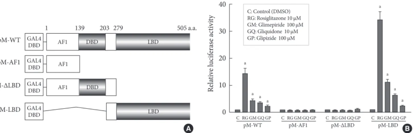
Target region of sulfonylureas in PPARγ
In order to identify the target domain of sulfonylureas in PPARγ, we prepared three constructs from wild type PPARγ (pM-WT). One was the AF1 region (pM-AF1), another lacked the ligand binding domain (pM-ΔLBD), and the other was the ligand binding domain (pM-LBD) (Fig. 2A). The increment of PPARγ transcriptional activity caused by sulfonylureas disap-
peared when pM-AF1 or pM-ΔLBD was transfected, regard- less of the type of sulfonylurea, whereas it was maintained in presence of pM-LBD (Fig. 2B). Based on these observations, it was suggested that the effect of sulfonylureas, like thiazolidin- ediones, on the PPARγ transcriptional activity involves bind- ing to the ligand binding domain of PPARγ.
Combinational treatment of sulfonylureas and rosiglitazone
We examined the effects of rosiglitazone in addition to each sulfonylurea on the PPARγ transcriptional activity. When glimepiride (100 μM), gliquidone (10 μM), or glipizide (100 μM) was administered with rosiglitazone (1 μM), sulfonylureas did not provide any additive effects on the PPARγ transcrip- tional activity of the cells compared to that achieved with rosi- glitazone alone. Interestingly, glimepiride reduced the effect of rosiglitazone on PPARγ transcriptional activity by about 50 percent, which was statistically significant, while gliquidone and glipizide showed no depletive influence (Fig. 3A).
Because glimepiride and gliquidone were more effective than was glipizide for stimulation of PPARγ transcriptional activity, we investigated the effects of combination treatments with
Relative luciferase activity
20
10
0 0.1 1 10 100 1,000
Concentration (μM)
Rosiglitazone Glimepiride Gliquidone Glipizide
Fig. 1. Peroxisome proliferator-activated receptor γ (PPARγ) transcriptional activity by thiazolidinediones (rosiglitazone) and sulfonylureas (glimepiride, gliquidone, and glipizide). Cos 7 cells were transfected with Gal4 tk-Luc, pCMV-β-gal, and pM-PPARγ and treated with rosiglitazone (1 to 10 μM), glimepiride (1 to 300 μM), gliquidone (1 to 100 μM) or glipi- zide (1 to 500 μM) for 24 hours. β-galactosidase activity was used for normalization of luciferase activity. The luciferase ac- tivity of the cells treated with DMSO was set to 1, and the oth- ers were expressed as relative values. Data represent the mean±
standard error of the mean (SEM) (n=3).
rosiglitazone in the presence of different sulfonylurea concen- trations using glimepiride and gliquidone. In the combined treatment, glimepiride inhibited the effect of rosiglitazone in a dose-dependent manner. Under constant rosiglitazone treat- ment at a dose of 1 µM, when treated with glimepiride 1 µM,
luciferase activity showed no change compared to that of the control; at 10 µM, luciferase activity seemed to be lowered, but the effect was not statistically significant. In contrast, when combined with 100 µM glimepiride, the reduction in activity was significantly greater; gliquidone did not show any differ-
pM-WT
1 139 203 279 505 a.a.
pM-AF1 pM-∆LBD pM-LBD
GAL4DBD GAL4DBD GAL4DBD GAL4DBD
AF1 AF1 AF1
DBD
DBD
LBD
LBD
Fig. 2. Target regions of sulfonylureas in peroxisome proliferator-activated receptor γ (PPARγ). (A) Schematic diagram of PPARγ constructs. Three constructs from wild type PPARγ (pM-WT) were prepared. pM-ΔLBD lacked the ligand binding do- main, pM-AF1 carried only the AF1 region and pM-LBD had only the ligand binding domain. (B) Transcriptional activity ac- cording to PPARγ construct. pM-WT, pM-AF1, pM-ΔLBD, or pM-LBD were cotransfected with Gal4 tk-Luc and pCMV-β-gal into COS7 cells. Cells were treated with glimepiride, gliquidone, glipizide or rosiglitazone at indicated doses for 24 hours. The lu- ciferase activity of the cells treated with DMSO after overexpression of PPARγ wild type or its deletions, respectively, was set to 1, and other activities were expressed as relative values. Data represent the mean±standard error of the mean (SEM) (n=5). aP<0.05.
C RG GM GQ GP C RG GM GQ GP C RG GM GQ GP C RG GM GQ GP
pM-WT pM-AF1 pM-∆LBD pM-LBD C: Control (DMSO)
RG: Rosiglitazone 10 μM GM: Glimepiride 100 μM GQ: Gliquidone 10 μM GP: Glipizide 100 μM
40 30 20 10 0
Relative luciferase activity
a
a
a
a
a
a a a
B A
Fig. 3. Combination treatments of thiazolidinediones and sulfonylurea. (A) Combination treatments of thiazolidinediones and sulfonylurea. To examine the effects of rosiglitazone in combination with each sulfonylurea on the peroxisome proliferator-acti- vated receptor γ (PPARγ) transcriptional activity, COS7 cells were transfected with Gal4 tk-Luc, pCMV-β-gal, and pM or pM- PPARγ and treated with glimepiride (100 μM), gliquidone (10 μM), or glipizide (100 μM) plus rosiglitazone (1 μM). Data repre- sent the mean±standard error of the mean (SEM) (n=4). aP<0.05. (B) Combination treatment according to sulfonylurea dose.
Cos 7 cells were transfected with Gal4 tk-Luc, pCMV-β-gal, or pM-PPARγ and treated with rosiglitazone, glimepiride or gliqui- done at the indicated doses for 24 hours. Data represent mean±standard error of the mean (SEM) (n=4). aP<0.05.
C GM GQ GP C GM GQ GP DMSO Rosiglitazone 1μM
pM pM-PPARγ
C GM GQ GP C GM GQ GP DMSO Rosiglitazone 1μM C: Control (DMSO)
GM: Glimepiride 100 μM GQ: Gliquidone 10 μM GP: Glipizide 100 μM
20 15 10 5 0
Relative luciferase activity
a a
a
a
Control 1 10 100 Glimepiride
Rosiglitazone 1μM
1 10 30 (μM) Gliquidone
15 12 9 6 3 0
Relative luciferase activity
a
A B
ence in luciferase activity at any concentration, although gliq- uidone was similar or more effective than glimepiride at the indicated dose (Fig. 3B).
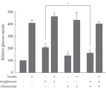
Effect of glimepiride on glucose uptake by thiazolidinediones
We evaluated the effects of rosiglitazone or glimepiride mono- therapy and the combined therapy of the two on insulin-stim- ulated glucose uptake in adipocytes. Each monotherapy and the combination therapy produced an increase in glucose up- take. However, the glucose uptake in co-treatment was less than that of rosiglitazone monotherapy (Fig. 4). It is supposed that the effect of rosiglitazone on glucose uptake was inhibited by glimepiride, and this seems to be related to the effect of PPARγ transcriptional activity.
DISCUSSION
There has been increasing interest in the effects of sulfonylurea on PPARγ transcriptional activity. The major findings of our experiments showed that sulfonylureas increased PPARγ tran- scriptional activity, and glimepiride showed dose-dependent
competition with rosiglitazone for PPARγ, whereas gliquidone and glipizide did not exhibit competitive behaviors.
It has been reported that some sulfonylureas enhance PPARγ transcriptional activity and, thus, target both sulfonylurea re- ceptors in the membranes of pancreatic β-cells and PPARγ in the nuclei of adipocytes [15-18]. Arrault et al. [17] explored the PPARγ-activating properties of a series of eight sulfonyl- ureas using transfection experiments with 293T cells and rosi- glitazone as a reference PPARγ agonist. They found that sec- ond generation sulfonylureas, including glimepiride, gliqui- done, and glibenclamide, stimulated PPARγ-activating prop- erties, but first generation agents, such as tolbutamide, chlor- propamide, tolazamide and gliclazide, in addition to the sec- ond generation agent glipizide, did not [17]. Their findings were consistent with our results and with previously published data by Fukuen et al. [15], with the exception of the glipizide results. In the present study and the work of Fukuen et al. [15], glipizide increased the PPARγ transcriptional activity.
Glipizide is known to exhibit a slightly different binding mode compared to those of glimepiride, gliquidone and glib- enclamide [17,18]; glipizide binds PPARγ more weakly than do the others. The strength of this binding is determined by the hydrogen bonds (H-bond) formed between the docked li- gand and the amino acids in PPARγ which form the binding pocket within the active site. The H-bonding pattern of glipi- zide is different from those of these other compounds [17].
Compared to glimepiride and gliquidone, which form more than two hydrogen bonds with the PPARγ ligand binding pocket, glipizide forms only one hydrogen bond and, thus, functions as a weak binder [17]. In this respect, glipizide showed weaker PPARγ agonistic activity than did the other second generation sulfonylureas. Additionally, glipizide may activate PPARγ in a tissue-specific manner; it showed PPARγ agonistic activity in CV-1 cells [18] and Cos7 cells but not in 293T cells [17].
Other authors have observed PPARγ agonistic activity of glimepiride at 1 and 10 μM [15,16] similar to our results. The mean maximal plasma concentration (Cmax) values for glimepiri- de can reach 1 μM when patients are treated with the suggest- ed maximum daily does of 8 mg [19,20]. For gliquidone, when treated with a 30 mg dose, Cmax is 1.2 μM, with a range from 0.2 to 0.4 μM [21]. In our experiment, gliquidone showed a PPARγ agonistic effect even when treated with 1 μM. Based on this result, glimepiride and gliquidone seem to be able to activate PPARγ at pharmacological concentrations. In addition, glimepiride, a
Insulin - + - + - + - +
Rosiglitazone - - + + - - + +
Glimepiride - - - - + + + +
500 400 300 200 100 0
Relative glucose uptake
b
b a
b
Fig. 4. Effects of glimepiride on rosiglitazone-induced glu- cose uptake. On day 8 of differentiation, 3T3-L1 adipocytes were treated with rosiglitazone (10 μM), glimepiride (100 μM) or both for 48 hours and with insulin for 30 minutes. Glucose uptake was measured using [3H]-deoxyglucose scintillation counting. The glucose uptake value of untreated cells was set to 100, and the others were relative values. Data represent the mean±standard error of the mean (SEM) (n=3). aP<0.05,
bP<0.05 compared to the control.
third generation sulfonylurea, demonstrated the ability to im- prove insulin sensitivity in a human study [22]. Insulin resis- tance estimated using homeostatic model assessment-insulin resistance (HOMA-IR) was significantly reduced in subjects with type 2 diabetes who took glimepiride, although glycemic control measured using HbA1c was unchanged [22]. Also, gliquidone was shown to be as potent as pioglitazone for in- ducing PPARγ target gene expression [18].
In the case of glipizide, which was able to weakly activate PPARγ at a dose of 10 μM in our experiment, Cmax values are 1.0±0.3 μM in subjects treated with a 5 mg dose [23]. Even in subjects administered the maximum daily dose of 40 mg, the concentration able to induce PPARγ agonistic activity is not achieved.
One of the novel findings of this study was that glimepiride reduced the effect of rosiglitazone at high concentrations, but gliquidone or glipizide did not. In our results, glimepiride sig- nificantly inhibits the effect of rosiglitazone on PPARγ tran- scriptional activity in a dose-dependent manner, and cell via- bility was not affected in the tested dose range (data not shown).
Our results are consistent with the results of a previous report [15].
Fukuen and co-workers performed competitive binding as- says using full-length PPARγ2 and [3H] rosiglitazone. The concentration-dependent displacement of [3H] rosiglitazone by glimepiride was observed; therefore, they concluded that glimepiride is in competition with rosiglitazone and activates PPARγ through direct association and could be considered as a partial agonist for PPARγ [15]. In the case of gliquidone, al- though it had stronger agonistic effects than did glimepiride, it did not reduce the effect of rosiglitazone even at 30 μM, the highest concentration tested.
The explanation for these differences is not clear. It is known that the TZD head-group forms hydrogen bonds with the PPARγ residues which form the loop structure within the ac- tive site [17,24]. Glimepiride generated H-bonds with Ser289, Ser342, and the Gly284, whereas gliquidone interacted with Gln286 and Ser342 [17]. Rosiglitazone and glimepiride shared Ser289, but gliquidone did not. Glipizide, which showed no competitive effect against rosiglitazone, was a weak binder com- pared to the other sulfonylureas. Further investigation about the exact mechanism is needed.
Though glimepiride inhibits the effect of rosiglitazone at a dose of 100 μM, competition between glimepiride and rosigli- tazone is not expected in clinical practice since Cmax can be 1
μM at the maximum daily dose of glimepiride (8 mg).
Although sulfonylurea drugs produce a glucose-lowering effect by stimulating insulin secretion in the pancreatic β-cells in an ATP-sensitive, K+ channel-dependent manner; they have also been proposed to have peripheral effects as an insulin sensitizer [22,25-27]. Glimepiride, the most recently developed sulfonylurea agent, induces glucose uptake in extrapancreatic tissue not only by increasing insulin secretion in the pancreat- ic β-cells, but also by stimulating GLUT1 and GLUT4 translo- cations in both normal and insulin-resistant states, indepen- dent of insulin [28,29]. In addition, although a peroxisome proliferator response element (PPRE) site in the GLUT4 gene has not been identified [30,31], PPARγ appears to increase GLUT4 [30-33]. In spite of that, the precise underlying mech- anism through which sulfonylureas and thiazolidinediones increase glucose uptake in extrapancreatic tissues has not been verified. It is suggested that PPARγ, activated by certain sulfo- nylureas, induces GLUT4 to be inserted into the plasma mem- brane through direct interaction of glimepiride with PPARγ molecules or through another indirect mechanism involving a definite signaling pathway that leads to increased glucose up- take.
Recently, the Food and Drug Administration (FDA) limited the use of rosiglitazone due to increased cardiovascular risk [34]. Sulfonylureas are also known to increase the risk of car- diovascular events [35,36], mostly attributed to their effect on myocardial ATP-sensitive K+ channels. Because some sulfo- nylureas activate PPARγ, one cannot exclude the possibility that increasing PPARγ activity might also contribute to sulfo- nylurea’s cardiovascular effect.
Our findings that sulfonylureas induce PPARγ transcrip- tional activity at clinically relevant concentrations will be help- ful for understanding the mechanisms of sulfonylureas in the treatment of subjects with type 2 diabetes mellitus.
CONFLICTS OF INTEREST
No potential conflict of interest relevant to this article was re- ported.
ACKNOWLEDGMENTS
This work was supported by MarineBio21, Ministry of Mari- time Affairs and Fisheries, Korea.
REFERENCES
1. Reaven GM. Banting lecture 1988: role of insulin resistance in human disease. Diabetes 1988;37:1595-607.
2. Taylor SI. Deconstructing type 2 diabetes. Cell 1999;97:9-12.
3. Olefsky JM. Treatment of insulin resistance with peroxisome proliferator-activated receptor gamma agonists. J Clin Invest 2000;106:467-72.
4. Tontonoz P, Spiegelman BM. Fat and beyond: the diverse biol- ogy of PPARgamma. Annu Rev Biochem 2008;77:289-312.
5. Panigrahy D, Singer S, Shen LQ, Butterfield CE, Freedman DA, Chen EJ, Moses MA, Kilroy S, Duensing S, Fletcher C, Fletcher JA, Hlatky L, Hahnfeldt P, Folkman J, Kaipainen A.
PPARgamma ligands inhibit primary tumor growth and me- tastasis by inhibiting angiogenesis. J Clin Invest 2002;110:923- 32.
6. Kliewer SA, Umesono K, Mangelsdorf DJ, Evans RM. Retinoid X receptor interacts with nuclear receptors in retinoic acid, thyroid hormone and vitamin D3 signalling. Nature 1992;355:
446-9.
7. Renaud JP, Moras D. Structural studies on nuclear receptors.
Cell Mol Life Sci 2000;57:1748-69.
8. Desvergne B, Wahli W. Peroxisome proliferator-activated re- ceptors: nuclear control of metabolism. Endocr Rev 1999;20:
649-88.
9. Nagy L, Tontonoz P, Alvarez JG, Chen H, Evans RM. Oxidized LDL regulates macrophage gene expression through ligand ac- tivation of PPARgamma. Cell 1998;93:229-40.
10. Schopfer FJ, Lin Y, Baker PR, Cui T, Garcia-Barrio M, Zhang J, Chen K, Chen YE, Freeman BA. Nitrolinoleic acid: an endoge- nous peroxisome proliferator-activated receptor gamma ligand.
Proc Natl Acad Sci U S A 2005;102:2340-5.
11. Lehrke M, Lazar MA. The many faces of PPARgamma. Cell 2005;123:993-9.
12. Lehmann JM, Lenhard JM, Oliver BB, Ringold GM, Kliewer SA. Peroxisome proliferator-activated receptors alpha and gamma are activated by indomethacin and other non-steroidal anti-inflammatory drugs. J Biol Chem 1997;272:3406-10.
13. Benson SC, Pershadsingh HA, Ho CI, Chittiboyina A, Desai P, Pravenec M, Qi N, Wang J, Avery MA, Kurtz TW. Identifica- tion of telmisartan as a unique angiotensin II receptor antago- nist with selective PPARgamma-modulating activity. Hyper- tension 2004;43:993-1002.
14. Schupp M, Janke J, Clasen R, Unger T, Kintscher U. Angioten- sin type 1 receptor blockers induce peroxisome proliferator-ac-
tivated receptor-gamma activity. Circulation 2004;109:2054-7.
15. Fukuen S, Iwaki M, Yasui A, Makishima M, Matsuda M, Shi- momura I. Sulfonylurea agents exhibit peroxisome prolifera- tor-activated receptor gamma agonistic activity. J Biol Chem 2005;280:23653-9.
16. Inukai K, Watanabe M, Nakashima Y, Takata N, Isoyama A, Sawa T, Kurihara S, Awata T, Katayama S. Glimepiride en- hances intrinsic peroxisome proliferator-activated receptor- gamma activity in 3T3-L1 adipocytes. Biochem Biophys Res Commun 2005;328:484-90.
17. Arrault A, Rocchi S, Picard F, Maurois P, Pirotte B, Vamecq J.
A short series of antidiabetic sulfonylureas exhibit multiple li- gand PPARgamma-binding patterns. Biomed Pharmacother 2009;63:56-62.
18. Scarsi M, Podvinec M, Roth A, Hug H, Kersten S, Albrecht H, Schwede T, Meyer UA, Rucker C. Sulfonylureas and glinides exhibit peroxisome proliferator-activated receptor gamma ac- tivity: a combined virtual screening and biological assay ap- proach. Mol Pharmacol 2007;71:398-406.
19. Langtry HD, Balfour JA. Glimepiride: a review of its use in the management of type 2 diabetes mellitus. Drugs 1998;55:563-84.
20. Malerczyk V, Badian M, Korn A, Lehr KH, Waldhausl W. Dose linearity assessment of glimepiride (Amaryl) tablets in healthy volunteers. Drug Metabol Drug Interact 1994;11:341-57.
21. von Nicolai H, Brickl R, Eschey H, Greischel A, Heinzel G, Konig E, Limmer J, Rupprecht E. Duration of action and phar- macokinetics of the oral antidiabetic drug gliquidone in pa- tients with non-insulin-dependent (type 2) diabetes mellitus.
Arzneimittelforschung 1997;47:247-52.
22. Inukai K, Watanabe M, Nakashima Y, Sawa T, Takata N, Tana- ka M, Kashiwabara H, Yokota K, Suzuki M, Kurihara S, Awata T, Katayama S. Efficacy of glimepiride in Japanese type 2 dia- betic subjects. Diabetes Res Clin Pract 2005;68:250-7.
23. Jaber LA, Ducharme MP, Edwards DJ, Slaughter RL, Grunberg- er G. The influence of multiple dosing and age on the pharma- cokinetics and pharmacodynamics of glipizide in patients with type II diabetes mellitus. Pharmacotherapy 1996;16:760-8.
24. Hiromori Y, Nishikawa J, Yoshida I, Nagase H, Nakanishi T.
Structure-dependent activation of peroxisome proliferator-ac- tivated receptor (PPAR) gamma by organotin compounds.
Chem Biol Interact 2009;180:238-44.
25. Mori RC, Hirabara SM, Hirata AE, Okamoto MM, Machado UF. Glimepiride as insulin sensitizer: increased liver and mus- cle responses to insulin. Diabetes Obes Metab 2008;10:596-600.
26. Rosskamp R, Wernicke-Panten K, Draeger E. Clinical profile
of the novel sulphonylurea glimepiride. Diabetes Res Clin Pract 1996;31 Suppl:S33-42.
27. Haupt A, Kausch C, Dahl D, Bachmann O, Stumvoll M, Har- ing HU, Matthaei S. Effect of glimepiride on insulin-stimulat- ed glycogen synthesis in cultured human skeletal muscle cells:
a comparison to glibenclamide. Diabetes Care 2002;25:2129-32.
28. Muller G, Wied S, Wetekam EM, Crecelius A, Unkelbach A, Punter J. Stimulation of glucose utilization in 3T3 adipocytes and rat diaphragm in vitro by the sulphonylureas, glimepiride and glibenclamide, is correlated with modulations of the cAMP regulatory cascade. Biochem Pharmacol 1994;48:985-96.
29. Muller G. The molecular mechanism of the insulin-mimetic/
sensitizing activity of the antidiabetic sulfonylurea drug Ama- ryl. Mol Med 2000;6:907-33.
30. Fernyhough ME, Okine E, Hausman G, Vierck JL, Dodson MV. PPARgamma and GLUT-4 expression as developmental regulators/markers for preadipocyte differentiation into an ad- ipocyte. Domest Anim Endocrinol 2007;33:367-78.
31. Ezaki O. Regulatory elements in the insulin-responsive glucose transporter (GLUT4) gene. Biochem Biophys Res Commun
1997;241:1-6.
32. McGowan KM, Long SD, Pekala PH. Glucose transporter gene expression: regulation of transcription and mRNA stability.
Pharmacol Ther 1995;66:465-505.
33. Hamm JK, el Jack AK, Pilch PF, Farmer SR. Role of PPAR gamma in regulating adipocyte differentiation and insulin-re- sponsive glucose uptake. Ann N Y Acad Sci 1999;892:134-45.
34. Kahn BB, McGraw TE. Rosiglitazone, PPARγ, and type 2 dia- betes. N Engl J Med 2010;363:2667-9.
35. Tzoulaki I, Molokhia M, Curcin V, Little MP, Millett CJ, Ng A, Hughes RI, Khunti K, Wilkins MR, Majeed A, Elliott P. Risk of cardiovascular disease and all cause mortality among patients with type 2 diabetes prescribed oral antidiabetes drugs: retro- spective cohort study using UK general practice research data- base. BMJ 2009;339:b4731.
36. Evans JM, Ogston SA, Emslie-Smith A, Morris AD. Risk of mortality and adverse cardiovascular outcomes in type 2 dia- betes: a comparison of patients treated with sulfonylureas and metformin. Diabetologia 2006;49:930-6.