대한소화기학회지 2005;45:328-334
서 론
는 살아 있는 미생물로서 복용하면 건강에 이로 Probiotics
운 효과가 있다.1 이 중에서 가장 많이 사용되는 것으로는 유산균 종류인lactobacilli, bifidobacteria와streptococci가 있 고 이외에도 비병원 대장균과 효모인Saccharomyces boulardii
대장암세포주 HT-29 세포에서 Saccharomyces boulardii 에 의한 Peroxisome Proliferator-activated Receptor- 발현의 회복 γ
경희대학교 의과대학 내과학교실 병리학교실, *
이상길․김효종․지성길*․장재영․남기덕․김남훈․주광로 동석호․김병호․장영운․이정일․장 린
Saccharomyces boulardii
Activates Expression of Peroxisome Proliferator-activated Receptor- in HT-29 Cells γ
Sang Kil Lee, M.D., Hyo Jong Kim, M.D., Sung Gil Chi, Ph.D.*, Jae Young Jang, M.D., Ki Deok Nam, M.D., Nam Hoon Kim, M.D.,
Kwang Ro Joo, M.D., Seok Ho Dong, M.D., Byung Ho Kim, M.D., Young Woon Chang, M.D., Joung Il Lee, M.D., and Rin Chang, M.D.
Departments of Internal Medicine and Pathology*, Kyung Hee University College of Medicine, Seoul, Korea
Background/Aims: Saccharomyces boulardii (S. boulardii) has been reported to be beneficial in the treatment of inflammatory bowel disease, however, little is known about its mechanism of action. Peroxisome proliferator- activated receptor- (PPAR- ) is recently found to regulate inflammation in intestinal epithelial cells. We hy γ γ - pothesized that the anti-inflammatory effects of S. boulardii are mediated, in part, through PPAR- . To test this γ hypothesis, we examined the ability of S. boulardii to modulate the expression of PPAR- in human colon cells. γ Methods: Effects of S. boulardii on survival and proliferation of HT-29 human colon cells were assessed by MTT and [
3H]thymidine incorporation assays. PPAR- expression was assessed by Western blot and RT-PCR. γ Induction of interleukin-8 (IL-8) expression by tumor necrosis factor-α (TNF-α), interleukin-1 (IL-1 ), or β β lipopolysaccharide (LPS) was assessed by RT-PCR. Results: S. boulardii did not affect viability and proliferation of HT-29 cells. S. boulardii up-regulated PPAR- expression at both mRNA and protein levels. Pretreatment of γ HT-29 cells with S. boulardii blocked PPAR- down-regulation by TNF- γ α, IL-1 , or LPS, whereas it amelio β - rated IL-8 response to these proinflammatory factors. Conclusions: S. boulardii stimulates PPAR-γ expression and reduces response of human colon cells to proinflammatory cytokines. (Korean J Gastroenterol 2005;45:
328-334)
Key Words: Saccharomyces boulardii; PPAR-γ
접수: 2004년 12월 6 ,일 승인: 2005년 2월 25일 연락처: 김효종, 120-752, 서울시 동대문구 회기 1동
경희의료원 내과
Tel: (02) 958-8199, Fax: (02) 968-1848 E-mail: [email protected]
Correspondence to: Hyo Jong Kim, M.D.
Department of Internal Medicine, Kyung Hee University College of Medicine, 1 Hoegi-dong, Dongdaemun-gu, Seoul 130-752, Korea Tel: +82-2-958-8199, Fax: +82-2-968-1848
E-mail: [email protected]
이상길 외11 .인 S. boulardii가 PPAR-γ와IL-8 발현에 미치는 영향 329
(S. boulardii) 등이 있다.S. boulardii는 호열 및 비병원 효모 로서 항생제 연관 설사 여행자 설사 등의 질환에 선택적으, 로 사용되고 있다.2 최근에는 염증성 장질환에서도 임상 효 과가 보고되었다.3,4Probiotics가 임상적으로 유용한 효과를 나타내기 위해서는 여러 가지 기전을 통할 것으로 생각한 다. Probiotics로 이용되는 다양한 비병원 균주들이 직접 혹 은 간접으로 병원균의 성장을 방해하거나,5-7점막장벽의 기 능을 강화시켜서 병원균이 침투하지 못하게 하거나,8 장상 피세포나 점막연관림프조직의 면역기능을 조절한다.9,10 최
근S. boulardii가 장상피세포의 염증반응을 조절하는 기전
에 대한 연구에서S. boulardii는 NF-κ 의 활성화를 억제하B 여 염증반응을 차단하였다.11 그러나S. boulardii가 다양한 염증반응의 경로 중에서 단지 NF-κ 에만 관계하는지는 확B 실하지 않다.
Peroxisome proliferator-activated receptor-γ (PPAR-γ 는 지) 방세포에서 세포 분화와 당 대사에 관여하는 핵수용체이 다.12PPAR-γ는 지방세포뿐만 아니라 림프구 간세포 근육, , 세포 유방 전립선 등에서도 발현이 되고 특히 장상피세포, , 에서도 고농도로 분포한다 정확한 기전이나 경로는 아직. 밝혀지지 않았으나 PPAR-γ의 배위자(ligand)는 장상피세포 와 면역세포에서 사이토카인과 케모카인 분비를 억제하여 염증반응을 억제함이 밝혀져, PPAR-γ가 장내 염증반응의 조절에서 중요한 역할을 할 것으로 추측된다.13-16이에 저자
들은 S. boulardii가 대장상피세포에서 항염증 작용을 나타
내는 데 있어서 PPAR-γ가 역할을 할 것으로 예측하고 이 를 알아보고자 하였다.
대상 및 방법
실험 세포 및 시약 1.
인체에서 기원한 대장암 상피세포주인HT-29은American 으로부터 구입 Type Culture Collection (Rockville, MD, USA)
하였으며, 100 IU/mL penicillin, 100μg/mL streptomycin및
우태아혈청이 포함된 배지
10% RPMI-1640 (Gibco-BRL, Gai- 를 이용하여
thersburg, MD, USA) 5% CO2, 37oC조건에서 배 양하였다 모든 실험은. HT-29세포를1×10⁴/well의 농도로 에 도포한 후 시 6 well plate (Coster, Cambridge, MA, USA) 24 간 후에 시행하였다. HT-29세포의 유전자 발현에 미치는 영향을 분석하기 위하여 tumor necrosis factor-α (TNF-α, Sigma Chemical Co., St. Louis, MO, USA) 20 ng/mL, in- terleukin-1β (IL-1β, Sigma) 20 ng/mL,Escherichia coli(sero-
의
type 0111: B4) lipopolysaccharide (LPS, Sigma) 20μg/mL를 각각 시간 처리한 후 세포를 회수하였다6 .
S. boulardii(비오플Ⓡ,건일제약 서울 는 동결 건조된 분, )
말형태로 제공받았으며 Sabouraud dextrose agar (Millipore, 를 이용하여
Billerica, MA, USA) 37oC에서 배양하였다 배양. 된S.boulardii는 원심 분리 후hemocytometer를 이용하여 균 수를 측정한 후에 일정량의phosphate buffered saline에 희석 하여 처리하였다.
2. S. boulardii가 HT-29 세포 생존율과 증식에 미치 는 영향
S. boulardii가HT-29세포의 생존율에 미치는 영향을 알 아보기 위해서 3-(4,5-dimethylthiazol-2-yl)-2,5-diphenyl-tetrazo-
를 시행하였다
lium bromide assay (MTT assay) . 1×10⁴/well 농도로HT-29세포를 분주하여12시간 배양한 후 항균제가 포함되지 않은 배양액으로 희석된S. boulardii를 농도별로 처리하여 일정시간 배양하였다 배양액을 제거한 후. 50μL 의MTT용액(2μg/mL)을 첨가하여 시간 동안 배양하였다4 . 상층액을 제거하고 50μ 의L dimethyl sulfoxide를 첨가한 후
분간 진탕하고 에서의 를 측정하였
10 570 nm optical density 다.
S. boulardii가HT-29세포의 증식에 미치는 영향을 알아 보기 위해서 [3H]thymidine incorporation assay를 시행하였다. 여러 농도의S. boulardii를 1×10⁴cell/well의HT-29세포에 처리한 후20시간 배양하였다. [3H]-labeled thymidine을1μ Ci/well농도로 시간 동안 처리한 후 흡착지에 부착시켜4 건 조시킨 후에 liquid scintillation counter (Perkin-Elmer, Boston,
를 이용하여 방사능을 측정하였다
MA, USA) .
3. S. boulardii가 PPAR-γ 및 IL-8 발현에 미치는 영향
분석 1) RT-PCR
을 이용하여 Trizol reagent (Invitrogen, Carlsbad, CA, USA)
세포로부터 를 추출하였다 합성을 위해
HT-29 RNA . cDNA
서 1μ 의g RNA에10 mM dithiothreitol, 50 mM Tris-HCl (pH 8.3), 75 mM KCl, 3 mM MgCl2, 1μM dNTPs, 10 U RNase inhibitor, random primer (0.05μ 와M) 250 U avian myeloblas- tosis virus-reverse transcriptase (Promega, Madison, WI, USA) 를 첨가하여 37oC에서 시간 동안 반응시켰다 역전사효소1 . 의 활성을 제거하기 위해 95oC에서 분간 방치하였다5 . PCR 분석에 사용한 유전자 특이primer의 염기서열은Table 1에 기술하였다. PCR 반응은 MgCl2이 포함된 PCR buffer II
를 이용하였으며
(Perkin-Elmer, Norwalk, CT, USA) 95oC에서 분
1 , 58oC에서 1분 그리고 72oC에서 1분의 조건으로 총 회의 증폭을 시행하였다 반응의 확인을 위해
32-38 . PCR
산물
PCR 10μ 를L 2% agarose gel을 이용하여 전기 영동하 였으며ethidium bromide로 염색하였다 각 샘플의. RT-PCR 산물은 동일한 샘플이 나타내는GAPDH유전자의RT-PCR
330 The Korean Journal of Gastroenterology: Vol. 45, No. 5, 2005
산물을 대조군으로 하여 분석하였다. RT-PCR은 각 샘플마 다 최소 회 이상 반복하였다3 .
2) Western blot assay
NaCl, 1 mM EDTA, 1 mM EGTA, 1% Triton, 2.5 mM sodium phosphate, 1 mM β-glycerolphosphate, 1 mM Na3VO4, 1μg/mL leupeptin,그리고1 mM phenylmethylsulfonyl fluo-
가 함유된 용해완충액에서 용해시키고 원심 분리하여
ride 상
층액을 취하였다 추출된 단백질은. 100oC에서 분간 가열한5 후10% sodium dodecyl sulfate가 포함된polyacrylamide gel을 이용하여 전기 영동하였으며 전개된 단백질은 polyvinylidene difluoride membrane (Millipore Corporation, Bedford, MA, USA) 에 이동시켰다. PPAR-γ 단백질은 anti-PPAR-γ 항체(Santa
와 결합시킨 후 Cruz Biotechnology, Santa Cruz, CA, USA) en- hanced chemical luminescence (ECL, Amersham, Piscataway, NJ,
방
USA) 법을 통해 검출하였다 동량의 단백이 전개되었는. 지의 여부는anti-GAPDH항체(Sigma)를 통하여 확인하였다.
결 과
1. S. boulardii가 HT-29 세포 활성과 증식에 미치는 영향
를 이용하여
MTT assay S. boulardii의 처리 농도 및 시간 에 따른HT-29세포활성의 변화 여부를 분석하였다 먼저. 처리시간에 따른 영향을 파악하기 위하여10 yeasts/cell의 농도로6-24시간 배양한 후 세포활성을 조사하였으나 처리 하지 않은 대조군에 비하여 뚜렷한 차이가 없었다(Fig. 1A).
S. boulardii의 처리농도에 따른 영향을 파악하기 위해 5-20 의 농도로 시간 배양한 경우에서도 뚜렷한 세포 yeasts/cell 24
활성의 변화는 없었다(Fig. 1B).S. boulardii가 세포증식에 영향을 미치는지를 파악하기 위하여HT-29세포의DNA합 성 변화 유무를 [3H]thymidine incorporation assay를 이용하여 조사하였다.S. boulardii가20 yeasts/cell의 농도로처리된 세포의 합성은 처리되지 않은 대조군과 비교할 HT-29 DNA
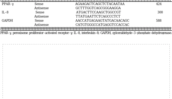
Table 1. Sequences of Primers Used for RT-PCR
Gene Primer Seqences (5' to 3') Size of PCR product (bp)
PPAR-γ Sense AGAAGACTCAGCTCTACAATAA 424
Antisense GCTTTGGTCAGCGGGAAGGA
IL-8 Sense ATGACTTCCAAGCTGGCCGT 300
Antisense TTATGAATTCTCAGCCCTCT
GAPDH Sense AACCATGAGAAGTATGACAACAGC 588
Antisense CATGTGGGCCATGAGGTCCACCAC
PPAR-γ, peroxisome proliferator-activated receptor-γ; IL-8, interleukin-8; GAPDH, gylceraldehyde-3-phosphate dehydrogenase.
Fig. 1. Effect ofSaccharomyces boulardiion viability of HT-29 cells. HT-29 cells were seeded at the density of 1×10⁴cells/well in 6-well plates and maintained in the medium with 10% FBS for 24 hr. According to various incubation time (A), the medium containing 10% FBS with various concentrations (B) ofS. boulardiiwas added to HT-29 cell. After 6-24 hr incubation, cell viability was determined by MTT assay. Data are the mean of triplicate assays and represent the relative viability compared to untreated controls.
Lee SK, et al.Saccharomyces boulardiiActivates Expression of Peroxisome Proliferator-activated Receptor-γ in HT-29 Cells 331
때 유의한 차이를 보이지 않았다(Fig. 2).이상의 결과를 통 하여S. boulardii는HT-29의 세포활성 및 세포증식에 영향 을 미치지 않음이 확인되었다.
2. S. boulardii가 HT-29 세포의 PPAR-γ 발현에 미 치는 영향
S. boulardii가 PPAR-γ 발현에 미치는 영향을 알아보기
위해서HT-29세포에20 yeasts/cell의 농도로 처리한 후에 을 이용해서
RT-PCR PPAR-γmRNA발현양상을 분석하였 다.S. boulardii처리 시간 후부터6 PPAR-γmRNA의 발현 이 증가하였으며 처리, 18시간 후 PPAR-γ mRNA 발현이 현저히 증가하였다(Fig. 3A). Western blot assay를 통하여 PPAR-γ 단백질의 발현양상을 분석한 결과 mRNA 발현증 가와 마찬가지로S. boulardii처리18시간 후부터PPAR-γ 단백질 발현이 현저히 증가하였다(Fig. 3B).
3. S. boulardii가 TNF-α, IL-1β, LPS에 의한 PPAR-γ 발현 억제 및IL-8 생성에 미치는 영향
PPAR-γ의 발현을 촉진하는 것으로 확인된S. boulardii가 TNF-α, IL-1β, LPS에 의한 PPAR-γ 발현 억제에 영향을 미치는지를 분석하였다. HT-29세포에TNF-α, IL-1β, LPS 각각을 6시간 동안 처리할 경우 PPAR-γ의 발현 감소와
의 발현 증가가 관찰되었다 그러나
IL-8 (Fig. 4). TNF-α, IL-1 β, LPS처리 시간 전에6 S. boulardii를20 yeasts/cell의 농도 로 전처리할 경우 TNF-α, IL-1β, LPS에 의한 PPAR-γ Fig. 2. Effect ofSaccharomyces boulardiion cell proliferation of
HT-29 cells. HT-29 cells were seeded at the density of 1×10⁴ cells/well in 6-well plates and maintained in the medium with 10% FBS for 24 hr. The cells were washed twice with PBS, and medium containing 10% FBS with various concentrations ofS.
boulardiiwas added and incubated for 20 hr. Cells were then pulse-labeled for 4 hr with 1 Ci/mL [3H]thymidine, and the radio- activity incorporated into trichloroacetic acid-precipitable materials was counted by a liquid scintillation counter. Data are the mean of duplicate assays.
Fig. 3. Induction of PPAR-γ expression bySaccharomyces boula- rdiiin HT-29 cells. After treatment ofSaccharomyces boulardii (20 yeasts/cell), PPAR-γ expression in HT-29 cell was measured by RT-PCR (A) and Western blot (B). GAPDH expression was used as control.
Fig. 4. Attenuation of TNF-α, IL-1β, or LPS-mediated suppres- sion of PPAR-γ expression bySacchromyces boulardiiin HT-29 cells. HT-29 cells were pre-incubated withSaccharomyces boulardii (20 yeasts/cell) for 6 hr before treatment with 20 ng/mL of TNF- α (A), 20 ng/mL of IL-1β (B), and 20μg/mL of LPS (C).
Cells were harvested for RT-PCR analysis of PPAR-γ and IL-8 mRNA expression. GAPDH expression was used as control.
332 대한소화기학회지 제: 45권 제 호5 , 2005
발현 감소가 현저히 억제되었고 발현
mRNA , IL-8 mRNA
증가도 현저히 차단되었다(Fig. 4).따라서S. boulardii는 인 체 대장암 세포주의 PPAR-γ 발현을 촉진할 뿐만 아니라 친염증 사이토카인에 의한 PPAR-γ 발현 억제 및IL-8발현 촉진을 억제하였다.
고 찰
이번 연구에서는probiotics의 일종인S. boulardii의 항염증 작용기전을 알아보기 위하여 대장암 세포주인HT-29를 이 용하여S. boulardii가 PPAR-γ 및IL-8발현에 미치는 영향 을 분석하였다 이번 연구 결과. S. boulardii는HT-29세포의 PPAR-γ 유전자의 발현을 증가시키고 친염증 사이토카인인 TNF-α, IL-1β와LPS에 의한IL-8생성과PPAR-γ 발현 억 제를 차단하였다 이러한 결과는 기존에 보고되었던. S. bou- lardii의 항염증 효과가S. boulardii의 PPAR-γ 발현 촉진기능 과 밀접하게 관련되어 있을 가능성을 강하게 시사한다.
S. boulardii는 여행자 설사 급성 위장염을 비롯한 다양한, 설사병에 사용되어 왔으나 가장 많이 사용된 것은 Clos- tridium difficile연관 질환이며 이에 대한 많은 임상연구와 동물실험의 결과가 보고되었다.17-19S. boulardii의 작용기전 에 대해서는 각각의 질환군에 따라서 독립된 연구 결과들로 설명이 되며 특히 장내 감염을 일으킬 수 있는 병원균에 대 한 작용기전은 각각의 균에 대해서 특이하다. Clostridium difficile은 독소A와 를 생성하여 장점막에 손상을 주고 염B 증반응을 유발하는데S. boulardii는 독소 를 단백질 분해A 시 키고 장점막에 결합하는 것을 막는54 KDa의serine protease 를 생성한다.20Vibrio cholerae의cholera toxin은adenylate 를 활성화시켜서 염화물과 중탄산염의 소실을 유발 cyclase
하여 설사를 일으킨다.S. boulardii는 콜레라 독소를 방해하 는120 kDa의 단백질을 생성한다.21이외에도S. boulardii는 병원균이 직접 점막세포에 붙지 못하도록 방해할 뿐만 아니 라tight junction 구조를 강화시키기도 한다.22
최근에S. boulardii는 감염 설사뿐만 아니라 염증성 장질
환에서도 시도되고 있다.S. boulardii는 크론병에서 질환의 활성도를 감소시키고 설사의 기간을 단축시켰고,3메살라친 단독요법보다는 관해 유지에 더 효과적이었다.4 궤양성 대 장염에서는 메살라친에 S. boulardii를 추가로 투여하였을 때 관해율이 약70%정도였다.23그러나 이러한 질환군에서
S. boulardii가 이로운 효과를 나타내는 기전에 대해서는 알
려진 것이 많지 않다 이번 연구 결과를 포함한 실험실 연구. 에서S. boulardii에 의한 항염증 작용은 일관된 결과를 보였 다 즉 친염증 사이토카인이나 독성 세균에 대한 염증 반응. 을S. boulardii가 감소시켰다.S. boulardii가 염증성 장질환 에서 항염증 작용을 매개하는 기전에 대해 최근 NF-κB경
로를 억제하여IL-8 생성을 차단하였다.24이번 연구에서도 S. boulardii는 TNF-α, IL-1β, LPS에 의한IL-8발현을 억제 하였고, PPAR-γ 유전자의 발현을 촉진함이 새롭게 발견되 었다.
PPAR-γ는 핵 수용체군의 하나로 지방세포의 분화와 세, 포자멸사 인슐린 감수성 및 포도당 항상성에 중요한 역할, 을 한다.12PPAR-γ는 대장상피세포에서 고농도로 발현되며 PPAR-γ+/-실험동물에서는 장염이 유발되어 PPAR-γ가 장 점막의 보존에 중요한 역할을 하는 것이 확인되었다.25,26 PPAR-γ 촉진제의 투여는 염증을 조절하는 것으로 알려져 염증반응이 병인의 주된 요소인 동맥경화증 염증성 장질환, 에서의 임상 적용이 고려되고 있다.27 특히 궤양성 대장염 환자의 조직에서는 PPAR-γ 발현이 감소되어 있고 장내 상 존하는 박테리아가 PPAR-γ의 발현을 조절할 수 있다.28이 러한 사실로 볼 때, PPAR-γ는 인체 내에서 염증 반응을 감 소시키는 방향으로 작용한다 이번 연구결과에서도. S.
boulardii는HT-29세포에서PPAR-γmRNA및 단백질 발현 을 증가시켰고, TNF-α, IL-1β, LPS에 의한PPAR-γ 발현 감소를 차단하였다 이 결과는 정상 상태나 염증이 유발된. 경우에서도S. boulardii가 PPAR-γ의 발현 증가에 기여함을 의미한다 이번 실험에서. PPAR-γ 억제제를 이용하거나 PPAR-γ 발현이 없는 동물모델에서의 실험을 병행하지는 못했으나S. boulardii는 TNF-α, IL-1 ,β LPS에 의한PPAR-γ 발현 억제를 차단함과 동시에IL-8생성을 억제하였다 이러. 한 사실은S. boulardii가 기존에 보고된 NF-κ 억제 또는B
와 와 같은
c-Jun N-terminal kinase (JNK) p38 kinase mito- 의 억제 경로
gen-activated protein kinase (MAPK) 25,26와 더불 어 PPAR-γ의 발현 증가를 통해서 항염증 작용을 할 가능 성을 강하게 시사한다 그러나 이번 연구에서는. S. boulardii 에 의한 PPAR-γ 발현 증가와 항염증 작용과의 관계를 직 접적으로 증명하지는 못하였으며 기존에 알려진, S. boul-
ardii의 작용기전과 PPAR-γ 발현 증가의 관계에 대해서도
알 수는 없다 그러므로 향후에. S. boulardii의 NF-κ 나B 억제 경로와
MAPK PPAR-γ의 발현 변화의 연관성에 대해 서도 추가 연구가 필요하리라 생각한다.
요 약
목적:S. boulardii는probiotics의 일종으로 여러 종류의 설 사병에 사용되고 있으며 작용기전이 정확히 알려져 있지는 않지만 염증성 장질환에서도 항염증 작용을 보인다. PPAR- γ는 지방세포에서 세포분화와 당대사에 관여하는 핵수용 체로 장상피세포에서 다량 발현이 되고 장내 염증 반응을, 조절한다 이에 저자들은. S. boulardii가 장상피세포에서 PPAR-γ의 발현에 미치는 영향을 보고자 하였다. 대상 및
이상길 외11 .인 S. boulardii가 PPAR-γ와IL-8 발현에 미치는 영향 333
방법 대장암 세포주인: HT-29에S. boulardii를 배양하여 처 리하였다.S. boulardii처리 후에HT-29세포의 생존율과 증 식은MTT assay와[3H]thymidine incorporation assay를 이용 해서 측정하였다.S. boulardii처리 후의 PPAR-γ 발현의 변 화는Western blot과RT-PCR을 이용하여 측정하였고, TNF- α, IL-1β, LPS로 자극한 후의PPAR-γ 및IL-8측정은RT-
을 이용하였다
PCR . 결과:S. boulardii처리는HT-29의 생존 율과 증식에 영향을 미치지 않았고, PPAR-γmRNA와 단백 발현을 증가시켰다.S. boulardii는 TNF-α, IL-1β와LPS에 의한IL-8생성을 억제함과 동시에PPAR-γ 발현 억제를 차 단하였다. 결론:S. boulardii는HT-29세포에서PPAR-γ의 발현을 증가시켰고, TNF-α, IL-1β와LPS에 의한IL-8생성 과 PPAR-γ의 발현 감소를 억제하였다.
색인단어: Saccharomyces boulardii, PPAR-γ
참고문헌
1. Fuller R. Probiotics in man and animals. J Appl Bacteriol 1989;66:365-378.
2. Goossens D, Jonkers D, Stobberingh E, van den Bogaard A, Russel M, Stockbrugger R. Probiotics in gastroenterology:
indications and future perspectives. Scand J Gastroenterol 2003;239:15-23.
3. Plein K, Hotz J. Therapeutic effects ofSaccharomyces boulardiion mild residual symptoms in a stable phase of Crohn's disease with special respect to chronic diarrhea pilot study. Z Gastroenterol 1993;31:129-134.
4. Guslandi M, Mezzi G, Sorghi M, Testoni PA.Saccharomyces boulardiiin maintenance treatment of Crohn's disease. Dig Dis Sci 2000;45:1462-1464.
5. Venturi A, Gionchetti P, Rizzello F, et al. Impact on the composition of the faecal flora by a new probiotic pre- paration: preliminary data on maintenance treatment of pa- tients with ulcerative colitis. Aliment Pharmacol Ther 1999;
13:1103-1108.
6. Mack DR, Michail S, Wei S, McDougall L, Hollingsworth MA. Probiotics inhibit enteropathogenicE. coliadherence in vitro by inducing intestinal mucin gene expression. Am J Physiol Gastrointest Liver Physiol 1999;276:941-950.
7. Flynn S, van Sinderen D, Thornton GM, Holo H, Nes IF, Collins JK. Characterization of the genetic locus responsible for the production of ABP-118, a novel bacteriocin produced by the probiotic bacteriumLactobacillus salivariussubsp.
salivarius UCC118. Microbiology 2002;148:973-984.
8. Madsen K, Cornish A, Soper P, et al. Probiotic bacteria
enhance murine and human intestinal epithelial barrier func- tion. Gastroenterology 2001;121:580-591.
9. Haller D, Bode C, Hammes WP, Pfeifer AM, Schiffrin EJ, Blum S. Non-pathogenic bacteria elicit a differential cytokine response by intestinal epithelial cell/leukocyte co-cultures. Gut 2000;47:79-87.
10. Helgeland L, Vaage JT, Rolstad B, Midtvedt T, Brandtzaeg P.
Microbial colonization influences composition and T-cell re- ceptor V beta repertoire of intraepithelial lymphocytes in rat intestine. Immunology 1996;89:494-501.
11. Neish AS, Gewirtz AT, Zeng H, et al. Prokaryotic regulation of epithelial responses by inhibition of IkappaB-alpha ubi- quitination. Science 2000;289:1560-1563.
12. Schoonjans K, Martin G, Staels B, Auwerx J. Peroxisome proliferator-activated receptors, orphans with ligands and functions. Curr Opin Lipidol 1997;8:159-166.
13. Ricote M, Li AC, Willson TM, Kelly CJ, Glass CK. The per- oxisome proliferator-activated receptor-gamma is a negative regulator of macrophage activation. Nature 1998;391:79-82.
14. Jiang C, Ting AT, Seed B. PPAR-gamma agonists inhibit pro- duction of monocyte inflammatory cytokines. Nature 1998;
391:8286.
15. Lefebvre M, Paulweber B, Fajas L, et al. Peroxisome proli- ferator-activated receptor gamma is induced during differen- tiation of colon epithelium cells. J Endocrinol 1999;162:
331-340.
16. Wu GD. Is there a role for PPAR gamma in IBD? Yes, no, maybe. Gastroenterology 2003;124:1538-1542.
17. McFarland LV, Surawicz CM, Greenberg RN, et al. A ran- domized placebo-controlled trial ofSaccharomyces boulardii in combination with standard antibiotics forClostridium difficile disease. JAMA 1994;271:1913-1918.
18. Surawicz CM, McFarland LV, Greenberg RN, et al. The search for a better treatment for recurrentClostridium difficile disease: use of high-dose vancomycin combined withSaccha- romyces boulardii.Clin Infect Dis 2000;31:1012-1017.
19. Toothaker RD, Elmer GW. Prevention of clindamycin-in- duced mortality in hamsters bySaccharomyces boulardii. Antimicrob Agents Chemother 1984;26:552-556.
20. Castagliuolo I, LaMont JT, Nikulasson ST, Pothoulakis C.
Saccharomyces boulardiiprotease inhibitsClostridium diffi- ciletoxin A effects in the rat ileum. Infect Immun 1996;
64:5225-5232.
21. Czerucka D, Rampal P. Effect ofSaccharomyces boulardiion cAMP- and Ca2+-dependent Cl secretion in T84 cells. Dig Dis Sci 1999;44:2359-2368.
22. Czerucka D, Dahan S, Mograbi B, Rossi B, Rampal P.Sacc-
334 The Korean Journal of Gastroenterology: Vol. 45, No. 5, 2005
haromyces boulardiipreserves the barrier function and modulates the signal transduction pathway induced in en- teropathogenicEscherichia coli-infected T84 cells. Infect Im- mun 2000;68:5998-6004.
23. Guslandi M, Giollo P, Testoni PA. A pilot trial ofSac- charomyces boulardiiin ulcerative colitis. Eur J Gastroenterol Hepatol 2003;15:697-698.
24. Sougioultzis S, Simeonidis S, Bhaskar KR, et al.Saccharo- myces boulardiiproduces a soluble factor that inhibits NF-kB mediated IL-8 gene expression. Gastroenterology 2003;125 (abstr):606A.
25. Nakajima A, Wada K, Miki H, et al. Endogenous PPAR gamma mediates anti-inflammatory activity in murine ische-
mia-reperfusion injury. Gastroenterology 2001;120:460-469.
26. Desreumaux P, Dubuquoy L, Nutten S, et al. Attenuation of colon inflammation through activators of the retinoid X receptor (RXR)/peroxisome proliferator-activated receptor gam- ma (PPARgamma) heterodimer. A basis for new therapeutic strategies. J Exp Med 2001;193:827-838.
27. Su CG, Wen X, Bailey ST, et al. A novel therapy for colitis utilizing PPAR-gamma ligands to inhibit the epithelial inflam- matory response. J Clin Invest 1999;104:383-389.
28. Dubuquoy L, Jansson EA, Deeb S, et al. Impaired expression of peroxisome proliferator-activated receptor gamma in ulce- rative colitis. Gastroenterology 2003;124:1265-1276.