Genipin Selectively Inhibits TNF-α-activated VCAM-1 But Not ICAM-1 Expression by Upregulation of PPAR-γ in Human Endothelial Cells
6
0
0
전체 글
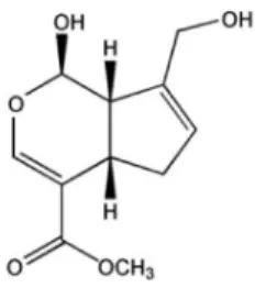
(2) 158. JS Hwa, et al. Fig. 1. Chemical structure of genipin.. PPAR-γ as well as inhibition of P-Akt and P-PKC which resulted in selective inhibition of expression of VCAM-1 but not ICAM-1 in HUVECs activated with TNF-α.. METHODS Materials Dulbecco's Modified Eagle Medium (DMEM), fetal bovine serum (FBS), and antibiotics (penicillin/streptomycin) were purchased from Gibco-BRL (Rockville, MD). Anti-ICAM-1, anti-VCAM-1, anti-PPAR-γ, anti-phospho-ERK1/2 and anti-β-actin antibodies were acquired from Santa Cruz Biotechnology (Santa Cruz, CA), anti-p-Akt and anti-p-PKC antibodies were obtained from Cell Signaling Technology (Beverly, MA). All other chemicals were supplied by Sigma-Aldrich (St. Louis, MO). Genipin was isolated from dried fruits of Gardenia jasminoides Ellis as described [9]. Cell culture Human umbilical endothelial cells (EA.hy 926 cells) were obtained from ATCC and grown in the DMEM supplemented with 10% FBS, 2 mM L-glutamine, 100 IU/ml penicillin, 10 μg/ml streptomycin. Cells were cultured in 100 mm dishes and grown in a humidified 5% CO2 incubator. 7 Cells were plated at a density of 1×10 cells per 100 mm dish. Cells were used between passage numbers 6 and 12. For adhesion assay, U937 human monocyte was obtained from Korea Cell Line Bank (KCLB, Seoul, Korea) and grown in RPMI 1640 supplemented with 10% FBS, 2 mM l-glutamine, 25 mM HEPES, 25 mM NaHCO3, 100 IU/ml penicillin, and 10 μg/ml streptomycin. MTT assay Cell viability was determined colorimetrically using the MTT assay. Cells in the exponential phase were seeded at 4 1×10 cells per well in 24-well plates. After different treatments, 20 μl of 5 mg/ml MTT solution was added to each well (0.1 mg/well), and wells were incubated for 4 h. The supernatants were aspirated, the formazan crystals in each well were dissolved in 200 μl of dimethyl sulfoxide for 30 o min at 37 C, and optical density at 570 nm was read on a Microplate Reader (Bio-Rad, Hercules, CA). Western blot analysis HUVECs were treated with TNF-α (10 ng/ml) and/or tested compound for 24 h. The cells were then washed two. times with cold PBS and lysed in RIPA buffer (PBS supplemented with 1% NP40, 0.5% sodium deoxycholate, 1 mmol/l phenylmethylsulfonyl fluoride, 1 μg/ml aprotinin, and 1 mmol/l sodium orthovanadate). The cell lysates were then o incubated at 4 C for 30 min, after which they were cleared by centrifugation at 10,000× g for 10 min. The protein concentration of each sample was determined using a BCA protein assay kit (Pierce, Rockford, IL). The proteins were then resolved by SDS-PAGE. The gels were transferred to polyvinylidene difluoride (PVDF) membranes by semidry electrophoretic transfer at 15 V for 60∼75 min. The PVDF o membranes were blocked overnight at 4 C in 5% bovine serum albumin (BSA). The cells were incubated with primary antibodies (PPAR-γ, ICAM-1, VCAM-1, p-ERK1/2, p-PKC, p-Akt, and β-actin) diluted 1:500 in Tris-buffered saline/ Tween 20 (TBS-T) containing 5% BSA for 2 h and then incubated with the secondary antibody at room temperature for 1 h. Anti-rabbit IgG was used as the secondary antibody (1:5,000 dilution in TBST containing 1% BSA). The signals were detected by ECL (Amersham, Piscataway, NJ). U937 cell adhesion assay Cells were seeded into two-well chamber slides 48 h before experiments. The medium was refreshed before stim7 ulation with TNF-α. U937 monocytes (3×10 ) were incubated in RPMI 1640 medium containing 2% FBS and 10 mg/ml of the fluorescent dye BCECF/AM (Boehringer, Mannheim, o Germany) at 37 C for 30 min, as described previously [7]. Fluorescence-labeled cells were pelleted and resuspended 5 (7.5×10 /ml) DMEM. Cells were washed three times with DMEM before addition of loaded cells and incubated at 37oC. After 30 min, cell suspensions were withdrawn and cells were gently washed with DMEM. Fluorescent images were selected using a high-resolution video camera (DXC-960MD; Sony) mounted on a BH-2 Olympus microscope (Melville, NY). We then picked images of 0.2 mm in width within these first selected areas and the immunoreactivity of these was measured using SigmaGel 1.0 (Jandel Scientific, Germany). Analyses were repeated three times over the same region and the results are the means of three independent experiments. ROS production assay The intracellular generation of ROS was monitored using dichlorofluorescein diacetate (DCFH-DA), a fluorescent dye. Inside the cells, this compound is oxidized by ROS to form a fluorescent carboxydichlorofluorescein. Briefly, cells were pretreated with different concentration of each compound during 30 min then incubated with or without TNF-α at 10 min, cells were suspended with 1xPBS then DCFH-DA was added at the final concentration of 10 μM. Fluorescence was monitored at the excitation and emission wavelength of 485 and 530 nm, respectively using a fluorescence plate reader (50 cycles per 20 s at 37oC). Data were expressed as relative changes to the initial fluorescence. Statistical analysis The data were expressed as the means±SD. ANOVA and Student's t-test were applied to determine the statistical significance with a p<0.05..
(3) Genipin Inhibits VCAM-1 in Human Endothelial Cells. RESULTS Cell viability Fig. 2 shows that anti-proliferative effect of genipin. When cell proliferation was measured by MTT assay, genipin did not show harmful effect from 1 to 50 μM concentration. However, at 100 μM it reduced cell survival rate about 15∼30%. Therefore, concentration was limited to maximum of 50 μM. Effects on ROS production To examine ROS production, cells were treated with genipin with various doses (1, 5, 10, 50 μM) along with TNF-α (10 ng/ml) which were incubated for 10 min. Fig. 3 shows that the TNF-α significantly increased production of ROS, which was significantly reduced by depending on genipin concentrations. Effects on TNF-α-induced VCAM-1 and ICAM-1 expression. 159. we asked whether genipin inhibits adhesion molecules in EC activated with TNF-α. As shown in Fig. 4, genipin significantly inhibited TNF-α-activated VCAM-1 but not ICAM-1 expression. Involvement of PPAR-γ in anti-inflammatory effect Then we asked whether the differential regulation of expression of adhesion molecules involves PPAR activation as suggested by Jackson et al [6]. Fig. 5 shows that genipin dose- and time-dependently increased PPAR-γ expression in HUVEC. Effect on phosphorylation of ERK1/ 2, Akt, and PKC In addition to PPAR-activators, signal molecules and kinases such as PI3K/Akt, PKC, and ERK1/2 inhibitors also showed differential effects on ICAM-1 and VCAM-1 expression [7,8]. Therefore, we explored the effect of genipin on the TNF-α-induced phosphorylation of these signal molecules. Fig. 6 shows that genipin significantly inhibited phosphorylation of PKC and Akt but not ERK.. Although genipin showed anti-inflammatory action [9,11], no report is available on adhesion molecules expression. So,. Fig. 2. Anti-proliferative effect of genipin in HUVECs. HUVECs were treated as indicated concentrations of genipin for 24 h. Cell proliferation was then assessed by MTT assay. The data were expressed as the means±SD of three independent experiments. ††p< 0.01 compared with control.. Fig. 3. Inhibition of ROS production in TNF-α-activated HUVECs. Different concentration of genipin was added 30 min prior to TNF-α and further incubated for 10 min. Cells were suspended and then DCFH-DA was added at the final concentration of 10 μM. Fluorescence was monitored at the excitation and emission wavelength of 485 and 530 nm, respectively using a fluorescence plate reader (50 cycles per 20 s at 37oC). Results were expressed as relative changes to the initial fluorescence. The data were expressed as the means± SD of three independent experiments. **p<0.01 compared with control. ††p<0.01, compared with TNF-α.. Fig. 4. Preferential inhibition of TNF-α-mediated induction of VCAM-1 over ICAM-1. HUVECs were pretreated with genipin for 1 h and then treated with TNF-α for 6 h. The protein level of ICAM-1 and VCAM-1 was detected by Western blot analysis, as detailed in Materials and Methods. Data were confirmed by three independent experiments. The expression levels of ICAM-1, VCAM-1 protein were quantified by densitometer. Data are presented as means±SD from three independent experiments. Significance compared with control. **p<0.01 compared with control. ††p< 0.01, compared with TNF-α..
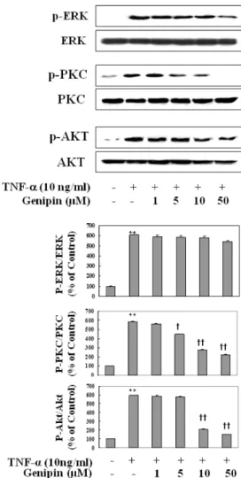
(4) 160. JS Hwa, et al. Fig. 5. Time- and concentration-dependent induction of PPAR-γ protein in TNF-α-induced HUVECs. Cells were treated with different time period with fixed concentration (upper) or different concentration (lower) of genipin. Proteins were isolated and subjected to Western blot for PPAR-γ expression. Data were confirmed by three independent experiments. **p<0.01 compared with control.. Inhibition of TNF-α stimulated adhesion of monocytes to HUVECs Finally, we asked whether genipin inhibits adhesion of monocytes to HUVEC due to inhibition of VCAM-1 expression. As shown in Fig. 7, adhesion of human monocytes (U937) to HUVEC increased by 6-fold after stimulation with TNF-α, which was significantly and concentration-dependently reduced by the presence of genipin.. DISCUSSION Although genipin showed antioxidant and anti-inflammatory action, no report is available so far on the expression of PPRA-γ induction and/or adhesion molecules in human ECs. Therefore, we investigated to gain scientific evidence why extract of fruit of gardenia (Gardenia jasminoides Ellis) has been used for chronic inflammatory disorders [10]. In the present study, we clearly demonstrated that genipin isolated from Gardenia jasminoides inhibited the expression of TNF-α-induced VCAM-1 protein but not ICAM-1 in EA, hy 926 cells. Previous studies have suggested genipin has remarkable anti-inflammatory and anti-angiogenic effect [11]. A critical step in initiation and progression of atherosclerosis is leukocyte infiltration through. Fig. 6. Effect on phosphorylation of ERK1/2, Akt, and PKC activation by TNF-α in HUVECs. Cells were pretreated with different concentration of genipin (1, 5, 10 and 50 μM) for 24 h, and then treated with TNF-α for 10 min for detection of phosphorERK1/2 and PKC or for 30 min for detection of phosphor-Akt. Cells were extracted and protein level was detected by Western blot analysis (upper). The blot was quantified by using densitometry and represented as % increase of control (lower). **p<0.01 compared with control. †p<0.05, ††p<0.01, compared with TNF-α, respectively. Data were confirmed by two independent experiments.. vascular endothelium into the vessel wall [12]. Inflammation of vascular EC causes induction of adhesion molecules and promotes leukocyte interaction [13]. Expression of adhesion molecules in EC is also essential for angiogensis in the vasculature. Can inhibition of VCAM-1 by genipin be responsible for traditional use of this chemical for anti-inflammatory and anti-angiogenic effect? If so, then, what is the possible mechanism for the selective inhibition of TNFα-induced expression of VCAM-1 in HUVEC by genipin? Given PPAR-γ activators can decrease expression of adhesion molecules in activated human endothelial cells [6,14,15] and PPAR-γ is expressed in vascular cells [16], we asked whether genipin induces PPAR-γ in HUVEC. We found that genipin up-regulated PPAR-γ expression in HUVEC by a concentration- and time-dependent manner. Although exact mechanism(s) by which genipin exerts selective in-.
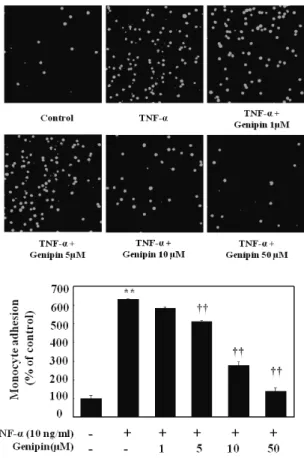
(5) Genipin Inhibits VCAM-1 in Human Endothelial Cells. Fig. 7. Effect on adhesion of monocytes to HUVECs stimulated with TNF-α. Cell were pretreated with different concentration of genipin (1, 5, 10 and 50 μM) and then stimulated with 10 ng/ml TNF-α for 6 h. Thereafter, cells were coincubated with fluorescent labeled monocytic cells for 30 min at 37oC. Monocyte adhesion was presented as images and a percentage of U937 cells bound to TNFα-untreated cells (control) Data represented mean±SD from three independent experiments. **p<0.01, compared with control, ††p< 0.01 compared with TNF-α, respectively.. hibition of TNF-α-induced VCAM-1 expression in HUVEC remain unclear. Induction of PPAR-γ, at least, can be a possible mechanism for differential inhibition. Or inhibition of phosphorylation of Akt and PKC could be involved in selective inhibition of VCAM-1 expression. These speculations come from our previous reports that PI3K/Akt and PKC signaling pathways are involved in VCAM-1 induction but not ICAM-1 in HUVEC [7,8]. Therefore, inhibition of phosphorylation of these signal molecules (Akt, PKC) can be another possible mechanism for this differential expression. As shown in the present study, phosphorylation of Akt and p-PKC by TNF-α was concentration- dependently inhibited by genipin. However, it remains further study as to the link between inhibition of these signal molecules and PPAR-γ induction. It seems likely that PKC and Akt signals are important regulators on differential regulation of adhesion molecules by genipin as triterpenoid glycosides do [5]. Lastly, some transcription factors such as GATA or IRF-1 [17,18] also can be target molecules for genipin. Since these transcription factors are not present in ICAM-1 promoters, so these can be target molecules by many medicinal compounds as we demonstrated that anthocyanins isolated. 161. from black soy bean coat selectively inhibited VCAM-1 but not ICAM-1 expression in TNF-α-activated HUVEC by inhibiting GATA-2, GTA-4, and IRF-1 [18]. It should be noted that TNF-α could activate NF-κB in endothelial cells via oxidative stress [19]. Genipin reduced TNF-α-induced ROS production in HUVECs by a fluorescent probe DCFH-DA assay. These results suggest that the inhibitory effect of genipin on adhesion molecule expressions may be due to ROS reduction possibly through inhibition of NF-kB activity, which needs further investigation. Finally, inhibitory effect on the adhesion of monocytic U937 cells to TNF-α-treated HUVEC was shown by genipin. This inhibitory effect on the adhesion of monocytes to vascular endothelium seems likely to contribute to the anti-atherosclerotic activity of genipin. Although expression of both VCAM-1 and ICAM-1 is upregulated in endothelial cells by TNF-α, it is indicated that VCAM-1 plays a dominant role in the initiation of atherosclerosis [20]. Thus, genipin may inhibit adhesion of monocytes without hampering on ICAM-1 expression in HUVECs by TNF-α. Even though we did not evaluate in vivo effect of genipin, Kim et al [9] nicely demonstrated that administration of genipin protected lipopolysaccharide-induced liver damage in mice, indicating that genipin showed anti-inflammatory action in vivo, too. We are under investigation this protective effect is due to PPAR-γ induction in vivo. In conclusion, we clearly demonstrated that genipin selectively inhibited VCAM-1 but not ICAM-1 expression in TNF-α-activated HUVEC via activating PPAR-γ. Although the precise mechanism of action awaits further investigation, we provide scientific evidence why extract of gardenia has been used in chronic inflammatory disorders [11]. Therefore, we propose that genipin can be used for the treatment of pathologic inflammatory disorders such as atherosclerosis.. ACKNOWLEDGEMENTS This work was supported by a grant from the Korea Food and Drug Administration for Studies on the Identification of the Efficacy of Biologically Active Components from Oriental Herbal Medicines.. REFERENCES 1. Ross R. Atherosclerosis--an inflammatory disease. N Engl J Med. 1999;340:115-126. 2. Fan J, Watanabe T. Inflammatory reactions in the pathogenesis of atherosclerosis. J Atheroscler Thromb. 2003;10:63-71. 3. Springer TA. Traffic signals for lymphocyte recirculation and leukocyte emigration: the multistep paradigm. Cell. 1994;76: 301-314. 4. Modur V, Zimmerman GA, Prescott SM, McIntyre TM. Endothelial cell inflammatory responses to tumor necrosis factor alpha. Ceramide-dependent and -independent mitogenactivated protein kinase cascades. J Biol Chem. 1996;271: 13094-13102. 5. Moon L, Ha YM, Jang HJ, Kim HS, Jun MS, Kim YM, Lee YS, Lee DH, Son KH, Kim HJ, Seo HG, Lee JH, Kim YS, Chang KC. Isoimperatorin, cimiside E and 23-O-acetylshengmanol-3xyloside from Cimicifugae rhizome inhibit TNF-α-induced VCAM-1 expression in human endothelial cells: involvement of PPAR-γ upregulation and PI3K, ERK1/2, and PKC signal pathways. J Ethnopharmacol. 2011;133:336-344. 6. Jackson SM, Parhami F, Xi XP, Berliner JA, Hsueh WA, Law.
(6) 162. 7.. 8.. 9. 10. 11.. 12. 13. 14.. JS Hwa, et al. RE, Demer LL. Peroxisome proliferator-activated receptor activators target human endothelial cells to inhibit leukocyte-endothelial cell interaction. Arterioscler Thromb Vasc Biol. 1999;19:2094- 2104. Nizamutdinova IT, Jeong JJ, Xu GH, Lee SH, Kang SS, Kim YS, Chang KC, Kim HJ. Hesperidin, hesperidin methyl chalone and phellopterin from Poncirus trifoliata (Rutaceae) differentially regulate the expression of adhesion molecules in tumor necrosis factor-alpha-stimulated human umbilical vein endothelial cells. Int Immunopharmacol. 2008;8:670-678. Tsoyi K, Kim WS, Kim YM, Kim HJ, Seo HG, Lee JH, Yun-Choi HS, Chang KC. Upregulation of PTEN by CKD712, a synthetic tetrahydroisoquinoline alkaloid, selectively inhibits lipopolysaccharide-induced VCAM-1 but not ICAM-1 expression in human endothelial cells. Atherosclerosis. 2009;207:412-419. Kim SJ, Kim JK, Lee DU, Kwak JH, Lee SM. Genipin protects lipopolysaccharide-induced apoptotic liver damage in D-galactosamine-sensitized mice. Eur J Pharmacol. 2010;635:188-193. Lee SJ, Oh PS, Lim KT. Hepatoprotective and hypolipidaemic effects of glycoprotein isolated from Gardenia jasminoides ellis in mice. Clin Exp Pharmacol Physiol. 2006;33:925-933. Koo HJ, Song YS, Kim HJ, Lee YH, Hong SM, Kim SJ, Kim BC, Jin C, Lim CJ, Park EH. Antiinflammatory effects of genipin, an active principle of gardenia. Eur J Pharmacol. 2004;495:201-208. Desideri G, Ferri C. Endothelial activation. Sliding door to atherosclerosis. Curr Pharm Des. 2005;11:2163-2175. Albelda SM, Smith CW, Ward PA. Adhesion molecules and inflammatory injury. FASEB J. 1994;8:504-512. Sakai S, Kawamata H, Kogure T, Mantani N, Terasawa K,. 15.. 16.. 17.. 18.. 19.. 20.. Umatake M, Ochiai H. Inhibitory effect of ferulic acid and isoferulic acid on the production of macrophage inflammatory protein-2 in response to respiratory syncytial virus infection in RAW264.7 cells. Mediators Inflamm. 1999;8:173-175. Pasceri V, Wu HD, Willerson JT, Yeh ET. Modulation of vascular inflammation in vitro and in vivo by peroxisome proliferator- activated receptor-gamma activators. Circulation. 2000;101:235-238. Blaschke F, Caglayan E, Hsueh WA. Peroxisome proliferatoractivated receptor gamma agonists: their role as vasoprotective agents in diabetes. Endocrinol Metab Clin North Am. 2006;35: 561-574. Umetani M, Mataki C, Minegishi N, Yamamoto M, Hamakubo T, Kodama T. Function of GATA transcription factors in induction of endothelial vascular cell adhesion molecule-1 by tumor necrosis factor-alpha. Arterioscler Thromb Vasc Biol. 2001;21:917-922. Nizamutdinova IT, Kim YM, Chung JI, Shin SC, Jeong YK, Seo HG, Lee JH, Chang KC, Kim HJ. Anthocyanins from black soybean seed coats preferentially inhibit TNF-alpha-mediated induction of VCAM-1 over ICAM-1 through the regulation of GATAs and IRF-1. J Agric Food Chem. 2009;57:7324-7330. Chen YH, Lin SJ, Chen YL, Liu PL, Chen JW. Antiinflammatory effects of different drugs/agents with antioxidant property on endothelial expression of adhesion molecules. Cardiovasc Hematol Disord Drug Targets. 2006;6:279-304. Cybulsky MI, Iiyama K, Li H, Zhu S, Chen M, Iiyama M, Davis V, Gutierrez-Ramos JC, Connelly PW, Milstone DS. A major role for VCAM-1, but not ICAM-1, in early atherosclerosis. J Clin Invest. 2001;107:1255-1262..
(7)
수치

관련 문서
