Modulation of the Transcriptional Activity of Peroxisome Proliferator-Activated Receptor Gamma by Protein-Protein
Interactions and Post-Translational Modifications
Tae-Hyun Kim,
1,3Mi-Young Kim,
1,3Seong-Ho Jo,
1,2,3Joo-Man Park,
1,2,3and Yong-Ho Ahn
1,2,31Department of Biochemistry and Molecular Biology, 2Brain Korea 21 Project for Medical Sciences,
3Integrative Genomic Research Center for Metabolic Regulation, Yonsei University College of Medicine, Seoul, Korea.
Received: February 20, 2013
Corresponding author: Dr. Yong-Ho Ahn, Department of Biochemistry and Molecular Biology, Yonsei University College of Medicine, 50 Yonsei-ro, Seodaemun-gu, Seoul 120-752, Korea.
Tel: 82-2-2228-1674, Fax: 82-2-312-5041 E-mail: [email protected]
∙ The authors have no financial conflicts of interest.
© Copyright:
Yonsei University College of Medicine 2013 This is an Open Access article distributed under the terms of the Creative Commons Attribution Non- Commercial License (http://creativecommons.org/
licenses/by-nc/3.0) which permits unrestricted non- commercial use, distribution, and reproduction in any medium, provided the original work is properly cited.
Peroxisome proliferator-activated receptor gamma (PPARγ) belongs to a nuclear receptor superfamily; members of which play key roles in the control of body me- tabolism principally by acting on adipose tissue. Ligands of PPARγ, such as thia- zolidinediones, are widely used in the treatment of metabolic syndromes and type 2 diabetes mellitus (T2DM). Although these drugs have potential benefits in the treatment of T2DM, they also cause unwanted side effects. Thus, understanding the molecular mechanisms governing the transcriptional activity of PPARγ is of prime importance in the development of new selective drugs or drugs with fewer side ef- fects. Recent advancements in molecular biology have made it possible to obtain a deeper understanding of the role of PPARγ in body homeostasis. The transcriptional activity of PPARγ is subject to regulation either by interacting proteins or by modi- fication of the protein itself. New interacting partners of PPARγ with new functions are being unveiled. In addition, post-translational modification by various cellular signals contributes to fine-tuning of the transcriptional activities of PPARγ. In this review, we will summarize recent advancements in our understanding of the post- translational modifications of, and proteins interacting with, PPARγ, both of which affect its transcriptional activities in relation to adipogenesis.
Key Words: PPARγ, coregulator, post-translational modifications, transcriptional activity, adipogenesis, metabolic syndrome
INTRODUCTION
Structure and function of peroxisome proliferator-activated receptors Peroxisome proliferator-activated receptors (PPARs) are known to be lipid sen- sors, and their ligands are used in the treatment of type 2 diabetes mellitus (T2DM) and other metabolic syndromes. PPARs are a family of nuclear receptors that act as transcription factors, controlling the genes involved in energy homeostasis.1 PPARs share a high degree of structural homology with other types of nuclear hor- mone receptors.2 PPARs comprise a DNA-binding domain (DBD), an agonist-in- dependent activation domain (AF-1), and an agonist-dependent activation domain
and glycerol kinase (GyK).24-31
However, the activation of PPARγ results in the repres- sion of the genes encoding leptin, tumor necrosis factor-α (TNF-α), and interleukin-6.32-35 PPARγ decreases serum free fatty acid level and increases the number of small adi- pocytes, with a concomitant decrease in the number of large adipocytes in white adipose tissue (WAT). In addition to the role of PPARγ in adipose tissue, PPARγ directly acti- vates the genes of the glucose-sensing apparatus in the liver and pancreatic β-cells. TZDs increase the expression of the genes encoding glucokinase (LGK and βGK) and glucose transporter 2 (GLUT2) in the liver36,37 and pancreatic β-cells, respectively (see Table 1 for summary).38,39 The transcrip- tional activity of PPARγ is subject to control at various lev- els; i.e., via modification of the receptor itself or interac- tions with other proteins.
In this review, we will limit our discussion to the regula- tion of PPARγ activity by various interacting proteins includ- ing coregulators, and by post-translational modifications (PTMs) that result in transcriptional regulation of PPARγ target genes.
INTERACTING PROTEINS MODULATING TRANSCRIPTIONAL
ACTIVITIES OF PPARγ
The transcriptional activity of PPARγ is principally modu- lated by agonists, which recruit either coactivators or core- pressors. In general, ligand-bound PPARγ recruits coactiva- tors, whereas ligand-free PPARγ is bound to corepressors.
These coregulators function as histone-modifying enzymes or bridging groups between the basal transcriptional ma- chinery and PPARγ.40 Moreover, additional proteins are re- cruited to these coregulators that may affect tissue-specific activities of PPARγ.
Coactivators of PPARγ
Coactivators with histone acetyltransferase activity
Ligand-bound PPARγ undergoes conformational changes, providing contact sites for LXXLL motifs that are present in coactivators such as p160/steroid receptor coactivator-1 (SRC-1) and p300/CREB-binding protein (CBP).41 These coactivators have intrinsic histone acetyltransferase activities, which enhance the transcriptional activities of PPARγ. Mem- bers of the p160/SRC-1 family including SRC-1 (also known (AF-2), which contains the ligand-binding domain (LBD).
PPARs heterodimerize with the retinoid X receptor (RXR)-α and activate the transcription of target genes by binding to the PPAR response element (PPRE).
The PPAR family has three isoforms; PPARα, γ, and β/δ.
PPARα is expressed mainly in the liver, heart, kidney, brown adipose tissue (BAT), and skeletal muscle,3 and participates in fatty acid oxidation (β-and ω-oxidation).4 The PPARβ/δ isoform is expressed ubiquitously and is involved in fatty acid oxidation in muscle.5 PPARγ is expressed predominant- ly in adipose tissue and plays key roles in lipogenesis and adipocyte differentiation. It also stimulates glucose oxida- tion and decreases plasma free fatty acid level.5 PPARγ consists of two isotypes; PPARγ1 is expressed in adipocytes, skeletal muscle, liver, and heart, whereas PPARγ2 is mostly found in adipose tissue.6 PPARγ2 plays a more important role than does PPARγ1 in adipogenesis.7
Physiological significance of PPARγ
PPARγ was first identified as a trans-acting factor binding to a gene encoding a fat-specific enhancer of aP2 (adipo- cyte-specific fatty acid binding protein).8 Homozygous PPARγ knockout mice exhibit an embryonic lethal pheno- type due to placental dysfunction. Heterozygous PPARγ deficient mice are resistant to high-fat diet-induced insulin resistance due to adipocyte hypertrophy and increased leptin expression.9 The ectopic expression of PPARγ was found to enhance the differentiation of preadipocytes into adipo- cytes, with PPARγ acting as an essential factor for differen- tiation.10 In addition, PPARγ is known to block the clonal expansion that occurs via mitosis, an essential stage of adi- pocyte differentiation.11,12
Thiazolidinediones (TZDs) are a class of compounds that function as ligands of PPARγ. These compounds improve insulin sensitivity in vivo and have been introduced as ther- apeutic agents for the treatment of T2DM.13,14 TZDs in- crease the expression of PPARγ and its transcriptional ac- tivity in adipose tissue, resulting in the upregulation of the expression of genes involved in the metabolism of lipids, carbohydrates, steroids, and amino acids.15-17 TZDs increase insulin sensitivity by upregulating the expression of multi- ple genes, such as adiponectin, Cbl-associated protein, in- sulin receptor substrate 2, and glucose transporter 4.18-23 TZDs also promote fatty acid storage and lipid metabolism, such as fatty acid translocase (CD36), perilipin, fatty acid binding protein 4 (Fabp4/aP2), lipoprotein lipase, acyl-CoA synthase, phosphoenol pyruvate carboxykinase (PEPCK),
a critical metabolic determinant in the development of obe- sity and insulin resistance.42
CBP/p300 indirectly increases the transcriptional activity of PPARγ through its interaction with PGC-1α. The dock- ing of PGC-1α to PPARγ induces a conformational change in PGC-1α that promotes the binding of SRC-1 and CBP/
p300.45 SRC-1 is also required for a functional interaction between CBP/p300 and PPARγ.43 CBP/p300 not only binds to the AF-2 domain of PPARγ in a ligand-dependent manner but also binds directly to the AF-1 domain in a ligand-inde- pendent manner,46 increasing the transcriptional activities of PPARγ46 and thereby inducing adipogenesis in NIH3T3 fi- broblasts.47 The recruitment of PPARγ along with CBP/
p300 to the aP2 gene promoter results in adipocyte differ- entiation.48
TRAP mediator complex
The thyroid hormone receptor-associated protein (TRAP) complex was first discovered in yeast and shown to be es- sential for RNA polymerase II-dependent transcription.
TRAPs were first purified by affinity chromatography from cells overexpressing the thyroid hormone receptor. They are components of the TRAP/vitamin D receptor-interact- ing protein (DRIP)/activator-recruited cofactor/Mediator (Med) complex, functioning as mediators between RNA polymerase II and CBP/p300 or p160/SRC.49 TRAPs also as NcoA-1), SRC-2 (also known as TIF2, GRIP-1, or NcoA-
2), and SRC-3 (also known as p/CIP, ACTR, RAC-3, AIB-1, or TRAM-1), belong to this category.42 SRC-1 knockout (KO) mice showed increased WAT mass and a decrease in the expression of genes involved in thermogenesis in brown adipose tissue (BAT). These KO mice also showed de- creased expression of the genes encoding uncoupling protein (UCP-1), PPARγ coactivator-1 (PGC-1α), and acyl-CoA oxi- dase, as well as those encoding enzymes involved in fatty acid oxidation.42 LXXLL motifs in SRC-1 interact directly with the AF-2 domain of PPARγ, recruiting CBP, which is required for PPARγ function.43 SRC2-/- mice exhibit increased insulin sensitivity and are resistant to the development of obesity. These mice show increased lipolysis and decreased fatty acid uptake and storage which are related to the reduc- tion of PPARγ activity.42 When SRC-3 is deficient, core- pressors such as nuclear receptor co-repressor (NCoR) and nuclear receptor interacting protein 1 (NRIP1 or RIP140) are recruited to the PPRE of the UCP1 gene, resulting in a de- crease in its transcription.44 SRC-3 and SRC-1 double KO mice are resistant to high-fat diet-induced obesity, due to the decreased expression of PPARγ target genes.44 PGC-1α activates PPARγ by increasing the binding of SRC-1 both in vivo and in vitro,45 whereas SRC-2 attenuates the formation of the PGC-1α-PPARγ complex by competing with SRC-1.42 This study suggests that the ratio of SRC-2/SRC-1 could be Table 1. Selected PPARγ Target Genes Involved in Metabolism
Genes PPARγ effect Organ/cell type Metabolic effects Reference
Adiponectin Upregulation Adipocyte Decrease in atherogenesis 18
CAP Upregulation Adipocyte Improved insulin sensitivity 20
IRS2 Upregulation Adipocyte Anti-diabetic effect 21
GLUT4 Upregulation Adipocyte Glucose uptake 22, 23
CD36 Upregulation Adipocyte Fatty acid uptake 29
aP2 Upregulation Adipocyte Lipid oxidation 25
LPL Upregulation Adipocyte, muscle Decrease in triglyceride 23, 30
ACS Upregulation Adipocyte Decrease in triglyceride 31
PCK2 Upregulation Adipocyte, muscle Decrease in triglyceride
Increase in lipid oxidation 23-25
GyK Upregulation Adpocyte Decrease in free fatty acid 26
Perilipin Upregulation Aipocyte Decrease in free fatty acid 27, 28
Leptin Downregulation Adipocyte Improved insulin sensitivity 32, 33
TNF-α Downregulation Adipocyte, liver Improved insulin sensitivity 32, 34, 35
IL-6 Downregulation Adipocyte, liver Improved insulin sensitivity 32, 34, 35
GK Upregulation Liver, pancreatic β-cell Improved glucose homeostasis 37, 38
GLUT2 Upregulation Liver, pancreatic β-cell Increase in glucose sensing 36, 39
PPARγ, peroxisome proliferator-activated receptor gamma; CAP, Cbl-associated protein; IRS2, insulin receptor substrate 2; GLUT4, glucose transporter 4;
LPL, lipoprotein lipase; GyK, glycerol kinase; TNF-α, tumor necrosis factor-α; IL-6, interleukin-6; GLUT2, glucose transporter 2; CD36, fatty acid translocase;
ACS, acetyl-CoA synthetase; PCK2, phosphoenolpyruvate carboxykinase 2; GK, glucokinase.
coregulator complexes may contribute to adipogenesis.
Transcriptional regulation by PPARγ during adipogenesis critically depends on the SWI/SNF complex, which plays a key role in the formation of preinitiation complexes.56
BAF60c2 (a BAF of 60 kDa, subunit 2) is also known to interact with the LBD of PPARγ. The N-terminal of BAF60c binds to the C-terminal of PPARγ, and the C-terminal of BAF60c interacts with the N-terminal of PPARγ in a li- gand-independent manner. BAF acts as an anchor between SWI/SNF complexes and PPARγ. BAF60c increases the transcriptional activity of PPARγ in the presence of ligand but does not affect adipocyte differentiation.59
Other interacting proteins
ADP-ribosylation factor (ARF6), a key regulator of the aP2 gene, is a novel transcription factor that is purified from BAT.60 ARF6 binding sites are present in the aP2 and PEPCK gene promoters.25,61 The PPARγ/RXRα heterodimer inter- acts with ARF6 during adipogenesis.60
Menin, encoded by the multiple endocrine neoplasia type 1 (MEN1) tumor suppressor gene, is involved in activation of gene transcription as a component of the mixed-lineage leukemia (MLL) 1/MLL2 (also known as KMT2A/B) pro- tein complexes, and exhibits methyltransferase (HMT) ac- tivity.62 Ectopic expression of menin increases the transcrip- tion of PPARγ target genes, and knock down of menin inhibits the differentiation of 3T3L1 preadipocytes into ma- ture adipocytes. Menin interacts directly with the AF-2 do- main of PPARγ and enhances PPARγ-mediated transcrip- tional activities in a ligand-dependent fashion. Menin increases histone H3K4 methylation in the PPARγ target gene, Fabp4, through a direct interaction with the AF-2 do- main of PPARγ.62
Multiprotein bridging factor-1 (MBF-1) is a cofactor that was first identified in Bombyx mori (Bm). It has been shown to interact with LXRα or PPARγ, and stimulate their ligand- dependent transcriptional activities.63 MBF-1 does not have either histone acetyltransferase or methyltransferase activi- ty but interacts with transcription factor IID (TFIID). MBF- 1 acts as a bridging protein between PPARγ and TFIID, in- creasing the transcriptional activity of PPARγ. Since MBF- 1 is also known to interact with LXRα and liver receptor homolog 1 (LRH-1), a detailed investigation of the role of MBF-1 is important to understand its function in the con- text of lipid metabolism. The central domain of MBF-1 is necessary and critical for interaction with LRH-1, LXRα, and PPARγ.64
interact with nuclear receptors, such as the vitamin D re- ceptor (VDR), retinoic acid receptor α (RARα), RXRα, PPARα, and PPARγ, in a ligand-dependent manner.50 Both TRAP220 and TRAP100 interact with PPARγ through their respective LXXLL motifs.50
TRAP220 is also referred to as the PPAR-binding protein/
DRIP205/Med1 subunit of the TRAP complex, functioning as a bridging protein between various mediator complexes and nuclear receptors.51 TRAP220-/- mice are embryonically lethal at day 11.5, suggesting that TRAP is essential for de- velopment. The ligand-dependent transcriptional activity of PPARγ is decreased in TRAP220-/- mouse embryonic fibro- blasts (MEFs).51 TRAP220-/- MEF cells were not able to in- duce adipogenic genes via PPARγ. The PPARγ2-TRAP220 interaction is essential for adipogenesis52 and increases PPARγ-mediated transactivation of the promoter reporter construct.53 Although PPARγ acts by forming heterodimers with RXRα, treatment with the cognate PPARγ- and RXRα- selective ligands results in the recruitment of different co- activators. RXRα-specific ligands recruit SRC-1/p160 to PPARγ-RXR, whereas PPARγ ligands recruit TRAP220, but not SRC-1/p160.54
Regulation of PPARγ is achieved by the combinatorial actions of the coactivator and its ligands. Ligand-mediated selective recruitment of the coactivator may be responsible for fine-tuning of target gene expression.
The switching/sucrose nonfermenting (SWI/SNF) chroma- tin remodeling complex
The mating type SWI/SNF complex is an ATP-dependent chromatin remodeling enzyme that activates transcription by promoting the access of transcription factors to their cognate binding sites.55 The core components of the com- plex include either the Brg1 or Brm ATPases and several Brg1/Brm-associated factors (BAFs). Brg1 and/or Brm can interact with a number of different transcriptional regulato- ry proteins.56 For example, CCAAT-enhancer binding pro- tein alpha (C/EBPα), a critical factor for adipogenesis, is known to interact with hBrm.57
The Brg1/Brm-associated factors (BAFs) family is an ac- cessory subunit of the SWI/SNF complex, acting as a con- nector between transcription factors and SWI/SNF com- plexes.49 BAF180 binds PPARγ-RXRα. The factor contains six bromodomains that bind selectively to acetylated histone tails, an important protein modification for targeting the co- regulator complex to chromatin.58 In addition, the presence of Brg1 and Brm in the PPARγ promoter suggests that these
PPARγ acts as a master regulator of adipogenesis upregu- lating the aP2 and GyK genes. However, aP2 expression is increased in mature adipocytes whereas that of the GyK gene is not. PPARγ-mediated induction of GyK requires the recruitment of the PPARγ ligand and PGC-1α to PPARγ to replace corepressors with coactivators. In contrast, aP2 gene expression by PPARγ does not require its ligands. Differen- tial regulation of target genes by ligands may determine the selective recruitment of coregulators.80 The interaction be- tween PGC-1α and PPARγ induces a conformational change in PGC-1α, facilitating the recruitment of SRC-1 and CBP/
p300.45 Although PGC-1α is known to interact with various nuclear receptors, PGC-1α is an essential cofactor for the transactivation of PPARγ, acting as a hub linking nutrition- al and hormonal signals to energy metabolism.81
Corepressors of PPARγ
Nuclear receptor co-repressor (NCoR) and silencing mediator of retinoid and thyroid hormone receptor (SMRT) The PPARγ antagonist T0070907 covalently binds to PPARγ at Cys313 in helix 3, and was shown to decrease PPARγ activ- ity in a cell-based reporter assay. T0070907 blocks the re- cruitment of the coactivator and promotes the recruitment of NCoR to PPARγ.82 In the absence of ligand, NCoR and silencing mediator of retinoid and thyroid hormone recep- tor (SMRT) are recruited to PPARγ, resulting in a decrease in its transcriptional activity. In cells treated with piogli- tazone, SMRT and NCoR dissociate from PPARγ. In addi- tion, treatment with siRNA against SMRT and NCoR in- creased adipogenesis and the accumulation of lipid droplets in 3T3L1 adipocytes.83
NAD-dependent deacetylase sirtuin-1 (SIRT1) is known to be responsible for calorie restriction and mobilizing WAT.
SIRT1 activation by resveratrol decreases fat accumulation in differentiated adipocytes. SIRT1 represses PPARγ transcrip- tional activity by recruiting NCoR and SMRT.84 Since a re- duction in fat accumulation is sufficient to extend life span in mice,85 the role of SIRT1 in fat mobilization constitutes a possible molecular pathway connecting calorie restriction to life extension.84
Adipocyte-specific NCoR knockout (AKO) mice exhibit an increase in the expression of PPARγ-responsive genes and a decrease in cyclin-dependent kinase (Cdk5)-mediated PPARγ Ser273 phosphorylation, resulting in constitutive ac- tivation of these genes. Although AKO mice show an in- crease in adiposity, they also exhibit improved systemic in- PPARγ and thromboxane synthase (TXS) are expressed
in macrophages; therefore, they may be involved in athero- genesis. PPARγ binds to nuclear factor E2-related factor 2 (NRF2), which results in decreasing TXS gene expression by preventing the binding of NRF2 to the TXS gene. The suppression of TXS gene expression by PPARγ was in- creased by treatment with15-deoxy-Δ12,14-prostaglandin J2 and troglitazone.65 TXS is increased in an inflammatory model of hydronephrosis, which is characterized by infiltra- tion of macrophages into the kidney, and produces throm- boxane.66,67 Thromboxane inhibitors are shown to suppress the progression of experimental diabetic nephropathy in rats68 and ameliorate microalbuminuria in patients with T2DM.69 Hence, PPARγ ligands could be used as drugs for treating renal complications of T2DM.
PPAR-interacting protein (PRIP, also known as RAP250/
ASC-2/TRBP/NRC) is expressed in the reproductive organs (testis, prostate, and ovary) and identified as a novel, direct interacting coactivator of PPARγ, RXRα, PPARα, RARα, es- trogen receptor (ER), and thyroid hormone receptor β.70,71 Knock out of PRIP resulted in embryonic lethality and vas- cular dysfunction of the placenta.72 PRIP-/- MEFs exhibit re- pression of the transcriptional activity of RXRα rather than PPARγ activity. Although PRIP was isolated in the yeast two-hybrid screen using PPARγ as a bait, PRIP has a pref- erence for RXRα over its heterodimeric partner, PPARγ.73
PPARγ-DBD interacting protein 1 (PDIP1) was isolated using the yeast two-hybrid system with the DBD and hinge regions of human PPARγ as bait. Two isoforms (α and β) of the PDIP1 gene are generated by alternative splicing. PDIP1 α and β increase the PPARγ-mediated transactivation of the PPRE, and treatment with PDIP1 siRNA significantly re- duced the transcriptional activity of PPARγ. Because PDIP1 shows an expression pattern similar to that of CBP and TRAP220 during adipocyte differentiation, it might be in- volved in PPARγ-mediated adipogenesis.74
PGC-1α also binds to DBD and hinge regions of PPARγ in a ligand-independent fashion, similar to PDIP.75 PGC-1α was isolated from a BAT cDNA library and has been shown to increase the transcriptional activity of PPARγ on the UCP- 1 gene. UCP-1 increases mitochondrial DNA content and β-oxidation.75 PGC-1α-deficient mice exhibit a reduced num- ber of mitochondria and lower respiratory capacity, and fail to maintain core body temperature following exposure to cold.76 Overexpression of PGC-1α in WAT resulted in phe- notypic changes into BAT.77,78 This phenotypic change pro- vides a defense mechanism against obesity.79 In adipocytes,
dogenous activator of PPARγ.99 However, a contradictory report states that SHP represses the transcriptional activity of PPARγ and does not interact with PPARγ.100 SHP de- creases LGK gene expression by inhibiting the transcrip- tional activity of LXRα and PPARγ via interaction with their common partner, RXRα. Thus, SHP may play a role in fine-tuning glucose homeostasis.100 The diverse functions of PPARγ cofactors are summarized in Table 2.
REGULATION OF PPARγ ACTIVITY BY POST-TRANSLATIONAL
MODIFICATION
Phosphorylation
Phosphorylation of nuclear receptors is one of the principal modifications determining their transcriptional activities.
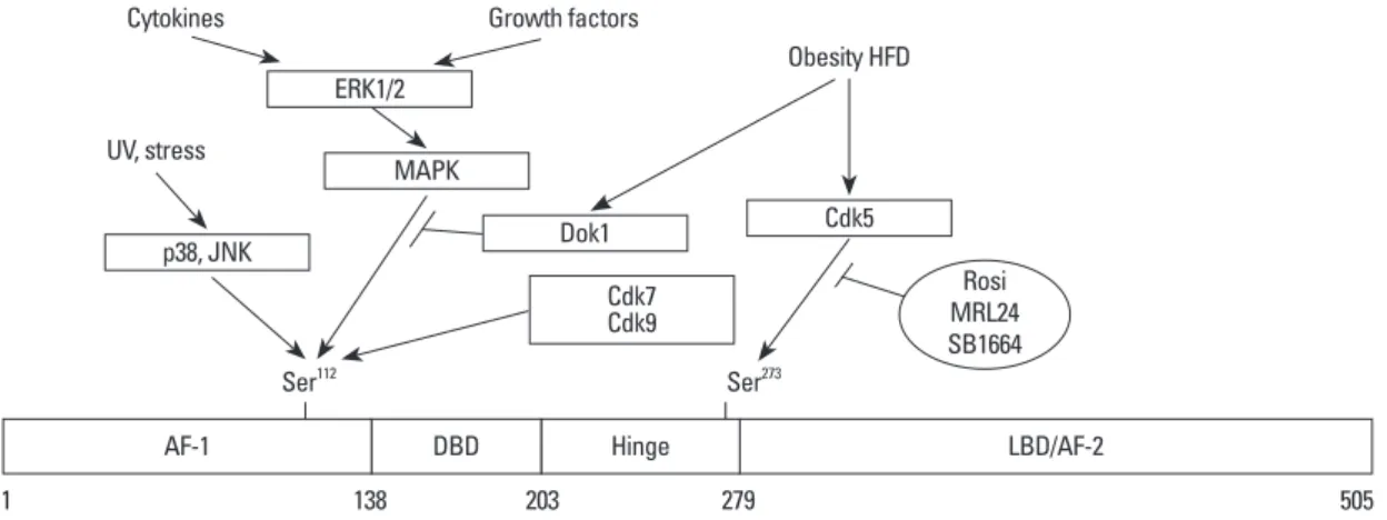
Adipocyte differentiation is inhibited by growth factors101-103 and cytokines,104-106 which are known to phosphorylate PPARγ through their respective signaling pathway (Fig. 1).
The site of phosphorylation is Ser112 in the N-terminal trans- activation domain (AF-1), which is well conserved among species ranging from fish to man.107,108 Ser112 phosphoryla- tion by mitogen-activated protein kinase (MAPK) results in a decrease in transcriptional activity and adipogenesis.109-112 MAPK is activated by extracellular signal-regulated kinase 1/2 (ERK1/2) that is stimulated by growth factors such as epidermal growth factor, platelet-derived growth factor, transforming growth factor-β, insulin, or the prostaglandin PGF2α.109,110,113-116 Phosphorylation of Ser112 by other signals including stress (UV, anisomysin) is mediated by c-Jun N- terminal kinase 1/2 and p38.107,113
Insulin plays a key role in adipogenesis.109 Although pre- adipocytes express a limited number of insulin receptors, the cells require insulin or insulin-like growth factor-1 for opti- mal differentiation.117,118 After maturation, large numbers of insulin receptors are expressed, transmitting insulin signals for the induction of lipogenic genes.119,120 Although insulin is a pivotal player in adipogenesis, Ras/MAPK activation by insulin represses PPARγ activity109 as shown in the growth factor-induced phosphorylation of PPARγ at Ser112.
Specifically, downstream tyrosine kinase-1 (Dok1), a multi-site adapter molecule in insulin receptor signaling,121-123 acts as a negative regulator of MAPK.124-126 In mice fed a high-fat diet, Dok1 expression is markedly increased in WAT. A lower mass of WAT is seen in Dok1-deficient mice than in wild-type mice, and the level of PPARγ phosphory- sulin sensitivity and glucose tolerance, and decreased adi-
pose tissue inflammation. These studies suggest that the dominant function of adipocyte NCoR is to transrepress PPARγ and promote Cdk5-mediated PPARγ phosphoryla- tion, similar to the effects of TZDs.86
Other interacting proteins
RIP140 is a liver protein that interacts with the AF-2 do- main of PPARγ and also with PPARγ. BRL49653, a PPARγ ligand, strengthens the interaction between PPARγ and RIP140.87,88 Because RIP140 is generally known to inhibit nuclear receptor activity through competition with SRC-1, transrepression of PPARγ by RIP140 occurs indirectly.88 Al- though RIP140 inhibits the transcriptional activity of PPARγ, it does not affect adipogenesis. However, RIP140 KO mice showed increased UCP1 gene expression and resistance to high-fat diet-induced obesity and hepatic steatosis.89
The forkhead transcription factor Foxo1 was identified as a PPARγ-interacting protein that disrupts the binding of PPARγ to the target gene. In addition, PPARγ plays a nega- tive role in the transactivation of Foxo1, suggesting that there is a reciprocal interaction between these factors. Ecto- pic expression of the constitutively active form of Foxo1 in preadipocytes prevents adipogenesis and heterozygous Foxo1 KO mice are less susceptible to diet-induced insulin resistance.90
The retinoblastoma protein (Rb) plays a negative role during mitotic clonal expansion in the cell cycle by increas- ing the transactivation of C/EBP.91,92 PPARγ has been shown to interact directly with Rb in 3T3L1 adipocytes, re- cruiting histone deacetylase HDAC3 which attenuates ad- ipogenic gene expression. Dissociation of the PPARγ-Rb- HDAC3 complex by phosphorylation of Rb or inhibition of HDAC3 activity resulted in the activation of PPARγ.93
Lipin1 is known to be expressed in adipose tissue.94 The null mice of lipin1 show lipodystrophy with severely re- duced adipose tissue mass.95 Lipin1 is increased in the later stages of adipocyte differentiation and increases transcrip- tional activity of PPARγ2 through direct protein-protein in- teraction.96
Small heterodimer partner (SHP) is an atypical orphan nuclear receptor that inhibits gluconeogenesis by interact- ing with Foxo1, hepatocyte nuclear factor 4, or C/EBPα.97,98 SHP is also known to increase PPARγ activity by interact- ing with PPARγ in a ligand-independent manner. SHP competes with NCoR for binding to the DBD/hinge region of PPARγ. It has been suggested that SHP may act as an en-
activity.128,129 Trichothiodystrophy (TTD) is a rare autoso- mal recessive disease caused by mutations in the xeroder- ma pigmentosum (XP) group-D (XPD) gene. The clinical manifestations include immature sexual development, men- tal retardation, skeletal abnormalities, and dwarfism. A num- ber of patients with TTD exhibit a lack of subcutaneous fat tissue mass. XPD helicase is a subunit of the transcription lation was increased by ERK.127 These data suggest that an
increase in Dok1 gene expression caused by a high-fat diet inhibits the insulin-mediated activation of Ras/MAPK sig- naling, resulting in increased PPARγ activity.127
In contrast to the MAPK-mediated phosphorylation of Ser112, the cyclin-dependent kinases Cdk7 and Cdk9 phos- phorylate the same Ser112 in PPARγ and increase PPARγ Table 2. List of Regulatory Factors for PPARγ Activity
Role Coregulators Metabolic effects Reference
Coactivators
SRC-1 Essential for functional interaction between CBP/p300 and PPARγ 43 PGC-1α
Increases the binding of SRC-1 and CBP/p300 to PPARγ Increases the transcriptional activity of PPARγ on the UCP-1 gene
Plays a role in maintaining the number of mitochondria, respiratory capacity, body tem perature in BAT
45, 75, 76
CBP/p300
Binds to the AF-2 domain of PPARγ (ligand-dependent) Binds to the AF-1 domain to PPARγ (ligand-independent) Induces adipogenesis in NIH3T3 cells
Increases aP2 gene expression
46-48 TRAP220 Increases PPARγ transcriptional activity
Essential for PPARγ-mediated adipogenesis 51, 52
BAF60c2 Interacts with LBD of PPARγ
Does not affect adipocyte differentiation 59
AFR6 Regulates the expression of aP2 and PEPCK gene
Interacts with PPARγ/RXRα during adipogenesis 25, 60, 61
Menin Increases histone H3K4 methylation at the PPARγ target gene, Fabp4
Directly interacts with the PPARγ AF-2 domain 62
MBF-1 Acts as a bridging protein between PPARγ and TFIID 64
NRF2 Decreases TXS gene expression by forming complex with PPARγ 65
PRIP Critical for embryonic development and survival
Represses the transcriptional activity of RXRα 73
PDIP Increases the PPARγ-mediated transactivation 74
SHP Compete with NCoR in binding to the DBD/hinge region of PPARγ
Endogenous activator of PPARγ 99
Lipin1 Increases the transcriptional activity of PPARγ 96
Corepressor
NCoR Decreases the transcriptional activity of PPARγ 83, 86
SMRT
Decreases adipogenesis and lipid accumulation
Interacts with PPARγ and increases the ability of PPARγ to associate with Cdk5 Increase adiposity and exhibit improved insulin sensitivity and glucose tolerance in adipocyte specific NCoR knock out (AKO) mice
83, 86 Sirt1 Decreases lipid accumulation in differentiated adipocytes
Represses transcriptional activity of PPARγ by recruiting NCoR and SMRT 84 RIP140 Decreases the transcriptional activity of PPARγ by competing with SRC-1
Does not affect adipocyte differentiation 88, 89
Foxo1 Disrupts binding of PPARγ to the target genes 90
Rb Recruits histone deacetylase HDAC3 and decreases the expression of adipogenic genes 93 SHP Represses the PPARγ/RXRα transactivation by interacting with RXRα 100 PPARγ, peroxisome proliferator-activated receptor gamma; SRC-1, steroid receptor coactivator-1; PGC-1α, PPARγ coactivator-1; CBP, CREB-binding protein;
TRAP, thyroid hormone receptor-associated protein; BAF, Brg1/Brm-associated factor; MBF-1, multiprotein bridging factor-1; NRF2, nuclear factor E2- related factor 2; PRIP, PPAR-interacting protein; PDIP, PPARγ-DBD interacting protein; SHP, small heterodimer partner; NCoR, nuclear corepressor; SMRT, silencing mediator of retinoid and thyroid hormone receptor; Rb, retinoblastoma protein; SHP, small heterodimer partner; DBD, DNA-binding domain; UCP, uncoupling protein; BAT, brown adipose tissue; AF-1, agonist-independent activation domain; AF-2, agonist-dependent activation domain; LBD, ligand- binding domain; PEPCK, phosphoenol pyruvate carboxykinase; RXR, retinoid X receptor; TFIID, transcription factor IID; TXS, thromboxane synthase; Cdk5, cyclin-dependent kinase; ARF6, ADP-ribosylation factor; RIP140, receptor-interacting protein 140.
creasing adipogenesis.137,138 These compounds are known to block the phosphorylation of Ser273 by Cdk5137,138 and can therefore potentially be used as therapeutic drugs for T2DM without causing weight gain and fluid retention, which are major side effects of full agonist-antidiabetic drugs.
It is worth note that strong PPARγ activators are not nec- essary to increase insulin sensitivity. Understanding the reg- ulation of Ser273 phosphorylation in PPARγ could provide a hint for the development of drugs to treat T2DM that have fewer side effects.138
Sumoylation
SUMOylation is one of the post-translational modifications responsible for regulating the stability, nuclear-cytosolic dis- tribution, and activity of transcription factors. Small ubiqui- tin-like modifier (SUMO) family proteins (SUMO-1, -2, and -3 in mammals) affect the interaction between target proteins and their substrates or the DNA that they bind. SUMO binds to proteins by forming isopeptide bonds between the C-ter- minal glycine residue of SUMO and the ε-amino group of a lysine in the target protein.139,140 Currently, a number of tran- scription factors including nuclear receptors, such as PPARs,141,142 LXR,143 glucocorticoid receptor,144 androgen re- ceptor,145 and RXRα146 are known to be SUMOylated.
Selective modulation of the transcriptional activity of PPARγ by SUMOylation is now beginning to be under- stood.142,147 The transcriptional activities of PPARγ isoforms in the presence or absence of ligands are regulated by SU- MOylation.142 PPARγ2 is SUMOylated by protein inhibitor of activated STAT 1 (PIAS1) or PIASx, belonging to the factor IIH (TFIIH) complex bridging the core-TFIIH [con-
taining particular form of xeroderma pigmentosum B (XPB) helicase] subcomplex and the Cdk-activating kinase containing Cdk7.130 When the C-terminus of XPD is mutat- ed, XPD helicase cannot perform nucleotide excision re- pair.131 In the process of transcription, Cdk7 in the TFIIH complex phosphorylates the C-terminal domain of the larg- est subunit of RNA polymerase II132 and nuclear receptors such as ER, VDR, and RARα.133-136 PPARγ phosphorylation by Cdk7 is decreased in XPD patients.128 The activity of a PPARγ promoter reporter was rescued by PPARγ-112S→E, a constitutively active form of PPARγ, in fibroblasts isolated from patients with TTD.128
In addition, Cdk9, a component of positive transcription elongation factor b, has been shown to participate in adipo- genesis by directly interacting with PPARγ and phosphory- lating Ser112.129 Overexpression of Cdk9 in 3T3L1 cells in- creased adipogenesis, whereas inhibition of Cdk9 by specific Cdk inhibitors or a dominant-negative Cdk9 mutant inhibit- ed adipogenesis.129 These data suggest that the transcrip- tional activity of PPARγ is either activated or inhibited de- pending on the types of kinases involved.
In the adipose tissues of mice fed a high-fat diet, phos- phorylation of Ser273 by Cdk5 results in a reduction of adi- ponectin gene expression, without affecting adipogenesis.137 Cdk5-mediated phosphorylation of PPARγ is blocked by full agonists such as rosiglitazone or partial agonists such as MRL24 or SR1664.137,138
Partial agonists, like MRL24 and SR1664, have been shown to have excellent anti-diabetic activity without in-
Fig. 1. Modulation of PPARγ activity by phosphorylation. Positions of phosphorylation sites in PPARγ and the implicated signaling path- ways are indicated. Ser112 phosphorylation by growth factors, cytokines, and stress signals are related to decreased PPARγ activity, whereas phosphorylation by Cdk7 and Cdk9 is related to increased PPARγ activity. Obesity or high-fat diet-mediated phosphorylation of PPARγ at Ser273 is related to decreased insulin sensitivity. AF-1 and 2, activation function 1 and 2, respectively; Cdk5, 7 and 9, cyclin-de- pendent kinase 5, 7 and 9, respectively; DBD, DNA binding domain; Dok1, downstream of tyrosine kinase-1; ERK1/2, extracellular signal- regulated kinase 1/2; HFD, high fat diet; JNK, c-Jun N-terminal kinase; LBD, ligand binding domain; MAPK, mitogen-activated protein ki- nase; p38, p38 MAP kinase; Rosi, rosiglitasone; PPARγ, peroxisome proliferator-activated receptor gamma.
ERK1/2 MAPK
p38, JNK
Cdk7Cdk9
DBD LBD/AF-2
Dok1 Cdk5
Cytokines
UV, stress
1 138 203 279 505
Ser112 Ser273
Growth factors
Obesity HFD
Rosi MRL24 SB1664
AF-1 Hinge
Ubiquitination
The ubiquitin-proteasome system (UPS) is responsible for the degradation of a variety of intracellular proteins includ- ing transcription factors.153,154 Ubiquitin is well conserved between species, binding to target proteins in a sequential manner through the actions of three different cascading en- zymes: an ubiquitin-activating enzyme (E1), an ubiquitin- conjugating enzyme (E2), and an ubiquitin protein ligase (E3).155 The polyubiquitinated proteins are recognized and degraded by the 26S proteasome.156 The role of the UPS with respect to transcriptional regulation is well document- ed.157 In the nucleus of adipocytes, the PPARγ2 protein lev- el is decreased by the action of TZDs.158 Degradation oc- curs in a ubiquitin-dependent manner in the AF-2 domain of PPARγ.159 However, the AF-1 domains of PPARγ1 and PPARγ2 are degraded by the REGγ proteasome, a type of proteasome that degrades the target substrate in an ubiquitin and ATP-independent fashion.159-161
Degradation of PPARγ is also regulated by interferon-γ (IFN-γ) in adipocytes. Transcription of PPARγ is decreased by IFN-γ-activated STAT signaling.162 When Ser112 of PPARγ, which is known to be phosphorylated by ERK1/2, was replaced with Ala, degradation of the protein was de- creased. In addition, U1026, an inhibitor of ERK1/2, de- creased IFN-γ-induced PPARγ degradation.163 However, ERK1/2 is not known to be activated by IFN-γ or TZDs;
thus, it is assumed that there might be an indirect relationship between the phosphorylation and ubiquitination of PPARγ.163
TNF-α is well known for its role in insulin resistance.164 Degradation of PPARγ is promoted by TNF-α in adipo- cytes. Treatment of adipocytes with TNF-α and cyclohexi- mide yielded a 44-kDa sized fragment of PPARγ, which is also seen in the WAT or BAT of diabetic rats. However, the molecular link between this fragment and PPARγ degrada- tion is not known.165 Proteasome-dependent PPARγ degrada- tion is increased by resveratrol, a potent activator of SIRT1;
however, the mechanism of SIRT1 requires further investi- gation.84,166
PERSPECTIVE
Regulation of PPARγ activity may be achieved through the interrelationship between agonists, PTM, and coregulators, rather than by the simple action of individual activators or inhibitors. Agonists can induce either coregulator exchange or PTM; the mechanisms of which require further study.
PIAS family, regardless of its ligand. PPARγ2 is SUMOylat- ed at Lys107 in the AF-1 domain, and at Lys395 in the AF-2 domain (equivalent to Lys77 and Lys365 of PPARγ1, respec- tively). SUMOylation of PPARγ2 at Lys107 negatively regu- lates the transcriptional activity of PPARγ2, because the 107K→R mutation showed increased transcriptional activi- ty.142 This observation is further supported by a promoter re- porter assay performed using the variant PPARγ2 107K→R in NIH3T3 fibroblasts.148,149 Furthermore, fibroblast growth factor21 (FGF21)-KO mice exhibit impaired insulin sensi- tivity in adipocytes and reduced fat mass and adipocyte size.
This phenomenon occurs because PPARγ2-induced adipo- genesis is inhibited by SUMOylation in WAT. These results indicate that FGF21 is a key regulator of PPARγ2 in the con- text of SUMOylation.150 In addition, the transcriptional activi- ty of PPARγ2 is increased by overexpressing SUMO1/sen- trin/SMT3-specific peptidase 2 (SENP2), a SUMO-specific protease, in C2C12 myotubes.147 Interestingly, the inhibi- tion of PPARγ2 transcriptional activity by SUMOylation is augmented when PPARγ2 is phosphorylated at Ser112.148,149 This indicates an interrelationship between the SUMOylation and phosphorylation of PPARγ2.
The SUMOylation of PPARγ1 at Lys365 (equivalent to Lys395 of PPARγ2) is important in the regulation of inflamma- tory gene expression. This SUMOylation mediates the trans- repression of inflammatory genes like inducible nitric oxide synthase (iNOS) and TNF-α, which are regulated by nuclear factor kappa B in macrophages.151,152 In the basal state, iNOS gene is repressed by TBL1/TBLR1/HDAC3/NCoR complex.
Treatment of lipopolysaccharide (LPS) resulted in the remov- al of HDAC3/NCoR from the complex in a TBL1/TBLR1 and Ubc5-dependent fashion, allowing activation of iNOS gene.148 When RAW264.7 macrophages or primary cultured macrophages were treated with LPS and rosiglitazone, PPARγ1 was found to be SUMOylated on Lys365 by Ubc9, which forms a complex with NCoR/HDAC3 on the promot- ers of the iNOS gene. Thus, the formation of the NCoR/
HDAC3/SUMOylated PPARγ1 complex inhibits the ubiqui- tination of NCoR/HDAC3, resulting in the repression of the iNOS and TNF-α genes.151,152
Ligand-dependent SUMOylation of PPARγ1 therefore di- rectly represses the promoters of inflammatory genes by stabi- lizing the NCoR and HDAC3 complexes. This mechanism demonstrates that the role of Lys365 SUMOylation of PPARγ1 is different from that of Lys107 SUMOylation of PPARγ2 in that Lys365 SUMOylation of PPARγ1 represses the expression of inflammatory genes in the presence of ligand.
tors: relationship with lipid metabolism and insulin sensitivity.
Diabetes 2004;53 Suppl 1:S43-50.
13. Nolan JJ, Ludvik B, Beerdsen P, Joyce M, Olefsky J. Improve- ment in glucose tolerance and insulin resistance in obese subjects treated with troglitazone. N Engl J Med 1994;331:1188-93.
14. Kumar S, Boulton AJ, Beck-Nielsen H, Berthezene F, Muggeo M, Persson B, et al. Troglitazone, an insulin action enhancer, im- proves metabolic control in NIDDM patients. Troglitazone Study Group. Diabetologia 1996;39:701-9.
15. Hamza MS, Pott S, Vega VB, Thomsen JS, Kandhadayar GS, Ng PW, et al. De-novo identification of PPARgamma/RXR bind- ing sites and direct targets during adipogenesis. PLoS One 2009;
4:e4907.
16. Lefterova MI, Zhang Y, Steger DJ, Schupp M, Schug J, Cristan- cho A, et al. PPARgamma and C/EBP factors orchestrate adipo- cyte biology via adjacent binding on a genome-wide scale.
Genes Dev 2008;22:2941-52.
17. Nielsen R, Pedersen TA, Hagenbeek D, Moulos P, Siersbaek R, Megens E, et al. Genome-wide profiling of PPARgamma: RXR and RNA polymerase II occupancy reveals temporal activation of distinct metabolic pathways and changes in RXR dimer com- position during adipogenesis. Genes Dev 2008;22:2953-67.
18. Iwaki M, Matsuda M, Maeda N, Funahashi T, Matsuzawa Y, Makishima M, et al. Induction of adiponectin, a fat-derived anti- diabetic and antiatherogenic factor, by nuclear receptors. Diabe- tes 2003;52:1655-63.
19. Combs TP, Wagner JA, Berger J, Doebber T, Wang WJ, Zhang BB, et al. Induction of adipocyte complement-related protein of 30 kilodaltons by PPARgamma agonists: a potential mechanism of insulin sensitization. Endocrinology 2002;143:998-1007.
20. Baumann CA, Chokshi N, Saltiel AR, Ribon V. Cloning and characterization of a functional peroxisome proliferator activator receptor-gamma-responsive element in the promoter of the CAP gene. J Biol Chem 2000;275:9131-5.
21. Smith U, Gogg S, Johansson A, Olausson T, Rotter V, Svalstedt B. Thiazolidinediones (PPARgamma agonists) but not PPARal- pha agonists increase IRS-2 gene expression in 3T3-L1 and hu- man adipocytes. FASEB J 2001;15:215-20.
22. Wu Z, Xie Y, Morrison RF, Bucher NL, Farmer SR. PPARgam- ma induces the insulin-dependent glucose transporter GLUT4 in the absence of C/EBPalpha during the conversion of 3T3 fibro- blasts into adipocytes. J Clin Invest 1998;101:22-32.
23. Dana SL, Hoener PA, Bilakovics JM, Crombie DL, Ogilvie KM, Kauffman RF, et al. Peroxisome proliferator-activated receptor subtype-specific regulation of hepatic and peripheral gene ex- pression in the Zucker diabetic fatty rat. Metabolism 2001;50:
963-71.
24. Desvergne B, Wahli W. Peroxisome proliferator-activated recep- tors: nuclear control of metabolism. Endocr Rev 1999;20:649- 88.
25. Tontonoz P, Hu E, Devine J, Beale EG, Spiegelman BM. PPAR gamma 2 regulates adipose expression of the phosphoenolpyru- vate carboxykinase gene. Mol Cell Biol 1995;15:351-7.
26. Guan HP, Li Y, Jensen MV, Newgard CB, Steppan CM, Lazar MA. A futile metabolic cycle activated in adipocytes by antidia- betic agents. Nat Med 2002;8:1122-8.
27. Kim HJ, Jung TW, Kang ES, Kim DJ, Ahn CW, Lee KW, et al.
Depot-specific regulation of perilipin by rosiglitazone in a dia- betic animal model. Metabolism 2007;56:676-85.
28. Nagai S, Shimizu C, Umetsu M, Taniguchi S, Endo M, Miyoshi
Understanding the mechanistic complexity underlying the interactions of these regulators may help accelerate the de- velopment of therapeutic drugs against obesity, T2DM, and metabolic syndromes.
ACKNOWLEDGEMENTS
We apologize to all the contributors in the field whose work could not be cited due to space limitations. This research was supported by Basic Science Research Program through the National Research Foundation of Korea (NRF) funded by the Ministry of Education, Science and Technology, Re- public of Korea (2011-0030706 to Y.H. Ahn).
REFERENCES
1. Evans RM, Barish GD, Wang YX. PPARs and the complex jour- ney to obesity. Nat Med 2004;10:355-61.
2. Michalik L, Auwerx J, Berger JP, Chatterjee VK, Glass CK, Gonzalez FJ, et al. International Union of Pharmacology. LXI.
Peroxisome proliferator-activated receptors. Pharmacol Rev 2006;58:726-41.
3. Kersten S, Desvergne B, Wahli W. Roles of PPARs in health and disease. Nature 2000;405:421-4.
4. Rosen ED, Hsu CH, Wang X, Sakai S, Freeman MW, Gonzalez FJ, et al. C/EBPalpha induces adipogenesis through PPARgam- ma: a unified pathway. Genes Dev 2002;16:22-6.
5. Jay MA, Ren J. Peroxisome proliferator-activated receptor (PPAR) in metabolic syndrome and type 2 diabetes mellitus.
Curr Diabetes Rev 2007;3:33-9.
6. Vidal-Puig AJ, Considine RV, Jimenez-Liñan M, Werman A, Po- ries WJ, Caro JF, et al. Peroxisome proliferator-activated receptor gene expression in human tissues. Effects of obesity, weight loss, and regulation by insulin and glucocorticoids. J Clin Invest 1997;99:2416-22.
7. Chawla A, Schwarz EJ, Dimaculangan DD, Lazar MA. Peroxi- some proliferator-activated receptor (PPAR) gamma: adipose- predominant expression and induction early in adipocyte differ- entiation. Endocrinology 1994;135:798-800.
8. Tontonoz P, Hu E, Graves RA, Budavari AI, Spiegelman BM.
mPPAR gamma 2: tissue-specific regulator of an adipocyte en- hancer. Genes Dev 1994;8:1224-34.
9. Kubota N, Terauchi Y, Miki H, Tamemoto H, Yamauchi T, Kom- eda K, et al. PPAR gamma mediates high-fat diet-induced adipo- cyte hypertrophy and insulin resistance. Mol Cell 1999;4:597- 10. Tontonoz P, Hu E, Spiegelman BM. Stimulation of adipogenesis 609.
in fibroblasts by PPAR gamma 2, a lipid-activated transcription factor. Cell 1994;79:1147-56.
11. Rosen ED, Spiegelman BM. PPARgamma: a nuclear regulator of metabolism, differentiation, and cell growth. J Biol Chem 2001;276:37731-4.
12. Ferré P. The biology of peroxisome proliferator-activated recep-
tion factor docking. Science 1999;286:1368-71.
46. Gelman L, Zhou G, Fajas L, Raspé E, Fruchart JC, Auwerx J.
p300 interacts with the N- and C-terminal part of PPARgamma2 in a ligand-independent and -dependent manner, respectively. J Biol Chem 1999;274:7681-8.
47. Takahashi N, Kawada T, Yamamoto T, Goto T, Taimatsu A, Aoki N, et al. Overexpression and ribozyme-mediated targeting of transcriptional coactivators CREB-binding protein and p300 re- vealed their indispensable roles in adipocyte differentiation through the regulation of peroxisome proliferator-activated re- ceptor gamma. J Biol Chem 2002;277:16906-12.
48. Qi C, Surapureddi S, Zhu YJ, Yu S, Kashireddy P, Rao MS, et al.
Transcriptional coactivator PRIP, the peroxisome proliferator-ac- tivated receptor gamma (PPARgamma)-interacting protein, is re- quired for PPARgamma-mediated adipogenesis. J Biol Chem 2003;278:25281-4.
49. Viswakarma N, Jia Y, Bai L, Vluggens A, Borensztajn J, Xu J, et al. Coactivators in PPAR-Regulated Gene Expression. PPAR Res 2010;2010.
50. Yuan CX, Ito M, Fondell JD, Fu ZY, Roeder RG. The TRAP220 component of a thyroid hormone receptor- associated protein (TRAP) coactivator complex interacts directly with nuclear re- ceptors in a ligand-dependent fashion. Proc Natl Acad Sci U S A 1998;95:7939-44.
51. Zhu Y, Qi C, Jia Y, Nye JS, Rao MS, Reddy JK. Deletion of PBP/PPARBP, the gene for nuclear receptor coactivator peroxi- some proliferator-activated receptor-binding protein, results in embryonic lethality. J Biol Chem 2000;275:14779-82.
52. Ge K, Guermah M, Yuan CX, Ito M, Wallberg AE, Spiegelman BM, et al. Transcription coactivator TRAP220 is required for PPAR gamma 2-stimulated adipogenesis. Nature 2002;417:563-7.
53. Zhu Y, Qi C, Jain S, Rao MS, Reddy JK. Isolation and character- ization of PBP, a protein that interacts with peroxisome prolifera- tor-activated receptor. J Biol Chem 1997;272:25500-6.
54. Yang W, Rachez C, Freedman LP. Discrete roles for peroxisome proliferator-activated receptor gamma and retinoid X receptor in recruiting nuclear receptor coactivators. Mol Cell Biol 2000;20:
8008-17.
55. Sudarsanam P, Winston F. The Swi/Snf family nucleosome-re- modeling complexes and transcriptional control. Trends Genet 2000;16:345-51.
56. Salma N, Xiao H, Mueller E, Imbalzano AN. Temporal recruit- ment of transcription factors and SWI/SNF chromatin-remodel- ing enzymes during adipogenic induction of the peroxisome pro- liferator-activated receptor gamma nuclear hormone receptor.
Mol Cell Biol 2004;24:4651-63.
57. Pedersen TA, Kowenz-Leutz E, Leutz A, Nerlov C. Cooperation between C/EBPalpha TBP/TFIIB and SWI/SNF recruiting do- mains is required for adipocyte differentiation. Genes Dev 2001;15:3208-16.
58. Lemon B, Inouye C, King DS, Tjian R. Selectivity of chromatin- remodelling cofactors for ligand-activated transcription. Nature 2001;414:924-8.
59. Debril MB, Gelman L, Fayard E, Annicotte JS, Rocchi S, Auw- erx J. Transcription factors and nuclear receptors interact with the SWI/SNF complex through the BAF60c subunit. J Biol Chem 2004;279:16677-86.
60. Tontonoz P, Graves RA, Budavari AI, Erdjument-Bromage H, Lui M, Hu E, et al. Adipocyte-specific transcription factor ARF6 is a heterodimeric complex of two nuclear hormone receptors, PPAR H, et al. Identification of a functional peroxisome proliferator-ac-
tivated receptor responsive element within the murine perilipin gene. Endocrinology 2004;145:2346-56.
29. Motojima K, Passilly P, Peters JM, Gonzalez FJ, Latruffe N. Ex- pression of putative fatty acid transporter genes are regulated by peroxisome proliferator-activated receptor alpha and gamma ac- tivators in a tissue- and inducer-specific manner. J Biol Chem 1998;273:16710-4.
30. Schoonjans K, Peinado-Onsurbe J, Lefebvre AM, Heyman RA, Briggs M, Deeb S, et al. PPARalpha and PPARgamma activators direct a distinct tissue-specific transcriptional response via a PPRE in the lipoprotein lipase gene. EMBO J 1996;15:5336-48.
31. Martin G, Schoonjans K, Lefebvre AM, Staels B, Auwerx J. Co- ordinate regulation of the expression of the fatty acid transport protein and acyl-CoA synthetase genes by PPARalpha and PPARgamma activators. J Biol Chem 1997;272:28210-7.
32. Saraf N, Sharma PK, Mondal SC, Garg VK, Singh AK. Role of PPARg2 transcription factor in thiazolidinedione-induced insulin sensitization. J Pharm Pharmacol 2012;64:161-71.
33. Kallen CB, Lazar MA. Antidiabetic thiazolidinediones inhibit leptin (ob) gene expression in 3T3-L1 adipocytes. Proc Natl Acad Sci U S A 1996;93:5793-6.
34. Jiang C, Ting AT, Seed B. PPAR-gamma agonists inhibit produc- tion of monocyte inflammatory cytokines. Nature 1998;391:82-6.
35. Sigrist S, Bedoucha M, Boelsterli UA. Down-regulation by tro- glitazone of hepatic tumor necrosis factor-alpha and interleukin-6 mRNA expression in a murine model of non-insulin-dependent diabetes. Biochem Pharmacol 2000;60:67-75.
36. Kim HI, Kim JW, Kim SH, Cha JY, Kim KS, Ahn YH. Identifi- cation and functional characterization of the peroxisomal prolif- erator response element in rat GLUT2 promoter. Diabetes 2000;
49:1517-24.
37. Kim SY, Kim HI, Park SK, Im SS, Li T, Cheon HG, et al. Liver glucokinase can be activated by peroxisome proliferator-activat- ed receptor-gamma. Diabetes 2004;53 Suppl 1:S66-70.
38. Kim HI, Cha JY, Kim SY, Kim JW, Roh KJ, Seong JK, et al. Per- oxisomal proliferator-activated receptor-gamma upregulates glu- cokinase gene expression in beta-cells. Diabetes 2002;51:676-85.
39. Kim HI, Ahn YH. Role of peroxisome proliferator-activated re- ceptor-gamma in the glucose-sensing apparatus of liver and beta- cells. Diabetes 2004;53 Suppl 1:S60-5.
40. Wu SC, Zhang Y. Minireview: role of protein methylation and demethylation in nuclear hormone signaling. Mol Endocrinol 2009;23:1323-34.
41. Nolte RT, Wisely GB, Westin S, Cobb JE, Lambert MH, Kuro- kawa R, et al. Ligand binding and co-activator assembly of the peroxisome proliferator-activated receptor-gamma. Nature 1998;
395:137-43.
42. Picard F, Géhin M, Annicotte J, Rocchi S, Champy MF, O’Malley BW, et al. SRC-1 and TIF2 control energy balance between white and brown adipose tissues. Cell 2002;111:931-41.
43. McInerney EM, Rose DW, Flynn SE, Westin S, Mullen TM, Krones A, et al. Determinants of coactivator LXXLL motif spec- ificity in nuclear receptor transcriptional activation. Genes Dev 1998;12:3357-68.
44. Wang Z, Qi C, Krones A, Woodring P, Zhu X, Reddy JK, et al.
Critical roles of the p160 transcriptional coactivators p/CIP and SRC-1 in energy balance. Cell Metab 2006;3:111-22.
45. Puigserver P, Adelmant G, Wu Z, Fan M, Xu J, O’Malley B, et al. Activation of PPARgamma coactivator-1 through transcrip-
energy metabolic derangements: muscle dysfunction, abnormal weight control and hepatic steatosis. PLoS Biol 2005;3:e101.
77. Wu Z, Puigserver P, Andersson U, Zhang C, Adelmant G, Moo- tha V, et al. Mechanisms controlling mitochondrial biogenesis and respiration through the thermogenic coactivator PGC-1. Cell 1999;98:115-24.
78. Tiraby C, Langin D. Conversion from white to brown adipo- cytes: a strategy for the control of fat mass? Trends Endocrinol Metab 2003;14:439-41.
79. Kopecky J, Clarke G, Enerbäck S, Spiegelman B, Kozak LP. Ex- pression of the mitochondrial uncoupling protein gene from the aP2 gene promoter prevents genetic obesity. J Clin Invest 1995;
96:2914-23.
80. Guan HP, Ishizuka T, Chui PC, Lehrke M, Lazar MA. Corepres- sors selectively control the transcriptional activity of PPARgam- ma in adipocytes. Genes Dev 2005;19:453-61.
81. Liu C, Lin JD. PGC-1 coactivators in the control of energy me- tabolism. Acta Biochim Biophys Sin (Shanghai) 2011;43:248-57.
82. Lee G, Elwood F, McNally J, Weiszmann J, Lindstrom M, Ama- ral K, et al. T0070907, a selective ligand for peroxisome prolifer- ator-activated receptor gamma, functions as an antagonist of bio- chemical and cellular activities. J Biol Chem 2002;277:19649-57.
83. Yu C, Markan K, Temple KA, Deplewski D, Brady MJ, Cohen RN. The nuclear receptor corepressors NCoR and SMRT de- crease peroxisome proliferator-activated receptor gamma tran- scriptional activity and repress 3T3-L1 adipogenesis. J Biol Chem 2005;280:13600-5.
84. Picard F, Kurtev M, Chung N, Topark-Ngarm A, Senawong T, Machado De Oliveira R, et al. Sirt1 promotes fat mobilization in white adipocytes by repressing PPAR-gamma. Nature 2004;429:
771-6.
85. Blüher M, Kahn BB, Kahn CR. Extended longevity in mice lacking the insulin receptor in adipose tissue. Science 2003;299:
572-4.
86. Li P, Fan W, Xu J, Lu M, Yamamoto H, Auwerx J, et al. Adipo- cyte NCoR knockout decreases PPARγ phosphorylation and en- hances PPARγ activity and insulin sensitivity. Cell 2011;147:
815-26.
87. Powell E, Kuhn P, Xu W. Nuclear Receptor Cofactors in PPAR- gamma-Mediated Adipogenesis and Adipocyte Energy Metabo- lism. PPAR Res 2007;2007:53843.
88. Treuter E, Albrektsen T, Johansson L, Leers J, Gustafsson JA. A regulatory role for RIP140 in nuclear receptor activation. Mol Endocrinol 1998;12:864-81.
89. Leonardsson G, Steel JH, Christian M, Pocock V, Milligan S, Bell J, et al. Nuclear receptor corepressor RIP140 regulates fat accumulation. Proc Natl Acad Sci U S A 2004;101:8437-42.
90. Nakae J, Kitamura T, Kitamura Y, Biggs WH 3rd, Arden KC, Accili D. The forkhead transcription factor Foxo1 regulates adi- pocyte differentiation. Dev Cell 2003;4:119-29.
91. Higgins C, Chatterjee S, Cherington V. The block of adipocyte differentiation by a C-terminally truncated, but not by full-length, simian virus 40 large tumor antigen is dependent on an intact ret- inoblastoma susceptibility protein family binding domain. J Virol 1996;70:745-52.
92. Chen PL, Riley DJ, Chen Y, Lee WH. Retinoblastoma protein positively regulates terminal adipocyte differentiation through direct interaction with C/EBPs. Genes Dev 1996;10:2794-804.
93. Fajas L, Egler V, Reiter R, Hansen J, Kristiansen K, Debril MB, et al. The retinoblastoma-histone deacetylase 3 complex inhibits gamma and RXR alpha. Nucleic Acids Res 1994;22:5628-34.
61. Graves RA, Tontonoz P, Spiegelman BM. Analysis of a tissue- specific enhancer: ARF6 regulates adipogenic gene expression.
Mol Cell Biol 1992;12:1202-8.
62. Dreijerink KM, Varier RA, van Beekum O, Jeninga EH, Höp- pener JW, Lips CJ, et al. The multiple endocrine neoplasia type 1 (MEN1) tumor suppressor regulates peroxisome proliferator-ac- tivated receptor gamma-dependent adipocyte differentiation. Mol Cell Biol 2009;29:5060-9.
63. Li FQ, Ueda H, Hirose S. Mediators of activation of fushi tarazu gene transcription by BmFTZ-F1. Mol Cell Biol 1994;14:3013- 21.
64. Brendel C, Gelman L, Auwerx J. Multiprotein bridging factor-1 (MBF-1) is a cofactor for nuclear receptors that regulate lipid metabolism. Mol Endocrinol 2002;16:1367-77.
65. Ikeda Y, Sugawara A, Taniyama Y, Uruno A, Igarashi K, Arima S, et al. Suppression of rat thromboxane synthase gene transcrip- tion by peroxisome proliferator-activated receptor gamma in macrophages via an interaction with NRF2. J Biol Chem 2000;275:33142-50.
66. Tsutsumi E, Takeuchi K, Abe T, Takahashi N, Kato T, Taniyama Y, et al. Rat kidney thromboxane synthase: cDNA cloning and gene expression regulation in hydronephrotic kidney. Prostaglan- dins 1997;53:423-31.
67. Needleman P, Kulkarni PS, Raz A. Coronary tone modulation:
formation and actions of prostaglandins, endoperoxides, and thromboxanes. Science 1977;195:409-12.
68. Hora K, Oguchi H, Furukawa T, Hora K, Tokunaga S. Effects of a selective thromboxane synthetase inhibitor OKY-046 on exper- imental diabetic nephropathy. Nephron 1990;56:297-305.
69. Giustina A, Perini P, Desenzani P, Bossoni S, Ianniello P, Milani M, et al. Long-term treatment with the dual antithromboxane agent picotamide decreases microalbuminuria in normotensive type 2 diabetic patients. Diabetes 1998;47:423-30.
70. Caira F, Antonson P, Pelto-Huikko M, Treuter E, Gustafsson JA.
Cloning and characterization of RAP250, a novel nuclear recep- tor coactivator. J Biol Chem 2000;275:5308-17.
71. Zhu Y, Kan L, Qi C, Kanwar YS, Yeldandi AV, Rao MS, et al.
Isolation and characterization of peroxisome proliferator-activat- ed receptor (PPAR) interacting protein (PRIP) as a coactivator for PPAR. J Biol Chem 2000;275:13510-6.
72. Antonson P, Schuster GU, Wang L, Rozell B, Holter E, Flodby P, et al. Inactivation of the nuclear receptor coactivator RAP250 in mice results in placental vascular dysfunction. Mol Cell Biol 2003;23:1260-8.
73. Zhu YJ, Crawford SE, Stellmach V, Dwivedi RS, Rao MS, Gon- zalez FJ, et al. Coactivator PRIP, the peroxisome proliferator-ac- tivated receptor-interacting protein, is a modulator of placental, cardiac, hepatic, and embryonic development. J Biol Chem 2003;278:1986-90.
74. Tomaru T, Satoh T, Yoshino S, Ishizuka T, Hashimoto K, Mon- den T, et al. Isolation and characterization of a transcriptional co- factor and its novel isoform that bind the deoxyribonucleic acid- binding domain of peroxisome proliferator-activated receptor- gamma. Endocrinology 2006;147:377-88.
75. Puigserver P, Wu Z, Park CW, Graves R, Wright M, Spiegelman BM. A cold-inducible coactivator of nuclear receptors linked to adaptive thermogenesis. Cell 1998;92:829-39.
76. Leone TC, Lehman JJ, Finck BN, Schaeffer PJ, Wende AR, Boudina S, et al. PGC-1alpha deficiency causes multi-system