Comparison between Measured and Calculated Length of Side Branch Ostium in Coronary Bifurcation
Lesions with Intravascular Ultrasound
Hyeon Min Ryu,
1* Byeong-Keuk Kim,
2* Jung-Sun Kim,
2Young-Guk Ko,
2Donghoon Choi,
2Yangsoo Jang,
2,3and Myeong-Ki Hong
2,31Division of Cardiology, Gumi CHA University Medical Center, Gumi;
2Division of Cardiology, Severance Cardiovascular Hospital, Yonsei University College of Medicine, Seoul;
3Division of Cardiology, Severance Biomedical Science Institute, Yonsei University College of Medicine, Seoul, Korea.
Received: August 4, 2011 Revised: September 14, 2011 Accepted: September 30, 2011
Corresponding author: Dr. Myeong-Ki Hong, Division of Cardiology, Severance Cardiovascular Hospital, Severance Biomedical Science Institute, Yonsei University College of Medicine, 50 Yonsei-ro, Seodaemun-gu, Seoul 120-752, Korea.
Tel: 82-2-2228-8458, Fax: 82-2-393-2041 E-mail: [email protected]
*Hyeon Min Ryu and Byeong-Keuk Kim contributed equally to this work.
∙ The authors have no financial conflicts of interest.
© Copyright:
Yonsei University College of Medicine 2012 This is an Open Access article distributed under the terms of the Creative Commons Attribution Non- Commercial License (http://creativecommons.org/
licenses/by-nc/3.0) which permits unrestricted non- commercial use, distribution, and reproduction in any medium, provided the original work is properly cited.
Purpose: Accurate evaluation of side branch (SB) ostium could be critical to the treatment of bifurcation lesions. We compared measured and calculated values of side branch ostial length (SBOL) in coronary bifurcation lesions with intravascular ultrasound (IVUS). Materials and Methods: Pre-intervention and post-intervention IVUS was performed in 113 patients who underwent stent implantation of bifurca- tion lesions. For the IVUS longitudinal reconstruction of the bifurcation lesions, SBOL, SB diameter, and the angle between the distal portion of the main vessel (MV) and SB were directly measured. In addition, SBOL was calculated as: SB di- ameter/sin (angle between distal MV and SB). The relationship between measured and calculated SBOL was then evaluated. Results: The angled between the distal MV and SB were 57.3±12.4° at pre-intervention and 59.4±12.6° at post-intervention.
The mean measured and calculated SBOL values were 2.91±0.86 mm and 3.06±0.77 mm at pre-intervention and 2.79±0.82 mm and 2.92±0.69 mm at post-in- tervention, respectively. Differences between measured and calculated SBOL were 0.15±0.44 mm at pre-intervention and 0.13±0.41 mm at post-intervention. We found that calculated SBOL was correlated with measured SBOL (pre-intervention r=0.863, p<0.001; post-intervention r=0.868, p<0.001). Conclusion: There was a good correlation between measured and calculated SBOLs of the bifurcation lesions in IVUS longitudinal reconstruction. SBOL in the bifurcation lesions can therefore be estimated using the SB diameter and the angle between distal MV and SB.
Key Words: Coronary artery disease, ultrasonics, bifurcation
INTRODUCTION
Percutaneous coronary intervention (PCI) for coronary bifurcation lesions is tech- nically challenging and is associated with lower procedural success rates and worse clinical outcomes than PCI for non-bifurcation lesions.1 The use of coronary stents to treat coronary bifurcation lesions has led to a high incidence of side branch (SB) occlusion during PCI.2-4 Avoiding SB occlusion during PCI of bifur-
mg nitroglycerin using a motorized transducer pullback sys- tem (0.5 mm/s) and a commercial scanner (Boston Scientific Corp./Scimed, Natrick, MA, USA), comprising a rotating 30- or 40-MHz transducer within a 2.9- or 3.2-Fr imaging sheath.
The ultrasound catheter was advanced approximately 10 mm beyond the target lesion of the MV, and an imaging run was performed from beyond the target lesion to the aorto-ostial junction. Ultrasound images were recorded on a 1/2-in high- resolution s-VHS tape or compact disc for off-line analysis.
Quantitative analyses were performed with a computerized planimetry (Tape Measure, Indec Systems, Inc.; Mountain View, CA, USA) according to the criteria of the American College of Cardiology Clinical Expert Consensus Document on IVUS.12 For the longitudinal reconstruction of bifurcation lesions before and after stent implantation, we directly mea- sured the SBOL, proximal and distal MV diameters, SB di- ameter, and the angle between the distal portion of the MV and SB (Fig. 1). Diameters of the proximal and distal MV, SB diameter, and SBOL were determined by measuring maximum lumen diameters in the most normal-looking cross-sections within 10 mm proximal and distal of the le- sions. The reconstructed images in the section with the lon- gest SBOL and largest angle between the distal portion of the MV and SB were selected. The calculated value of SBOL cation lesions appears to be crucial for favorable in-hospital
outcomes. In addition, the reported restenosis rate remains as high as 15% to 25% in SB vessels, even in the era of drug-eluting stents.5 The predominant site of restenosis af- ter implantation of a drug-eluting stent is the SB ostium.6 Therefore, accurate evaluation of the SB ostium may be important for successful PCI of bifurcation lesions. Intra- vascular ultrasound (IVUS) is useful in determining ana- tomic configurations and determining treatment strategies for coronary bifurcation lesions.7-9 Previous studies reported that the use of IVUS improved long-term clinical outcomes in patients who underwent stent implantation for bifurca- tion lesions.10,11 In addition, it is not known which factors determine side branch ostium length (SBOL). In spite of the importance of accurate morphologic assessment of the SB ostium, detailed imaging is not always available with the current IVUS system, because the anatomic structures of the SB ostium can vary. In addition, for the perfect anal- ysis of SB, an IVUS evaluation of both the main branch and the SB, which is not always available in real world practice, is required. Under these circumstances, the devel- opment various methods for the evaluation of the SB osti- um, as well as the analysis of comparative data regarding the association of various parameters, and the final simple methods to evaluate the SB without the aid of IVUS are needed. Therefore, in the present study, we used IVUS to compare the measured and calculated values of SBOL in patients who underwent stent implantation of coronary bi- furcation lesions.
MATERIALS AND METHODS
From the IVUS database of our institute, we recruited 113 patients who had undergone drug-eluting stent implantation for coronary bifurcation lesions and had a SB diameter >2.5 mm and a diameter stenosis of SB <20%, as assessed by quantitative coronary angiography. Stent implantation was performed in the main vessel (MV), but not in the SB. We excluded patients for whom pre-intervention and post-inter- vention IVUS images of the MV were not available. Patients who underwent any intervention in the SB before or after stent implantation were also excluded. This study was ap- proved by the institutional review board of our institute, and written informed consent was obtained from each patient.
Pre-intervention and post-intervention IVUS imaging of the MV was performed after intracoronary administration of 0.2
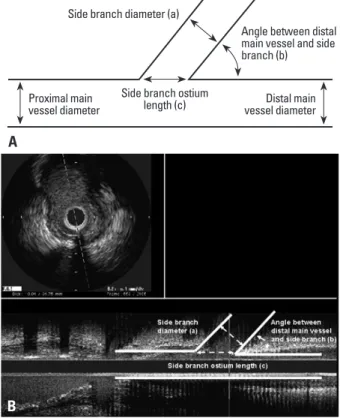
Fig. 1. Intravascular ultrasound (IVUS) measurements in this study are shown in a schematic diagram (A) and an actual IVUS image (B); side branch diameter (a), angle between distal main vessel and side branch (b), and side branch ostium length (c).
Side branch diameter (a)
Side branch ostium
length (c) Distal main vessel diameter Proximal main
vessel diameter
Angle between distal main vessel and side branch (b)
A
B
was obtained from the trigonometric function as follows: SB diameter/sin (angle between distal MV and SB).13 The corre- lation between the directly measured and calculated values of SBOL was then evaluated.
Analysis was performed using the Statistical Package for the Social Sciences (SPSS) software (version 15.0; SPSS, Inc., Chicago, IL, USA). Data are presented as mean±stan- dard deviation for continuous variables and as numbers (per- centage) for categorical variables. Simple Pearson’s correla- tion was used to evaluate the relationship between the two parameters. The paired t-test was used to compare continu- ous variables before and after implantation. p-values less than 0.05 were considered statistically significant.
RESULTS
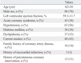
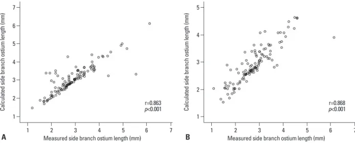
The baseline clinical characteristics of the patients are shown in Table 1, and the angiographic and procedural char- acteristics are shown in Table 2. Evaluation of the IVUS findings revealed that the angle between the distal MV and the SB was 57.3±12.4° at pre-intervention and 59.4±12.6° at post-intervention (Table 3). The mean measured and calcu- lated SBOL were 2.91±0.86 mm and 3.06±0.77 mm at pre- intervention and 2.79±0.82 mm and 2.92±0.69 mm at post- intervention, respectively. The differences between the measured and calculated SBOL were 0.15±0.44 mm at pre- intervention and 0.13±0.41 mm at post-intervention. We found that calculated SBOL was significantly correlated with measured SBOL (pre-intervention r=0.863, p<0.001;
post-intervention r=0.868, p<0.001) (Fig. 2).
DISCUSSION
This IVUS study with longitudinal reconstruction images Table 1. Baseline Clinical Characteristics
Values
Age (yrs) 62±10
Male sex, n (%) 88 (78)
Left ventricular ejection fraction, % 59.1±11.7 Acute coronary syndrome, n (%) 43 (38)
Hypertension, n (%) 63 (56)
Diabetes mellitus, n (%) 38 (34)
Dyslipidemia, n (%) 37 (33)
Current smoker, n (%) 48 (43)
Family history of coronary artery disease,
n (%) 16 (14)
History of myocardial infarction, n (%) 3 (3) History of percutaneous coronary
intervention, n (%) 12 (11)
SD, standard deviation.
Data are presented as mean±SD or number (percentage).
Table 2. Angiographic and Procedural Characteristics Values Extent of involved vessel, n (%)
Single-vessel disease 51 (45)
Two-vessel disease 40 (35)
Three-vessel disease 22 (20)
Bifurcation lesions, n (%)
Distal left main coronary artery 27 (24) Left anterior descending artery and
diagonal branch 81 (72)
Left circumflex artery and obtuse marginal
branch 5 (4)
Number of stents implanted 1.8±0.9
Diameter of stent implanted, mm 3.1±0.4 Length of stent implanted, mm 23.6±6.1 Types of stents, n (%)
Sirolimus-eluting stent 37 (33)
Paclitaxel-eluting stent 14 (12)
Zotarolimus-eluting stent 33 (29)
Everolimus-eluting stent 29 (26)
SD, standard deviation.
Data are presented as mean±SD or number (percentage).
Table 3. Intravascular Ultrasound Findings
Pre-stent Post-stent p value
Proximal MV diameter, mm 3.08±0.77 3.14±0.56 <0.001
Distal MV diameter, mm 2.33±0.55 2.40±0.44 <0.001
SB diameter, mm 2.49±0.66 2.44±0.63 0.129
Angle between distal MV and SB, degree 57.3±12.4 59.4±12.6 0.047
Measured SBOL, mm 2.91±0.86 2.79±0.82 0.010
Calculated SBOL, mm 3.06±0.77 2.92±0.69 0.001
Difference between measured and calculated SBOL, mm 0.15±0.44 0.13±0.41 0.550
MV, main vessel; SB, side branch; SBOL, side branch ostial length; SD, standard deviation.
Data are presented as mean±SD or number (percentage).
ber of patients was relatively small. Second, over-estimation or under-estimation of the angle between the distal MV and SB could lead to over-estimation or under-estimation of the calculated SBOL. Third, patients with bifurcation lesions showing significant stenosis or who had undergone any in- tervention in the SB were excluded. Therefore, the results of this study can not be applied to bifurcation lesions with sig- nificant stenosis in the SB. Fourth, IVUS imaging in the SB was not performed. In addition, the IVUS variables were en- tered as the values of measurement of the lumen, and were not reflective of remodeling of the SB by measurement of the external elastic membrane. Finally, there was no data re- garding SBOL and its related PCI outcomes and long-term clinical events. In the future, further study to evaluate the clinical implications of SBOL will be need.
In conclusion, this IVUS analysis showed that the value of SBOL obtained by a trigonometric method correlated well with directly measured SBOL in bifurcation lesions. There- fore, the SBOL in bifurcation lesions can be determined using SB diameter and the angle between the distal MV and SB.
ACKNOWLEDGEMENTS
This study was supported in part by a grant from the Korea Healthcare Technology R&D Project, Ministry for Health, Welfare & Family Affairs, Republic of Korea (No. A085012 and A102064), a grant from the Korea Health 21 R&D Proj- ect, Ministry of Health & Welfare, Republic of Korea (No.
A085136), and the Cardiovascular Research Center, Seoul, Korea.
demonstrated a good correlation between measured and calculated SBOL in bifurcation lesions. We found that the trigonometric method, using the SB diameter and the angle between the distal MV and SB used to calculate SBOL, provided a simple and accurate method for estimating the actual SBOL in bifurcation lesions.
Previous studies showed that SB occlusion occurred in 3.3% to 10.6% of cases following PCI in bifurcation lesions.14 SB occlusion is associated with decreased success and in- creased complication rates in bifurcation lesions compared to non-bifurcation lesions. Passing an additional guide wire into the occluded SB can salvage the branch vessel. There- fore, the SB ostium should be the main target during PCI of bifurcation lesions. IVUS is the standard method for quan- tifying coronary atherosclerosis and is commonly used to assess coronary lumen dimensions.15 Previous IVUS study demonstrated that SB occlusion after stent implantation in patients with bifurcation lesions is associated with ostial le- sions and plaque distribution.16 Accurate IVUS evaluation of the SB ostium could be useful for the stenting of bifurca- tion lesions. In the present study, we used IVUS longitudi- nal reconstruction to compare the directly measured SBOL and calculated SBOL in coronary bifurcation lesions and found a highly significant correlation between the measured and calculated values. Differences between the measured and calculated SBOL were only 0.15±0.44 mm at pre-inter- vention and 0.13±0.41 mm at post-intervention. The mea- sured SBOL was smaller than the calculated SBOL because there were fewer diseases of the SB in this study.
There are some limitations in this study. First, this study was a retrospective analysis at a single center, and the num-
Fig. 2. The correlation between the measured and calculated values of the side branch ostium length are shown; pre-intervention r=0.863, p<0.001 (A) and post-intervention r=0.868, p<0.001 (B).
1 1
2
2 3
4 3
5 6
4
7 5
Calculated side branch ostium length (mm) Calculated side branch ostium length (mm)
1 2 3 4 5 6 7 1 2 3 4 5 6 7
Measured side branch ostium length (mm) Measured side branch ostium length (mm)
A B
r=0.863
p<0.001 r=0.868
p<0.001
10. Park SJ, Kim YH, Park DW, Lee SW, Kim WJ, Suh J, et al. Im- pact of intravascular ultrasound guidance on long-term mortality in stenting for unprotected left main coronary artery stenosis. Circ Cardiovasc Interv 2009;2:167-77.
11. Kim JS, Hong MK, Ko YG, Choi D, Yoon JH, Choi SH, et al. Im- pact of intravascular ultrasound guidance on long-term clinical outcomes in patients treated with drug-eluting stent for bifurcation lesions: data from a Korean multicenter bifurcation registry. Am Heart J 2011;161:180-7.
12. Mintz GS, Nissen SE, Anderson WD, Bailey SR, Erbel R, Fitzger- ald PJ, et al. American College of Cardiology Clinical Expert Consensus Document on Standards for Acquisition, Measurement and Reporting of Intravascular Ultrasound Studies (IVUS). A re- port of the American College of Cardiology Task Force on Clini- cal Expert Consensus Documents. J Am Coll Cardiol 2001;37:
1478-92.
13. Lima T, Alves C, Funayama CA. Proposal for a trigonometric method to evaluate the abduction angle of the lower limbs in neo- nates. J Child Neurol 2008;23:1451-4.
14. Al Suwaidi J, Yeh W, Cohen HA, Detre KM, Williams DO, Hol- mes DR Jr. Immediate and one-year outcome in patients with cor- onary bifurcation lesions in the modern era (NHLBI dynamic reg- istry). Am J Cardiol 2001;87:1139-44.
15. Sano K, Mintz GS, Carlier SG, de Ribamar Costa J Jr, Qian J, Missel E, et al. Assessing intermediate left main coronary lesions using intravascular ultrasound. Am Heart J 2007;154:983-8.
16. Furukawa E, Hibi K, Kosuge M, Nakatogawa T, Toda N, Taka- mura T, et al. Intravascular ultrasound predictors of side branch occlusion in bifurcation lesions after percutaneous coronary inter- vention. Circ J 2005;69:325-30.
REFERENCES
1. Latib A, Colombo A, Sangiorgi GM. Bifurcation stenting: current strategies and new devices. Heart 2009;95:495-504.
2. Aliabadi D, Tilli FV, Bowers TR, Benzuly KH, Safian RD, Gold- stein JA, et al. Incidence and angiographic predictors of side branch occlusion following high-pressure intracoronary stenting.
Am J Cardiol 1997;80:994-7.
3. Fischman DL, Savage MP, Leon MB, Schatz RA, Ellis S, Cleman MW, et al. Fate of lesion-related side branches after coronary ar- tery stenting. J Am Coll Cardiol 1993;22:1641-6.
4. Pan M, Medina A, Suárez de Lezo J, Romero M, Melián F, Pav- lovic D, et al. Follow-up patency of side branches covered by in- tracoronary Palmaz-Schatz stent. Am Heart J 1995;129:436-40.
5. Hoye A, Iakovou I, Ge L, van Mieghem CA, Ong AT, Cosgrave J, et al. Long-term outcomes after stenting of bifurcation lesions with the “crush” technique: predictors of an adverse outcome. J Am Coll Cardiol 2006;47:1949-58.
6. Moussa I, Costa RA, Leon MB, Lansky AJ, Lasic Z, Cristea E, et al. A prospective registry to evaluate sirolimus-eluting stents im- planted at coronary bifurcation lesions using the “crush tech- nique”. Am J Cardiol 2006;97:1317-21.
7. Latib A, Colombo A. Bifurcation disease: what do we know, what should we do? JACC Cardiovasc Interv 2008;1:218-26.
8. Costa RA, Mintz GS, Carlier SG, Lansky AJ, Moussa I, Fujii K, et al. Bifurcation coronary lesions treated with the “crush” tech- nique: an intravascular ultrasound analysis. J Am Coll Cardiol 2005;46:599-605.
9. Mintz GS, Weissman NJ. Intravascular ultrasound in the drug- eluting stent era. J Am Coll Cardiol 2006;48:421-9.