INTRODUCTION
Esophageal cancer (EC) is a highly lethal and aggressive tumor worldwide. Around the world, there were 482300 new cases of EC and 406800 deaths in 2008.1,2 According to 2013 Annual Re-
port of Cancer Registration in China, it is the fourth leading cause of cancer-related death among male, and sixth among fe- male. EC can be divided into two types according to histology:
esophageal adenocarcinoma (EADC) and esophageal squa- mous cell carcinoma (ESCC). In China, ESCC accounts for most esophageal malignant tumors (about 87%). Although screening and multimodality therapy technology has greatly improved,3,4 the outcome for EC remains very poor; the 5-year overall sur- vival rate is below 15 %,5,6 emphasizing the need for early detec- tion and prognostic markers. Over the past decade, scientific re- search have worked on revealing the molecular and biological mechanism that lead to carcinoma, leading to extensive search for prognostic markers in EC. For example, the expression of epidermal growth factor receptor (EGFR), Her-2, heat shock proteins (HSPs), and P53 has been found to be associated with the prognosis of EC.7-10
The Prognostic Impact of Heat Shock Proteins Expression in Patients with Esophageal
Cancer: A Meta-Analysis
Xiao-wei Wang1*, Xin-hui Shi2*, Yu-suo Tong1, and Xiu-feng Cao3
1Department of Medical Oncology, Huai’an First People’s Hospital, Nanjing Medical University, Huai'an, Jiangsu;
2Department of Laboratory Medicine, Yancheng First People’s Hospital, Yancheng, Jiangsu;
3Department of Surgical Oncology, Nanjing First Hospital, Nanjing Medical University, Nanjing, Jiangsu, P.R. China.
Purpose: Heat shock proteins (HSPs) are highly conserved molecular chaperones. There are various studies that assess the prog- nostic value of HSPs in patients with esophageal cancer, but the conclusion remains controversial. This is the first meta-analysis study aiming to summarize the evidence on the suitability of HSPs to predict patients’ survival.
Materials and Methods: Searching PubMed, Web of science and Medline until May 31, 2014, data were compared for overall sur- vival in patients with down-regulated HSPs level with those with up-regulated level. We conducted a meta-analysis of 9 studies (801 patients) that correlated HSPs levels with overall survival. Data were synthesized with hazard ratios (HRs).
Results: The estimated risk of death was 2.93-fold greater in HSP27 negative patients than HSP27 positive patients [95% confi- dence interval (CI), 1.12–7.62]. When limited to esophageal squamous cell carcinoma (ESCC), the risk of death in HSP27 negative patients seemed more significant (HR, 3.90; 95% CI, 2.35–6.49). Decreased expression of HSP70 was also associated with worse survival in esophageal cancer (HR, 2.83; 95% CI, 1.90–4.23) and, when limited to ESCC, HR was 3.21 (95% CI, 1.94–5.30). Data col- lected, however, were not sufficient to determine the prognostic value of HSP90 in patients with ESCC nor esophageal adenocar- cinomas (EADC).
Conclusion: In this meta-analysis, reduced HSP27 and HSP70 expressions were associated with poor survival in patients with esophageal cancer, especially esophageal squamous cell carcinoma.
Key Words: HSP27, HSP70, HSP90, esophageal cancer, survival, meta-analysis Yonsei Med J 2015 Nov;56(6):1497-1502
http://dx.doi.org/10.3349/ymj.2015.56.6.1497 pISSN: 0513-5796 · eISSN: 1976-2437
Received: October 20, 2014 Revised: December 29, 2014 Accepted: February 3, 2015
Corresponding author: Dr. Xiu-feng Cao, Department of Surgical Oncology, Affili- ated Nanjing Hospital of Nanjing Medical University, 68 Changle Road, Nanjing 210006, Jiangsu, P.R. China.
Tel: 86-13951618052, Fax: 86-2552269924, E-mail: [email protected]
*Xiao-wei Wang and Xin-hui Shi contributed equally to this work.
•The authors have no financial conflicts of interest.
© Copyright: Yonsei University College of Medicine 2015
This is an Open Access article distributed under the terms of the Creative Com- mons Attribution Non-Commercial License (http://creativecommons.org/ licenses/
by-nc/3.0) which permits unrestricted non-commercial use, distribution, and repro- duction in any medium, provided the original work is properly cited.
The HSPs family is molecular chaperone and biochemical regulator, which functions to mediate cell growth, apoptosis, protein homeostasis, and cellular targets of peptides.11 It is a highly conserved cellular proteins group which is upregulated under stress conditions, including thermal, oxyradical, and in- flammatory stress.11,12 They are classified into six major family members based on their molecular size: HSP100, HSP90, HSP70, HSP60, HSP40, and small HSPs.13-15 Excessive expres- sion of HSPs in a wide range of human tumors have been re- ported, including breast, endometrial, ovarian, colon, lung, and prostate.16 Expression levels of HSPs are also reportedly altered, either increasing or decreasing, during malignant transforma- tion.17 Studies have also shown that HSPs expressions are close- ly related with prognosis of carcinoma.16,18
A question arises whether these findings justify the use of HSPs detection, in a routine clinical setting, as a prognostic in- dicator in patients with EC. In our study, we conducted a sys- tematic review on HSP27, HSP70, and HSP90, which are three main members of HSPs family, and meta-analysis to estimate the prognostic importance of HSPs expression for survival among patients with EC.
MATERIALS AND METHODS
Search strategy
Two authors (Wang and Shi) conducted a systematic literature
search independently in PubMed, Web of Science and Med- line. Searches were applied from the day of establishment of the database to May 31, 2014. The following MeSH headings, keywords, and text words were used: 1) esophageal cancer OR esophageal neoplasms OR oesophageal neoplasms OR oe- sophageal cancer; 2) heat shock protein OR HSPs. We also used the references cited in the identified studies to complete the search.
Inclusion and exclusion criteria
Studies enrolled in this meta-analysis should meet the following criteria: 1) diagnosis of EC in humans was proven; 2) the expres- sion of HSPs in the primary EC tissue was measured by any methods available; 3) data reported were related to the prognos- tic value; 4) survival rate, survival curve or sufficient data were provided for estimating an hazard ratio (HR) with 95% confi- dence interval (CI); 5) the follow-up time should be more than 5 years; 6) the study was published in English. The major exclu- sion criteria were: 1) study without extractable data, 2) case re- ports, editorials, commentaries, reviews or abstracts only, and 3) for duplicate publications and the smaller dataset.
Data extraction
Two investigators (Wang and Shi) reviewed all eligible studies and extracted data independently and resolved controversies by discussion. The name of the first author, year of publication, origin of country, histology, disease stage, number of eligible Table 1. Main Characteristics and Results of the Eligible Studies
First author (yrs)
Origin
country Histology Stage I+II (%)
No. of
patientsMale (%) Median/
mean age, yrs
Method Cutoff HSPs negative/low (%)
Survival
analysis HR (95% CI) Kawanishi,
et al. (1999)24 Japan ESCC 63.7 102 82.4 61.7 IHC 80%
staining
HSP27: 20.4
HSP70: 68.9 Multivariate HSP27: 8.74 (3.11–24.58) HSP70: 3.25 (0.6–17.6) Shiozaki,
et al. (2000)25 Japan ESCC 67.5 77 NR NR IHC 10%
staining
HSP27: 11.7
HSP70: 20.8 Multivariate HSP27: 5.1 (2.39–10.9) HSP70: 3.17 (1.53–6.55) Nakajima,
et al. (2002)23 Japan ESCC 58.1 62 85.5 61.3 IHC 20%
staining HSP27: 50 Multivariate HSP27: 1.47 (0.6–3.58) Noguchi,
et al. (2002)26 Germany ESCC 52.1 71 88.7 63.8 IHC 50%
staining HSP70: 32.4 Univariate HSP70: 3.26 (1.51–7.06) Faried,
et al. (2004)28 Japan ESCC 61.8 123 86.2 61.2 IHC 40%
staining HSP90: 49.6 Multivariate HSP90: 0.93 (0.54–1.63) Nakajima,
et al. (2009)17 Japan Esophageal
cancer 61.6 125 86.4 62.1 IHC 40%
staining HSP70: 48.8 Multivariate HSP70: 2.26 (1.17–4.39) Wu,
et al. (2009)29 Sweden Esophageal
cancer 31.7 82 69.5 NR IHC 75%
staining HSP90: 28.0 Univariate HSP90: 1.11 (0.67–1.84) Berg,
et al. (2011)10 Germany EADC NR 87 91 63 RPPA NR HSP27: 81.6 Multivariate HSP27: 0.9 (0.24–3.44) Huang,
et al. (2014)30 China ESCC 64.2 72 30.9 58.3 IHC 80%
staining HSP90: 12.3 Univariate HSP90: 0.72 (0.23–2.27) NR, not reported; HR, hazard ratio; CI, confidence interval; ESCC, esophageal squamous cell carcinoma; EADC, esophageal adenocarcinoma; IHC, immunohisto- chemistry; RPPA, reverse phase protein arrays; HSP, heat shock protein.
Esophageal cancer includes ESCC and EADC.
patients, gender, median age, test method, cutoff value, HSPs negativity, and survival results from each study were recorded (Table 1). Differences in the extraction of data were checked to- gether by the two authors. If data from any of the above catego- ries were not reported in the primary article, items were treated as “not reported.” We did not contact the author to request the information.
Statistical analysis
For appropriate HSPs evaluation in a single study, the summary HR and their 95% CIs were combined to present the value re- ported in the study using methods described by Parmar, et al.19 We used HR calculations spreadsheet provided by Tierney, et al.20 were to calculate the estimation of HR from published summary statistics or data extracted from Kaplan-Meier curves.
Cochran’s test (I2 statistics) was used to assess the heteroge- neity of included studies. When heterogeneity was detected, a random effect model was applied; otherwise, a fixed effect model was used.21
The combined HRs were estimated using forest plots graphi- cally. An observed HR of more than 1 implied a worse survival
for the HSPs-negative, low HSPs expression group relative to the HSPs-positive, or high expression group, and if the 95% CI line of a study across the invalid line, it means that the study was not statistically significant. The study was considered statis- tically significant if the 95% CI did not overlap 1 (p≤0.05). Hori- zontal lines represent 95% CIs. Boxes represent the HR point estimate, and its area is proportional to the weight of the study.
The diamond represents overall summary estimate, with the CI represented by its width. The unbroken vertical line was set at the null value (HR, 1.0). Assessment of publication bias was conducted using Funnel plot asymmetry.22 All the statistical analyses were conducted using Review Manager 5.2 (The Nor- dic Cochrane Centre, The Cochrane Collaboration, 2012).
RESULTS
Characteristics of the studies
As the search flow diagram shows (Fig. 1), we identified the ab- stracts and titles of 248 primary studies for initial review using the search strategies as described. After exclusion of articles that were duplicate or not clearly relevant to our meta-analysis, we identified 56 candidate studies for full-text review. Upon further review, 4 review articles and 37 articles were eliminated because of having no survival data for meta-analysis. In the re- maining 15 articles, 3 were not focused on the HSP members which we cared, 2 duplicate data and 1 was about gastric esoph- ageal. Finally, 9 articles were selected.
These studies included three HSP members; 510,23-25 studies for HSP27, 617,24-26 for HSP70, and 327-29 for HSP90. The histopa- thology in most of the included studies was ESCC, while 1 study showed EADC and 2 studies showed EC (both ESCC and EADC). The studies were conducted in different countries; 5 of 9 studies were conducted in Japan, 2 in Germany, 1 in China, and 1 study in Sweden. The total number of patients was 801 and ranged from 62 to 125 patients per study (median, 89). The characteristics of the eligible studies are summarized in Table 1.
Fig. 1. Search strategy flowchart.
248 studies identified using described search strategies
56 reviewed for full text
192 studies excluded for:
133 duplicate data
59 clearly not relevant according to titles or abstracts
47 studies excluded for:
4 review articles 37 without survival data
3 for GRP78, GP96, HSPA2 respectively 2 duplicate data
1 for gastric and gastroesophageal carcinomas
9 studies included in our meta-analysis
Table 3. Meta-Analysis: HR Value of HSPs in Esophageal Cancer Subgroups According to Histology
No. of studies Patients HR (95% CI) Model Heterogeneity test (Chi2, I2, p value)
HSP27 in ESCC 3 241 3.90 (2.35–6.49) Random 7.35, 73%, 0.03
HSP70 in ESCC 3 250 3.21 (1.94–5.30) Fixed 0.00, 0%, 1.00
HSP90 in ESCC 2 195 0.89 (0.54–1.46) Fixed 0.16, 0%, 0.69
HR, hazard ratio; CI, confidence interval; HSP, heat shock protein; ESCC, esophageal squamous cell carcinoma.
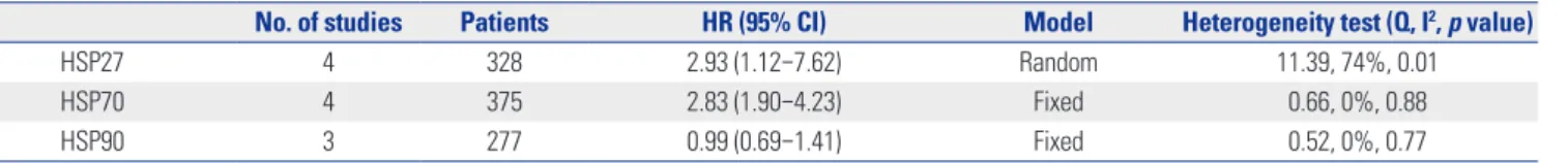
Table 2. Meta-Analysis: HR Value of HSPs in Esophageal Cancer
No. of studies Patients HR (95% CI) Model Heterogeneity test (Q, I2, p value)
HSP27 4 328 2.93 (1.12–7.62) Random 11.39, 74%, 0.01
HSP70 4 375 2.83 (1.90–4.23) Fixed 0.66, 0%, 0.88
HSP90 3 277 0.99 (0.69–1.41) Fixed 0.52, 0%, 0.77
HR, hazard ratio; CI, confidence interval; HSP, heat shock protein.
Meta-analysis results
The results of the meta-analysis are represented in Table 2 and 3, Figs. 2, 3, and 4. For studies evaluating HSP27 levels in EC (Fig.
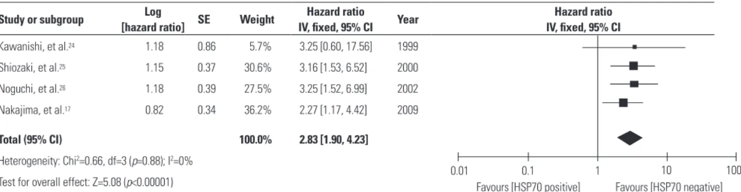
2), the combined HRs were 2.93 (95% CI, 1.12–7.62). The results were significantly heterogeneous across these studies (p=0.01), and therefore, a random effect model was used in meta-analy- sis. For studies in ESCCs subgroup, the combined HRs were 3.90 (95% CI, 2.35–6.49) using a random-effect model. The re- sults demonstrate that the reduced HSP27 expression was a significant prognostic factor in ESCC patients. For studies eval- uating HSP70 levels in EC (Fig. 3), the test of heterogeneity was not significant, and a fixed-effect model was used in meta- analysis. The pooled HRs estimate for survival in these studies evaluating HSP70 levels in EC were 2.83 (95% CI, 1.90–4.23).
However, when we limited the analysis to the 3 studies with ESCC, the combined HR was 3.21 (95% CI, 1.94–5.30), indicat- ing that HSP70-positive patients had a more favorable progno- sis than HSP70-negative patients. The combined HRs of HSP90 showed that the expression of HSP90 could not act as an effec- tive prognostic marker in EC (HR: 0.99, 95% CI: 0.69–1.41, Z=0.07, p=0.95).
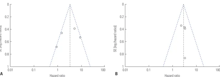
Potential publication bias
The possibility of publication bias was assessed with Begg’s funnel plots.22 The shape of the funnel plots did not reveal any evidence of asymmetry (Fig. 5).
Study or subgroup Log
[hazard ratio] SE Weight Hazard ratio
IV, random, 95% CI Year Hazard ratio
IV, random, 95% CI Kawanishi, et al.24 2.17 0.53 24.6% 8.76 [3.10, 24.75] 1999
Shiozaki, et al.25 1.63 0.39 28.3% 5.10 [2.38, 10.96] 2000 Nakajima, et al.23 0.38 0.46 26.4% 1.46 [0.59, 3.60] 2002 Berg, et al.10 -0.1 0.68 20.7% 0.90 [0.24, 3.43] 2011
Total (95% CI) 100.0% 2.93 [1.12, 7.62]
Heterogeneity: Tau2=0.69; Chi2=11.39, df=3 (p=0.010); I2=74%
Test for overall effect: Z=2.20 (p=0.03) 0.01
Favours [HSP27 positive] Favours [HSP27 negative]
0.1 1 10 100
Fig. 2. Meta-analysis (forest plot) of the 5 eligible studies assessing HSP27 in esophageal cancer. SE, standard error; CI, confidence interval; HSP, heat shock protein.
Study or subgroup Log
[hazard ratio] SE Weight Hazard ratio
IV, fixed, 95% CI Year Hazard ratio
IV, fixed, 95% CI Kawanishi, et al.24 1.18 0.86 5.7% 3.25 [0.60, 17.56] 1999
Shiozaki, et al.25 1.15 0.37 30.6% 3.16 [1.53, 6.52] 2000 Noguchi, et al.26 1.18 0.39 27.5% 3.25 [1.52, 6.99] 2002 Nakajima, et al.17 0.82 0.34 36.2% 2.27 [1.17, 4.42] 2009
Total (95% CI) 100.0% 2.83 [1.90, 4.23]
Heterogeneity: Chi2=0.66, df=3 (p=0.88); I2=0%
Test for overall effect: Z=5.08 (p<0.00001) 0.01
Favours [HSP70 positive] Favours [HSP70 negative]
0.1 1 10 100
Fig. 3. Meta-analysis (forest plot) of the 6 eligible studies assessing HSP70 in esophageal cancer. SE, standard error; CI, confidence interval; HSP, heat shock protein.
Study or subgroup Log
[hazard ratio] SE Weight Hazard ratio
IV, fixed, 95% CI Year Hazard ratio
IV, fixed, 95% CI Faried, et al.27 -0.07 0.28 41.9% 0.93 [0.54, 1.61] 2004
Wu, et al.28 0.1 0.26 48.6% 1.11 [0.66, 1.84] 2009
Huang, et al.29 -0.33 0.59 9.4% 0.72 [0.23, 2.29] 2014
Total (95% CI) 100.0% 0.99 [0.69, 1.41]
Heterogeneity: Chi2=0.52, df=2 (p=0.77); I2=0%
Test for overall effect: Z=0.07 (p=0.95) 0.01
Favours [HSP90 positive] Favours [HSP90 negative]
0.1 1 10 100
Fig. 4. Meta-analysis (forest plot) of the 3 eligible studies assessing HSP90 in esophageal cancer. SE, standard error; CI, confidence interval; HSP, heat shock protein.
not been published, which leading to unavoidable publication bias. Second, this meta-analysis was limited to English articles, which leading to potential language bias. Third, studies enrolled in our meta-analysis used IHC to detect HSP level, which repre- sent potential selection bias. Cutoff values for HSP expression differed in the percentage cell staining. Fourth, the estimated data that we obtained were not adjusted for other variables such as age, gender, histologic grade, and tumor stage. This may cause variability in assessing these variables between studies.
Finally, there still might be a little error when the approximate calculation method was used to estimate the HR values, al- though 2 investigators calculated them separately.
In conclusion, our results suggest that reduced HSP27 and HSP70 expressions may be associated with a poor prognosis in patients with EC, thus warranting further definitive investiga- tions into the potential clinical usefulness of HSP expression in ECs. It also appears worthwhile to prospectively validate if HSP27 and HSP70 expression used as prognostic markers could improve the outcomes of patients with EC, especially those with ESCC when integrated into clinical decision making.
ACKNOWLEDGEMENTS
We thank all our colleagues at the Department of Medical On- cology, Huai’an First People’s Hospital, Nanjing Medical Uni- versity.
REFERENCES
1. Jemal A, Bray F, Center MM, Ferlay J, Ward E, Forman D. Global cancer statistics. CA Cancer J Clin 2011;61:69-90.
2. Mohamed A, El-Rayes B, Khuri FR, Saba NF. Targeted therapies in metastatic esophageal cancer: advances over the past decade.
Crit Rev Oncol Hematol 2014;91:186-96.
3. Quiros RM, Bui CL. Multidisciplinary approach to esophageal and gastric cancer. Surg Clin North Am 2009;89:79-96, viii.
4. Chen M, Cai E, Huang J, Yu P, Li K. Prognostic value of vascular endothelial growth factor expression in patients with esophageal cancer: a systematic review and meta-analysis. Cancer Epidemiol
DISCUSSION
HSPs are highly conserved molecular chaperones, which are also referred to as stress proteins. It has been suggested that ‘on- cogenic stress’ such as acidosis, hypoxia or hypothermia induc- es up-regulated expression of HSPs that assist in the recovery from stress by either repairing damaged proteins or by degrad- ing them, which evokes a DNA damage response network that delays or prevents cancer at the beginning of tumorigenesis, thereby restoring protein homoeostasis and promoting cell sur- vival.30,31 Furthermore, molecular chaperones have been shown to influence tumour growth, differentiation and resistance to ra- dio- and chemotherapy treatment, and they may have a signifi- cant impact on the survival of patients with cancer.32,33
Many studies have shown that HSPs were related to cell pro- liferation and apoptosis. In the process of tumor formation, some tumors with expression of HSP27 and/or HSP70 ap- peared loss of differentiation ability, metastasis, and poor prog- nosis. In esophageal carcinoma patients, however, down-regu- lated expression of HSPs was associated with poor prognosis. In order to find an explanation for the observation, we summa- rized as follows: 1) it may be secondary to fundamental histo- logic differences between squamous cell carcinoma and adeno- carcinoma, such as rectal or gastric cancer; 2) HSPs may be expressed continually because normal esophageal squamous epithelia are frequently exposed to agents such as heat or chem- icals, so that HSPs can play roles in protecting cells; 3) HSPs ex- pression is known to be correlated negatively with lymph node metastasis and depth of invasion. This may indicate that reduc- tion of HSPs expression causes the tumor cells to proliferate; 4) There was a significant correlation between HSPs expression and lymphocyte infiltration, and this may indicate that HSPs expression promotes host immunity.17,23-26 Thus, the patients with HSPs positive tumors tend to have a better prognosis than those with HSPs negative tumors.
This meta-analysis has some limitations. First, there may be some reports with negative or controversial results that have
Fig. 5. Begg’s funnel plot for publication bias. (A) Funnel plot of HSP27. (B) Funnel plot of HSP70. SE, standard error.
0 0.2 0.4 0.6 0.8
1
Hazard ratio
0.01 0.1 1 10 100
SE [log (hazard ratio)]
A
0 0.2 0.4 0.6 0.8
1
Hazard ratio
0.01 0.1 1 10 100
SE [log (hazard ratio)]
B
Biomarkers Prev 2012;21:1126-34.
5. Ferlay J, Shin HR, Bray F, Forman D, Mathers C, Parkin DM. Esti- mates of worldwide burden of cancer in 2008: GLOBOCAN 2008.
Int J Cancer 2010;127:2893-917.
6. Xu YW, Peng YH, Chen B, Wu ZY, Wu JY, Shen JH, et al. Autoanti- bodies as potential biomarkers for the early detection of esopha- geal squamous cell carcinoma. Am J Gastroenterol 2014;109:36-45.
7. Kaneko K, Kumekawa Y, Makino R, Nozawa H, Hirayama Y, Kogo M, et al. EGFR gene alterations as a prognostic biomarker in ad- vanced esophageal squamous cell carcinoma. Front Biosci (Land- mark Ed) 2010;15:65-72.
8. Delektorskaya VV, Chemeris GY, Zavalishina LE, Ryazantseva AA, Grigorchuk AY, Kononets PV, et al. Squamous cell carcinoma of the esophagus: evaluation of the status of epidermal growth factor re- ceptors (EGFR and HER-2) by immunohistochemistry and in situ hybridization. Bull Exp Biol Med 2010;149:615-20.
9. Langer R, Ott K, Specht K, Becker K, Lordick F, Burian M, et al. Pro- tein expression profiling in esophageal adenocarcinoma patients indicates association of heat-shock protein 27 expression and che- motherapy response. Clin Cancer Res 2008;14:8279-87.
10. Berg D, Wolff C, Langer R, Schuster T, Feith M, Slotta-Huspenina J, et al. Discovery of new molecular subtypes in oesophageal adeno- carcinoma. PLoS One 2011;6:e23985.
11. Morimoto RI. Cells in stress: transcriptional activation of heat shock genes. Science 1993;259:1409-10.
12. Argon Y, Simen BB. GRP94, an ER chaperone with protein and peptide binding properties. Semin Cell Dev Biol 1999;10:495-505.
13. Langer T, Lu C, Echols H, Flanagan J, Hayer MK, Hartl FU. Succes- sive action of DnaK, DnaJ and GroEL along the pathway of chap- erone-mediated protein folding. Nature 1992;356:683-9.
14. Hoe KL, Won M, Chung KS, Jang YJ, Lee SB, Kim DU, et al. Isola- tion of a new member of DnaJ-like heat shock protein 40 (Hsp40) from human liver. Biochim Biophys Acta 1998;1383:4-8.
15. Liu Y, Zhou J, Zhang C, Fu W, Xiao X, Ruan S, et al. HLJ1 is a novel biomarker for colorectal carcinoma progression and overall patient survival. Int J Clin Exp Pathol 2014;7:969-77.
16. Ciocca DR, Calderwood SK. Heat shock proteins in cancer: diag- nostic, prognostic, predictive, and treatment implications. Cell Stress Chaperones 2005;10:86-103.
17. Nakajima M, Kato H, Miyazaki T, Fukuchi M, Masuda N, Fukai Y, et al. Tumor immune systems in esophageal cancer with special ref- erence to heat-shock protein 70 and humoral immunity. Antican- cer Res 2009;29:1595-606.
18. Lebret T, Watson RW, Molinié V, O’Neill A, Gabriel C, Fitzpatrick JM, et al. Heat shock proteins HSP27, HSP60, HSP70, and HSP90:
expression in bladder carcinoma. Cancer 2003;98:970-7.
19. Parmar MK, Torri V, Stewart L. Extracting summary statistics to
perform meta-analyses of the published literature for survival end- points. Stat Med 1998;17:2815-34.
20. Tierney JF, Stewart LA, Ghersi D, Burdett S, Sydes MR. Practical methods for incorporating summary time-to-event data into meta- analysis. Trials 2007;8:16.
21. DerSimonian R, Laird N. Meta-analysis in clinical trials. Control Clin Trials 1986;7:177-88.
22. Egger M, Davey Smith G, Schneider M, Minder C. Bias in meta- analysis detected by a simple, graphical test. BMJ 1997;315:629-34.
23. Nakajima M, Kuwano H, Miyazaki T, Masuda N, Kato H. Signifi- cant correlation between expression of heat shock proteins 27, 70 and lymphocyte infiltration in esophageal squamous cell carcino- ma. Cancer Lett 2002;178:99-106.
24. Kawanishi K, Shiozaki H, Doki Y, Sakita I, Inoue M, Yano M, et al.
Prognostic significance of heat shock proteins 27 and 70 in patients with squamous cell carcinoma of the esophagus. Cancer 1999;85:
1649-57.
25. Shiozaki H, Doki Y, Kawanishi K, Shamma A, Yano M, Inoue M, et al.
Clinical application of malignancy potential grading as a prognostic factor of human esophageal cancers. Surgery 2000;127:552-61.
26. Noguchi T, Takeno S, Shibata T, Uchida Y, Yokoyama S, Müller W.
Expression of heat shock protein 70 in grossly resected esopha- geal squamous cell carcinoma. Ann Thorac Surg 2002;74:222-6.
27. Faried A, Sohda M, Nakajima M, Miyazaki T, Kato H, Kuwano H.
Expression of heat-shock protein Hsp60 correlated with the apop- totic index and patient prognosis in human oesophageal squa- mous cell carcinoma. Eur J Cancer 2004;40:2804-11.
28. Wu X, Wanders A, Wardega P, Tinge B, Gedda L, Bergstrom S, et al.
Hsp90α is expressed and represents a therapeutic target in human oesophageal cancer using the inhibitor 17-allylamino-17-deme- thoxygeldanamycin. Br J Cancer 2009;100:334-43.
29. Huang T, Chen S, Han H, Li H, Huang Z, Zhang J, et al. Expression of Hsp90α and cyclin B1 were related to prognosis of esophageal squamous cell carcinoma and keratin pearl formation. Int J Clin Exp Pathol 2014;7:1544-52.
30. Bartkova J, Horejsí Z, Koed K, Krämer A, Tort F, Zieger K, et al. DNA damage response as a candidate anti-cancer barrier in early hu- man tumorigenesis. Nature 2005;434:864-70.
31. Jolly C, Morimoto RI. Role of the heat shock response and molecu- lar chaperones in oncogenesis and cell death. J Natl Cancer Inst 2000;92:1564-72.
32. Lee AS. GRP78 induction in cancer: therapeutic and prognostic implications. Cancer Res 2007;67:3496-9.
33. Khalil AA, Kabapy NF, Deraz SF, Smith C. Heat shock proteins in oncology: diagnostic biomarkers or therapeutic targets? Biochim Biophys Acta 2011;1816:89-104.