High A20 expression negatively impacts
survival in patients with breast cancer
Chang Ik Yoon1☯, Sung Gwe AhnID2☯, Soong June Bae2, Yun Jin Shin3, Chihwan Cha2, So
Eun ParkID2, Ji-Hyung Lee4, Akira Ooshima5, Hye Sun LeeID6, Kyung-Min Yang5,
Seong-Jin Kim5,7, Seok Hee Park4, Joon Jeong
ID2*
1 Department of Surgery, St Mary’s Hospital, Catholic University College of Medicine, Seoul, Korea, 2 Department of Surgery, Gangnam Severance Hospital, Yonsei University College of Medicine, Seoul, Republic of Korea, 3 Gangnam Severance Hospital, Yonsei University College of Medicine, Seoul, Republic of Korea, 4 Department of Biological Sciences, Sungkyunkwan University, Suwon, Gyeonggi-do, Republic of Korea, 5 Precision Medicine Research Center, Advanced Institutes of Convergence Technology,
6 Biostatistics Collaboration Unit, Yonsei University College of Medicine, Seoul, Republic of Korea, 7 Department of Transdisciplinary Studies, Graduate School of Convergence Science and Technology, Seoul National University, Suwon, Kyunggi-do, Korea
☯These authors contributed equally to this work.
Abstract
Background
A20 protein has ubiquitin-editing activities and acts as a key regulator of inflammation and immunity. Previously, our group showed that A20 promotes tumor metastasis through multi-monoubiquitylation of SNAIL1 in basal-like breast cancer. Here, we investigated survival outcomes in patients with breast cancer according to A20 expression.
Patients and methods
We retrospectively collected tumor samples from patients with breast cancer. Immunohis-tochemistry (IHC) with an A20-specific antibody was performed, and survival outcomes were analyzed.
Results
A20 expression was evaluated in 442 patients. High A20 expression was associated with advanced anatomical stage and young age. High A20 expression showed significantly infe-rior recurrence-free-survival and overall-survival (P<0.001 and P<0.001, respectively). Mul-tivariate analysis showed that A20 was an independent prognostic marker for RFS (HRs: 2.324, 95% CIs: 1.446–3.736) and OS (HRs: 2.629, 95% CIs: 1.585–4.361). In human epi-dermal growth factor receptor 2 (HER2)-positive and triple negative breast cancer (TNBC) subtypes, high A20 levels were associated with poor OS.
Conclusion
We found that A20 expression is a poor prognostic marker in breast cancer. The prognostic impact of A20 was pronounced in aggressive tumors, such as HER2-positive and TNBC
a1111111111 a1111111111 a1111111111 a1111111111 a1111111111 OPEN ACCESS
Citation: Yoon CI, Ahn SG, Bae SJ, Shin YJ, Cha C,
Park SE, et al. (2019) High A20 expression negatively impacts survival in patients with breast cancer. PLoS ONE 14(8): e0221721.https://doi. org/10.1371/journal.pone.0221721
Editor: Kelly A. Avery-Kiejda, The University of
Newcastle, AUSTRALIA
Received: May 2, 2019 Accepted: August 13, 2019 Published: August 26, 2019
Copyright:© 2019 Yoon et al. This is an open access article distributed under the terms of the
Creative Commons Attribution License, which permits unrestricted use, distribution, and reproduction in any medium, provided the original author and source are credited.
Data Availability Statement: All relevant data are
within the manuscript and its Supporting Information files.
Funding: This research was supported by the Basic
Science Research Program through the NRF, funded by the Ministry of Science, ICT, & Future Planning (NRF-2016R1D1A1A09917675 to SGA), and grant from the National R&D Program for Cancer Control, Ministry of Health & Welfare, Republic of Korea (1520120). The funders had no role in study design, data collection and analysis,
subtypes. Our findings suggested that A20 may be a valuable target in patients with aggres-sive breast cancer.
Background
Breast cancer is the most common cancer in women worldwide, and 1.5 million women are diagnosed each year. With the development of improved treatment modalities and new drugs, the survival rate has improved, and the annual breast cancer mortality rate has decreased by almost 2.3% [1].
Despite advances in systemic therapy, including targeted therapies, such as anti-estrogen and anti-human epidermal growth factor receptor-2 (HER2) therapies, treatment outcomes for metastatic cancer remain poor. To improve treatment outcomes for patients with breast cancer, there is an urgent need to identify novel targets that play an integral role in the meta-static cascade.
The A20 protein, which is also called tumor necrosis factorα-induced protein 3
(TNFAIP3), acts as a key regulator of inflammation and immunity since it stimulates anti-apo-ptotic signaling pathway through suppression of TNF signal pathway and regulates nuclear factor-κB signaling [2–5]. Recent studies have also shown that A20 protein has ubiquitin (Ub)-editing activities, including deubiquitylase [6–8] and E3 ubiquitin ligase activities [9], and can function as a ubiquitin-binding protein [10]. Although the roles of A20 in inflammation and immune responses are well understood, its functions in cancer are still unclear. However, sev-eral studies in tumor cell lines, such as hepatocellular carcinoma, glioma stem cells and breast cancer, have suggested that A20 plays a role in carcinogenesis by preventing apoptosis [11–
14].
Recently, our group reported that the A20 protein plays an oncogenic role in mediating epi-thelial mesenchymal transition (EMT), which is strongly associated with cancer progression characteristics, such as invasion and metastasis [15]. Our study showed that A20 protein pro-moted metastasis in triple negative breast cancer (TNBC) through multi-monoubiquitylation of Snail1 [16]. We found, throughin vitro and in vivo experiments, that the A20 protein was
more highly expressed in aggressive tumors and that A20 upregulation promoted metastasis. However, the clinical characteristics of A20 expression in breast cancer have been poorly explored.
The aim of this study was to evaluate the association between A20 expression and survival outcomes in patients with breast cancer. We also investigated the prognostic impact of A20 expression according to immunohistochemistry (IHC)-based molecular subtypes.
Patients and methods
Patients
We retrospectively collected tumor tissues from patients undergoing surgery for breast cancer at Gangnam Severance Hospital in Seoul, Korea between January 1996 and December 2014. All the study subjects were diagnosed with stage I–III primary breast cancer. These patients underwent adjuvant treatments according to standard protocols. Clinical data were collected, including age at surgery, histologic grade (HG), lymph node status, estrogen receptor (ER) sta-tus, progesterone receptor (PR) stasta-tus, HER2 stasta-tus, lymphovascular invasion (LVI), Ki67 lev-els, treatment modality, breast cancer recurrence, and death.
decision to publish, or preparation of the manuscript.
Competing interests: The authors have declared
TNM stage was determined according to the American Joint Committee on Cancer (AJCC), 7thedition. Tumor grade was determined using the modified Scarff-Bloom-Richard-son grading system. Before February 1999, ER and PR status were assessed using a ligand bind-ing assay, and tumors were considered positive if the score was >10 fmol/mg.
The study protocol was approved by the institutional review board of Gangnam Severance Hospital.
Ethics approval and consent to participate
Our study itself was conducted as human-specimen subject research and was approved by the institutional review board (IRB) review (Local IRB number: 3-2018-0067). All procedures per-formed in studies involving human participants were in accordance with the ethical standards of the institutional and/or national research committee and with the 1964 Helsinki declaration and its later amendments or comparable ethical standards. The need for informed consent was waived under the approval of the IRB due to the retrospective design.
Immunohistochemistry (IHC) and molecular subtyping
As previously described [17], we evaluated ER, PR, HER2, and Ki67 expression using the fol-lowing antibodies, ER (1:100 clone 6F11; Novocastra, Newcastle upon Tyne, UK), PR (clone 16; Novocastra), HER2 (4B5 rabbit monoclonal antibody; Ventana Medical Systems, Tucson, AZ, USA), and Ki-67 (MIB-1; Dako, Glostrup, Denmark). ER- and PR-positive IHC expres-sion was defined according to the modified Allred system: positive, Allred score 3–8 and nega-tive, Allred score 0–2. HER2 status was re-evaluated according to American Society of Clinical Oncology/College of American Pathologists guideline [18]. HER2 status was considered posi-tive if the score was 3+, and was considered negaposi-tive with a score of 0 or 1+. Tumors with a score of 2+ underwent FISH or SISH analysis, according to the manufacturer’s instructions (PathVysion kit; Vysis, Downers Grove, IL, USA or HER2 inform; Ventana). Ki-67 expression is presented as the percentage (range 0–100%) of positive tumor cells.
For the molecular subtyping, the following definitions were used: i) Luminal/HER2 nega-tive: ER positive and/or PR positive and HER2 negative; ii) HER2 posinega-tive: HER2 positive regardless of ER and PR status; and iii) TNBC: ER negative, PR negative, and HER2 negative.
Tissue microarray and IHC staining to evaluate A20 expression
Tissue microarray (TMA) paraffin blocks were generated as previously described using an Accu Max Array tissue-arraying instrument (Petagen, Inc., Seoul, Korea) [17]. For IHC, each TMA slide was stained with a rabbit monoclonal anti-A20 antibody (ab92324, 1:200; Abcam) and counter-stained with hematoxylin. After staining, A20 expression, assessed as cytoplasmic staining, was scored by an experienced pathologist (A.O.) using a microscope (400× magnifi-cation). Positive A20 expression in tumor cytoplasm was defined when the percentage of stained cells was equal to more than 60%. Finally, a score of 2+ or 3+ was defined as high A20 expression, whereas a score of 0 or 1+ was defined as low A20 expression (S1 Fig). The IHC results were interpreted blindly, without any information regarding clinical parameters or outcomes.
Statistical analysis
Continuous variables were compared using Student’s t-test or Mann-Whitney test. Categorical variables were compared using the Chi-square test or Fisher’s exact test. Overall survival (OS) was defined as the period from primary curative surgery to the last follow-up or death from
any cause. Recurrence free survival (RFS) was defined the period from primary curative sur-gery to the date of any recurrence (loco-regional or distant metastasis), death due to any cause or the last follow-up. Data for patients who did not have an event of interest were censored at the date of the last follow-up. Kaplan-Meier survival curves were compared using the log-rank test. Clinicopathologic factors associated with survival outcomes were analyzed using the Cox proportional hazard model. The variables used in the multivariate analysis were those that showed statistical significance in the univariate analysis for OS and RFS.
To compare the predictive power of the Cox proportional hazard model with and without A20, we applied the Harrell’s c-statistic to obtain concordance index (c-index) [19]. It is calcu-lated to measure the concordance for time-to event data, in which increasing values between 0.5 and 1.0 indicated improved prediction. For model comparison, the bootstrapping method was used with resampling 1,000 times. We also performed net reclassification improvement (NRI) and integrated discrimination improvement (IDI) to evaluate improvement of discrimi-nating ability when new factors were added in the survival model [20]. In NRI and IDI, a significant improvement is recognized in the prediction model when their values are greater than 0.
These analyses were performed using SPSS version 23 (SPSS; Chicago, IL, USA) and R ver-sion 3.4.2 (www.R-project.org). Statistical significance was defined as aP value less than 0.05,
at a 95% confidence interval (CI) not including 1.
Results
Patient characteristics based on A20 expression
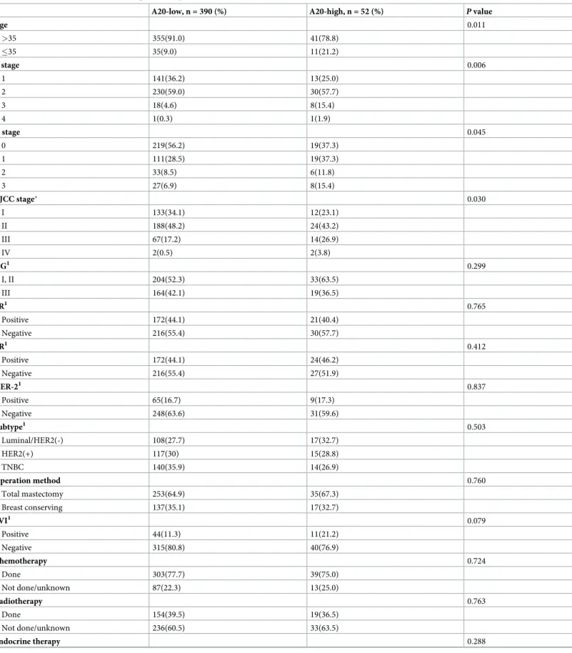
This study included 442 patients, with a median age of 47 (22–86) years. Of the 442 patients, 390 (88.2%) had low A20 expressing tumors, and 52 (11.8%) had high A20 expressing tumors. Clinical characteristics were compared based on A20 expression inTable 1. High A20 expres-sion was associated with anatomical tumor stage; A20 was highly expressed in advanced T stage (P = 0.006), N stage (P = 0.045), and AJCC stage tumors (P = 0.030). In addition, high
A20 expression was associated with younger age at surgery (P = 0.011). However, it was not
related to tumor grade (P = 0.299) or receptor status (ER; P = 0.765, PR; P = 0.412, HER2; P = 0.837). In addition, there was no statistical difference in adjuvant treatment, such as
che-motherapy (P = 0.724), radiotherapy (P = 0.763) or endocrine therapy (P = 0.288).
We classified patients into three subtypes based on ER, PR, and HER2 status; 125 patients were of the luminal/HER2-negative subtype, 132 were of the HER2-positive subtype, and 154 were of the TNBC subtype. There were no significant associations between A20 expression and breast cancer subtype (P = 0.503).
Prognostic impact of A20 expression
At a median follow-up time of 92.5 months (1–258), 88 mortality events had occurred. There were a total of 110 recurrence events, of which 29 were locoregional recurrences, 97 were dis-tant metastases, and 16 were both locoregional recurrences and disdis-tant metastases. Of 110 recurrence cases, there were 72 mortality events from breast cancer.
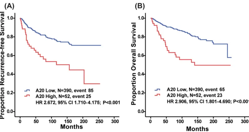
High A20 expression was significantly predictive of decreased RFS (P < 0.001, log-rank
test;Fig 1A). Patients with high A20 expression also showed reduced OS (P < 0.001, log-rank
test;Fig 1B). Younger age at surgery (�35 years old), advanced T stage, advanced N stage, LVI and A20 (HR 2.672, 95% CI 1.710–4.175,P<0.001) were significant factors in the univariate
analysis of RFS (Table 2). A20 (HR 2.906, 95% CI 1.801–4.690,P<0.001) was still a significant
Table 1. Clinical characteristics according to A20 expression.
A20-low, n = 390 (%) A20-high, n = 52 (%) P value
Age 0.011 >35 355(91.0) 41(78.8) �35 35(9.0) 11(21.2) T stage 0.006 1 141(36.2) 13(25.0) 2 230(59.0) 30(57.7) 3 18(4.6) 8(15.4) 4 1(0.3) 1(1.9) N stage 0.045 0 219(56.2) 19(37.3) 1 111(28.5) 19(37.3) 2 33(8.5) 6(11.8) 3 27(6.9) 8(15.4) AJCC stage� 0.030 I 133(34.1) 12(23.1) II 188(48.2) 24(43.2) III 67(17.2) 14(26.9) IV 2(0.5) 2(3.8) HG1 0.299 I, II 204(52.3) 33(63.5) III 164(42.1) 19(36.5) ER1 0.765 Positive 172(44.1) 21(40.4) Negative 216(55.4) 30(57.7) PR1 0.412 Positive 172(44.1) 24(46.2) Negative 216(55.4) 27(51.9) HER-21 0.837 Positive 65(16.7) 9(17.3) Negative 248(63.6) 31(59.6) Subtype1 0.503 Luminal/HER2(-) 108(27.7) 17(32.7) HER2(+) 117(30) 15(28.8) TNBC 140(35.9) 14(26.9) Operation method 0.760 Total mastectomy 253(64.9) 35(67.3) Breast conserving 137(35.1) 17(32.7) LVI1 0.079 Positive 44(11.3) 11(21.2) Negative 315(80.8) 40(76.9) Chemotherapy 0.724 Done 303(77.7) 39(75.0) Not done/unknown 87(22.3) 13(25.0) Radiotherapy 0.763 Done 154(39.5) 19(36.5) Not done/unknown 236(60.5) 33(63.5) Endocrine therapy 0.288 (Continued )
When adjusted for age, T stage, N stage, and LVI using the Cox proportional hazards regression model, high A20 expression was significantly associated with RFS and OS (RFS;
Table 2, HR 2.324, 95% CI 1.446–3.736,P<0.001,Table 3; OS, HR 2.629, 95% CI 1.585–4.361,
P<0.001).
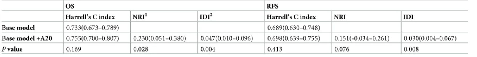
To evaluate an improvement of predicting ability between multivariate models with and without A20, we calculated c-index, NRI, and IDI. The addition of A20 to base model increased c-indices for both OS and RFS without a statistical significance (0.733 to 0.755 for OS; 0.689 to 0.698 for RFS;Table 4). At a median follow-up 93 months, the addition of A20 Table 1. (Continued)
A20-low, n = 390 (%) A20-high, n = 52 (%) P value
Done 153(39.2) 16(30.8)
Not done/unknown 237(60.8) 36(69.2)
Trastuzumab 0.235
Done 15(3.8) 0
Not done/unknown 375(96.2) 52(100)
ER, estrogen receptor; PR, progesterone receptor; HER-2, human epidermal growth factor receptor-2; HG, histologic grade; TNBC, triple negative breast cancer; LVI, Lymphovascular invasion
�AJCC stage was performed based on 7th edition 1
Missing value 2
HER-2 positive was defined by 3 positive on immunohistochemistry or amplification on fluorescencein situ hybridization
https://doi.org/10.1371/journal.pone.0221721.t001
Fig 1. Kaplan-Meier plots of recurrence free survival (RFS) and overall survival (OS) according to A20 expression level. (a) RFS (A20 Low, M = 390, event 85; A20
High, N = 52, event 25; HR 2.672, 95% CI 1.710–4.175;P<0.001, respectively, log-rank test) and (b) OS differed significantly according to A20 expression level (A20
Low, N = 390, event 65; A20 High, N = 52, event 23, HR 2.906, 95% CI 1.801–4.690;P<0.001).
improved a discriminating ability for OS because NRI and IDI were 0.230 and 0.047 (P = 0.028
andP = 0.004, respectively;Table 4). At a median follow-up 83 months, a model with A20 showed a superior discrimination by IDI (0.030,P = 0.008) whereas a NRI for this model was
greater than 0 (0.151) but not significant (P = 0.076).
Prognostic impact of A20 expression in aggressive subtypes
Survival outcomes based on A20 expression were compared in each breast cancer subtype. RFS differed significantly according to A20 expression in the HER2 subtype (Fig 2B; HR 5.751, 95% CI 2.683–12.330,P < 0.001), but it did not differ in the other subtypes (Fig 2A, Luminal/ Table 2. Hazard ratios (HRs) and 95% confidential intervals (CIs) for recurrence free survival (RFS).
Univariate analysis Multivariate analysis
HRs (95% CI) P value HRs (95% CI) P value
Age <0.001 <0.001 >35 1 1 �35 0.355(0.227–0.555) 0.416(0.256–0.674) T stage 0.001 0.514 1 1 1 2 1.5661.005–2.442) 0.048 1.293(0.801–2.085) 0.293 3 4.196(2.162–8.142) <0.001 1.740(0.833–3.635) 0.141 N stage <0.001 <0.001 0 1 1 1 1.694(1.066–2.690) 0.026 1.339(0.809–2.214) 0.256 2 3.702(2.111–6.492) <0.001 2.938(1.601–5.393) 0.001 3 5.313(3.057–9.232) <0.001 3.969(2.068–7.619) <0.001 LVI <0.001 0.272 Negative 1 1 Positive 2.377(1.468–3.848) 1.349(0.791–2.300) HG 0.990 I and II 1 III 1.001(0.825–1.216) A20 <0.001 <0.001 Low 1 1 High 2.672(1.710–4.175) 2.324(1.446–3.736) ER 0.535 Negative 1 Positive 1.127(0.772–1.646) PR 0.535 Negative 1 Positive 0.887(0.607–1.296) HER2 0.862 Negative 1 Positive 0.954(0.560–1.624) TNBC vs non-TNBC 0.842 non-TNBC 1 TNBC 1.042(0.692–1.570)
LVI, Lymphovascular invasion; HG, histologic grade; ER, estrogen receptor; PR, progesterone receptor; HER-2, human epidermal growth factor receptor-2; TNBC, triple negative breast cancer
Table 3. Hazard ratios (HRs) and 95% confidential intervals (CIs) for Overall survival (OS).
Univariate analysis Multivariate analysis
HR (95% CI) P value HR (95% CI) P value
Age 0.001 0.042 >35 1 1 �35 0.429(0.260–0.708) 0.536(0.325–0.978) T stage <0.001 0.073 1 1 1 2 1.677(0.995–2.828) 0.052 1.340(0.763–2.355) 3 5.751(2.836–11.662) <0.001 2.123(0.955–4.718) N stage <0.001 <0.001 0 1 1 1 1.460(0.845–2.524) 0.175 1.128(0.619–2.053) 2 3.847(2.086–7.093) <0.001 2.894(1.483–5.648) 3 6.566(3.672–11.741) <0.001 4.067(2.027–8.162) LVI <0.001 0.116 Negative 1 1 Positive 3.079(1.833–5.172) 1.595(0.891–2.856) HG 0.492 I and II 1 III 1.080(0.867–1.344) A20 <0.001 <0.001 Low 1 1 High 2.906(1.801–4.690) 2.629(1.585–4.361) ER 0.292 Negative 1 Positive 0.793(0.515–1.221) PR 0.330 Negative 1 Positive 0.808(0.526–1.241) HER2 0.831 Negative 1 Positive 0.934(0.497–1.754) TNBC vs non-TNBC 0.857 Non-TNBC 1 TNBC 1.045(0.645–1.693)
LVI, Lymphovascular invasion; HG, histologic grade; ER, estrogen receptor; PR, progesterone receptor; HER-2, human epidermal growth factor receptor-2; TNBC, triple negative breast cancer
https://doi.org/10.1371/journal.pone.0221721.t003
Table 4. Evaluation of Cox proportional hazard model using Harrell’s C index, NRI and IDI.
OS RFS
Harrell’s C index NRI1 IDI2 Harrell’s C index NRI IDI
Base model 0.733(0.673–0.789) 0.689(0.630–0.748)
Base model +A20 0.755(0.700–0.807) 0.230(0.051–0.380) 0.047(0.010–0.096) 0.698(0.639–0.755) 0.151(-0.034–0.261) 0.030(0.004–0.067)
P value 0.169 0.028 0.004 0.413 0.076 0.008
NRI, net reclassification improvement; IDI, integrated discrimination improvement
HER2 negative, HR 1.689, 95% CI 0.738–3.869,P = 0.209;Fig 2C, TNBC, HR 1.905, 95% CI 0.737–4.928,P = 0.175). In addition, decreased OS was noted in the high A20-expressing
HER2--positive subtype (Fig 2E; HR 4.440, 95% CI 1.902–10.365,P < 0.001) and TNBC subtypes subtype
(Fig 2F; HR 2.842, 95% CI 1.060–7.617,P = 0.030) of breast cancer, but it was not observed in the
luminal/HER2-negative subtype (Fig 2D; HR 1.970, 95% CI 0.795–4.884,P = 0.135).
Univariate analysis of OS and RFS in each subtype was performed using the Cox-regression model. In HER2-positive subtype, A20 expression was a significant factor for prediction of both OS (Table 5; HR 3.987, 95% CI 1.628–9.760,P = 0.002) and RFS (Table 5; HR 5.276, 95% CI 2.395–11.620,P<0.001). For the TNBC subtype, A20 expression was a significant factor for
prediction of OS (Table 5; HR 2.842, 95% CI 1.060–7.617,P = 0.038).
Discussion
In this study, we found that A20 expression was an independent prognostic factor for breast cancer. Compared to patients with low expressing tumors, patients with high A20-Fig 2. Kaplan-Meier plots of RFS and OS according to A20 expression level in each subtype. RFS differed significantly according to A20 expression in the HER2
subtype (Fig B; HR 5.751, 95% CI 2.683–12.330, P < 0.001, respectively, log-rank test), but it did not differ in the other subtypes (Fig A, Luminal/HER2 negative, HR 1.689, 95% CI 0.738–3.869, P = 0.209; Fig C, TNBC, HR 1.905, 95% CI 0.737–4.928, P = 0.175). OS differed significantly according to A20 expression in the HER2 and TNBC subtypes (Fig E; HER2, HR 4.440, 95% CI 1.902–10.365, P < 0.001,Fig 2F; TNBC, HR 2.842, 95% CI 1.060–7.617, P = 0.030), but it did not differ in luminal/ HER2 negative (Fig D; HR 1.970, 95% CI 0.795–4.884, P = 0.135).
expressing tumors showed poorer RFS and OS (RFS, HR: 2.324, 95% CI: 1.446–3.736; OS, HR: 2.629, 95% CI: 1.585–4.361). Although A20 expression was not associated with specific sub-types, the prognostic impact of A20 expression was more pronounced in aggressive subsub-types, such as HER2-positive and TNBC subtypes. It might be implicated that A20 has clinical value as a novel target for improving treatment outcomes in patients with aggressive breast cancer, such as HER2-positive subtype.
Earlier studies on A20 in inflammatory and autoimmune diseases showed that A20 is asso-ciated with a complex regulator of ubiquitylation of canonical NF-κB and cell-survival signals A20: linking a complex regulator of ubiquitylation to immunity and human disease [2–10,21,
22]. In contrast, the role of A20 in malignant disease has not been extensively explored. Several genetic studies have reported that deletion or mutation of A20 is associated with hematologic malignancies, such as multiple lymphoma, MALT lymphoma, Hodgkin’s disease, and follicu-lar lymphoma, which are characterized by NF-κB signal activation [23–26]. Other studies have suggested that high A20 expression was associated with tumor survival and growth in solid tumors such as malignant glioma and breast cancer [11–14]. Previous studies on the role of A20 in malignancies suggest that it acts as a tumor suppressor or enhancer depending on the cell type.
In addition to the above findings, A20 has also been reported to have a novel role in breast cancer; A20 promotes tumor progression and activates EMT signals, mainly by affecting SNAIL1 stabilization [16]. EMT signaling, which is associated with the capacity to migrate to distant organs [15], is frequently activated in TNBC [27,28]. A mechanism by which the EMT pathway is activated via A20 elevation [16] might partly explain our findings: poor survival outcomes in TNBC patients with high A20 expression. Further studies of EMT features, such as high vimentin expression or E-cadherin loss, in TNBC with high A20 expression levels are warranted.
Intriguingly, we also found that high A20 expression is a poor prognostic marker in patients with HER2-positive breast cancer. Several studies have provided evidence that upregulation of NF-κB signaling in HER2-positive breast cancer is associated with resistance to various thera-pies, including anti-HER2 therapy [29,30]. Although A20 is a negative regulator of NF-κB
sig-naling in inflammation and immune response, a previous study by Lerebours et al. showed that A20 is involved in upregulation of the NF-κB pathway in inflammatory breast cancer (IBC) [31]. Although HER2 status was not reported in the study, it has been noted that 30~50% of IBCs have HER2 overexpression [32–34], suggesting that A20 expression might be associated with NF-κB pathway activation in HER2-positive breast cancer. Further studies on the association between A20 and NF-κB are warranted in HER2-positive cancer.
The biology underlying poor outcomes in patients with high A20-expressing HER2-positive breast cancer is also supported by a mechanism of A20-mediated TGF-β-activation. Previous Table 5. Univariate analysis on A20 in each subtype.
OS RFS
HR (95% CI) P value HR (95% CI) P value
Luminal/HER2 negative 1.970(0.795–4.884) 0.143 1.689(0.738–3.869) 0.215
HER2-positive 3.987(1.628–9.760) 0.002 5.276(2.395–11.620) <0.001
TNBC 2.842(1.060–7.617) 0.038 1.905(0.737–4.927) 0.184
HR, hazard ratio; CI, confidential interval; HER-2, human epidermal growth factor receptor-2; TNBC, triple negative breast cancer
studies have shown that HER2 stimulates TGF-β-mediated EMT, contributing to the aggres-sive behavior of HER2-positive breast cancer [35–37]. Our previous study also provided evi-dence that A20 elevates TGF-β signaling, suggesting that A20 could promote a TGF-β-meditated EMT pathway and tumor aggressiveness in HER2-positive breast cancer.
Other studies have reported that A20 overexpression was associated with endocrine ther-apy, chemotherther-apy, and radiotherapy treatment failure. A preclinical study of MCF7 cells demonstrated that tamoxifen-resistant cancer cells showed A20 overexpression [14]; however, there was no prognostic influence of A20 expression in patients with luminal/HER2-negative breast cancer. In addition, another study suggested that A20 reduced the efficacy of radiother-apy and chemotherradiother-apy via the regulation of anti-apoptotic mechanisms [38].
Our study had several limitations. First, it was a retrospective study with a small sample size, and the enrolled patients received uncontrolled adjuvant treatments. Particularly, most patients (88.5%) with HER2-positive tumors did not receive anti-HER2 therapy (S2 Fig) because most of the HER2-positive samples were collected prior to 2010 when the use of tras-tuzumab was not reimbursed by the national insurance system in Korea. In addition, there were higher proportions of HER2-positivie and TNBC subtypes in our study than there are in global breast cancer populations, because of selection bias during sample storage and preserva-tion. Despite these limitations, our study comprehensively showed the clinical outcomes based on A20 expression among patients with breast cancer and demonstrated A20 expression as an independent prognostic factor. Furthermore, prognostic discrimination based on A20 expres-sion was pronounced in aggressive tumors, such as HER2-positive and TNBC subtypes. Our findings suggest that A20 may be a valuable target in patients with aggressive breast cancer.
Conclusion
In conclusion, we found that A20 expression is a poor prognostic marker in breast cancer. The prognostic impact of A20 was pronounced in aggressive tumors, such as HER2-positive and TNBC subtypes. Our findings suggested that A20 may be a valuable target in patients with aggressive breast cancer
Supporting information
S1 Fig. Immunohistochemical analysis of A20 expression. A20 expression was evaluated in high-power fields (400× magnification) by an experienced pathologist (A.O.) (a) Negative for A20. (b) 1+ for A20. (c) 2+ for A20. (d) 3+ for A20.
(TIF)
S2 Fig. Kaplan-Meier plots of RFS and OS for HER2 subtype breast cancer according to trastuzumab use. (a) RFS and (b) OS did not differ significantly according to trastuzumab use (P = 0.050 and P = 0.102, respectively, log-rank test).
(TIF)
Author Contributions
Conceptualization: Chang Ik Yoon, Sung Gwe Ahn, Joon Jeong.
Data curation: Chang Ik Yoon, Soong June Bae, Yun Jin Shin, Chihwan Cha, So Eun Park. Formal analysis: Chang Ik Yoon, Soong June Bae, Ji-Hyung Lee, Hye Sun Lee, Seong-Jin Kim,
Seok Hee Park.
Investigation: Chang Ik Yoon, Sung Gwe Ahn, Ji-Hyung Lee, Akira Ooshima, Kyung-Min Yang.
Methodology: Chang Ik Yoon, Sung Gwe Ahn, Hye Sun Lee. Project administration: Chang Ik Yoon, Sung Gwe Ahn.
Resources: Chang Ik Yoon, Sung Gwe Ahn, Akira Ooshima, Joon Jeong. Software: Chang Ik Yoon, Hye Sun Lee.
Supervision: Sung Gwe Ahn, Joon Jeong. Validation: Sung Gwe Ahn.
Visualization: Chang Ik Yoon, Sung Gwe Ahn.
Writing – original draft: Chang Ik Yoon, Sung Gwe Ahn.
Writing – review & editing: Chang Ik Yoon, Sung Gwe Ahn, Yun Jin Shin, Joon Jeong.
References
1. Jemal A, Siegel R, Ward E, Murray T, Xu J, Thun MJ. Cancer statistics, 2007. CA: a cancer journal for clinicians. 2007; 57(1):43–66. Epub 2007/01/24. PMID:17237035.
2. Catrysse L, Vereecke L, Beyaert R, van Loo G. A20 in inflammation and autoimmunity. Trends in immu-nology. 2014; 35(1):22–31. Epub 2013/11/20.https://doi.org/10.1016/j.it.2013.10.005PMID:
24246475.
3. Lee EG, Boone DL, Chai S, Libby SL, Chien M, Lodolce JP, et al. Failure to regulate Tinduced NF-kappaB and cell death responses in A20-deficient mice. Science (New York, NY). 2000; 289 (5488):2350–4. Epub 2000/09/29.https://doi.org/10.1126/science.289.5488.2350PMID:11009421; PubMed Central PMCID: PMC3582399.
4. Ma A, Malynn BA. A20: linking a complex regulator of ubiquitylation to immunity and human disease. Nature reviews Immunology. 2012; 12(11):774–85. Epub 2012/10/13.https://doi.org/10.1038/nri3313
PMID:23059429; PubMed Central PMCID: PMC3582397.
5. Musone SL, Taylor KE, Lu TT, Nititham J, Ferreira RC, Ortmann W, et al. Multiple polymorphisms in the TNFAIP3 region are independently associated with systemic lupus erythematosus. Nature genetics. 2008; 40(9):1062–4. Epub 2009/01/24.https://doi.org/10.1038/ng.202PMID:19165919; PubMed Cen-tral PMCID: PMC3897246.
6. Boone DL, Turer EE, Lee EG, Ahmad RC, Wheeler MT, Tsui C, et al. The ubiquitin-modifying enzyme A20 is required for termination of Toll-like receptor responses. Nature immunology. 2004; 5(10):1052– 60. Epub 2004/08/31.https://doi.org/10.1038/ni1110PMID:15334086.
7. Komander D, Barford D. Structure of the A20 OTU domain and mechanistic insights into deubiquitina-tion. The Biochemical journal. 2008; 409(1):77–85. Epub 2007/10/27.https://doi.org/10.1042/ BJ20071399PMID:17961127.
8. Lin SC, Chung JY, Lamothe B, Rajashankar K, Lu M, Lo YC, et al. Molecular basis for the unique deubi-quitinating activity of the NF-kappaB inhibitor A20. Journal of molecular biology. 2008; 376(2):526–40. Epub 2008/01/01.https://doi.org/10.1016/j.jmb.2007.11.092PMID:18164316; PubMed Central PMCID: PMC2346432.
9. Wertz IE, O’Rourke KM, Zhou H, Eby M, Aravind L, Seshagiri S, et al. De-ubiquitination and ubiquitin ligase domains of A20 downregulate NF-kappaB signalling. Nature. 2004; 430(7000):694–9. Epub 2004/07/20.https://doi.org/10.1038/nature02794PMID:15258597.
10. Bosanac I, Wertz IE, Pan B, Yu C, Kusam S, Lam C, et al. Ubiquitin binding to A20 ZnF4 is required for modulation of NF-kappaB signaling. Molecular cell. 2010; 40(4):548–57. Epub 2010/11/26.https://doi. org/10.1016/j.molcel.2010.10.009PMID:21095585.
11. Dong B, Lv G, Wang Q, Wei F, Bellail AC, Hao C, et al. Targeting A20 enhances TRAIL-induced apopto-sis in hepatocellular carcinoma cells. Biochemical and biophysical research communications. 2012; 418(2):433–8. Epub 2012/01/31.https://doi.org/10.1016/j.bbrc.2012.01.056PMID:22285182. 12. Guo Q, Dong H, Liu X, Wang C, Liu N, Zhang J, et al. A20 is overexpressed in glioma cells and may
serve as a potential therapeutic target. Expert opinion on therapeutic targets. 2009; 13(7):733–41. Epub 2009/06/06.https://doi.org/10.1517/14728220903045018PMID:19492975.
13. Hjelmeland AB, Wu Q, Wickman S, Eyler C, Heddleston J, Shi Q, et al. Targeting A20 decreases glioma stem cell survival and tumor growth. PLoS biology. 2010; 8(2):e1000319. Epub 2010/02/27.https://doi. org/10.1371/journal.pbio.1000319PMID:20186265; PubMed Central PMCID: PMC2826371. 14. Vendrell JA, Ghayad S, Ben-Larbi S, Dumontet C, Mechti N, Cohen PA. A20/TNFAIP3, a new
estro-gen-regulated gene that confers tamoxifen resistance in breast cancer cells. Oncogene. 2007; 26 (32):4656–67. Epub 2007/02/14.https://doi.org/10.1038/sj.onc.1210269PMID:17297453. 15. Thiery JP, Acloque H, Huang RY, Nieto MA. Epithelial-mesenchymal transitions in development and
disease. Cell. 2009; 139(5):871–90. Epub 2009/12/01.https://doi.org/10.1016/j.cell.2009.11.007PMID:
19945376.
16. Lee JH, Jung SM, Yang KM, Bae E, Ahn SG, Park JS, et al. A20 promotes metastasis of aggressive basal-like breast cancers through multi-monoubiquitylation of Snail1. Nature cell biology. 2017; 19 (10):1260–73. Epub 2017/09/12.https://doi.org/10.1038/ncb3609PMID:28892081.
17. Ahn SG, Dong SM, Oshima A, Kim WH, Lee HM, Lee SA, et al. LOXL2 expression is associated with invasiveness and negatively influences survival in breast cancer patients. Breast cancer research and treatment. 2013; 141(1):89–99. Epub 2013/08/13.https://doi.org/10.1007/s10549-013-2662-3PMID:
23933800.
18. Wolff AC, Hammond ME, Schwartz JN, Hagerty KL, Allred DC, Cote RJ, et al. American Society of Clin-ical Oncology/College of American Pathologists guideline recommendations for human epidermal growth factor receptor 2 testing in breast cancer. Journal of clinical oncology: official journal of the Amer-ican Society of Clinical Oncology. 2007; 25(1):118–45. Epub 2006/12/13.https://doi.org/10.1200/jco. 2006.09.2775.xPMID:17159189.
19. Harrell FE Jr., Lee KL, Mark DB. Multivariable prognostic models: issues in developing models, evaluat-ing assumptions and adequacy, and measurevaluat-ing and reducevaluat-ing errors. Statistics in medicine. 1996; 15 (4):361–87. Epub 1996/02/28.https://doi.org/10.1002/(SICI)1097-0258(19960229)15:4< 361::AID-SIM168>3.0.CO;2-4PMID:8668867.
20. Uno H, Tian L, Cai T, Kohane IS, Wei LJ. A unified inference procedure for a class of measures to assess improvement in risk prediction systems with survival data. Statistics in medicine. 2013; 32 (14):2430–42. Epub 2012/10/06.https://doi.org/10.1002/sim.5647PMID:23037800; PubMed Central PMCID: PMC3734387.
21. Dixit VM, Green S, Sarma V, Holzman LB, Wolf FW, O’Rourke K, et al. Tumor necrosis factor-alpha induction of novel gene products in human endothelial cells including a macrophage-specific chemo-taxin. The Journal of biological chemistry. 1990; 265(5):2973–8. Epub 1990/02/15. PMID:2406243. 22. Jaattela M, Mouritzen H, Elling F, Bastholm L. A20 zinc finger protein inhibits TNF and IL-1 signaling.
Journal of immunology (Baltimore, Md: 1950). 1996; 156(3):1166–73. Epub 1996/02/01. PMID:
8557994.
23. Compagno M, Lim WK, Grunn A, Nandula SV, Brahmachary M, Shen Q, et al. Mutations of multiple genes cause deregulation of NF-kappaB in diffuse large B-cell lymphoma. Nature. 2009; 459 (7247):717–21. Epub 2009/05/05.https://doi.org/10.1038/nature07968PMID:19412164; PubMed Central PMCID: PMC2973325.
24. Kato M, Sanada M, Kato I, Sato Y, Takita J, Takeuchi K, et al. [Frequent inactivation of A20 through gene mutation in B-cell lymphomas]. [Rinsho ketsueki] The Japanese journal of clinical hematology. 2011; 52(6):313–9. Epub 2011/07/09. PMID:21737982.
25. Novak U, Rinaldi A, Kwee I, Nandula SV, Rancoita PM, Compagno M, et al. The NF-{kappa}B negative regulator TNFAIP3 (A20) is inactivated by somatic mutations and genomic deletions in marginal zone lymphomas. Blood. 2009; 113(20):4918–21. Epub 2009/03/05. https://doi.org/10.1182/blood-2008-08-174110PMID:19258598; PubMed Central PMCID: PMC2686142.
26. Schmitz R, Hansmann ML, Bohle V, Martin-Subero JI, Hartmann S, Mechtersheimer G, et al. TNFAIP3 (A20) is a tumor suppressor gene in Hodgkin lymphoma and primary mediastinal B cell lymphoma. The Journal of experimental medicine. 2009; 206(5):981–9. Epub 2009/04/22.https://doi.org/10.1084/jem. 20090528PMID:19380639; PubMed Central PMCID: PMC2715030.
27. Foulkes WD, Smith IE, Reis-Filho JS. Triple-negative breast cancer. The New England journal of medi-cine. 2010; 363(20):1938–48. Epub 2010/11/12.https://doi.org/10.1056/NEJMra1001389PMID:
21067385.
28. Perou CM. Molecular stratification of triple-negative breast cancers. The oncologist. 2011; 16 Suppl 1:61–70. Epub 2011/02/10.https://doi.org/10.1634/theoncologist.2011-S1-61PMID:21278442. 29. Bailey ST, Miron PL, Choi YJ, Kochupurakkal B, Maulik G, Rodig SJ, et al. NF-kappaB
activation-induced anti-apoptosis renders HER2-positive cells drug resistant and accelerates tumor growth. Molecular cancer research: MCR. 2014; 12(3):408–20. Epub 2013/12/10. https://doi.org/10.1158/1541-7786.MCR-13-0206-TPMID:24319068; PubMed Central PMCID: PMC4026253.
30. Biswas DK, Martin KJ, McAlister C, Cruz AP, Graner E, Dai SC, et al. Apoptosis caused by chemothera-peutic inhibition of nuclear factor-kappaB activation. Cancer research. 2003; 63(2):290–5. Epub 2003/ 01/25. PMID:12543776.
31. Lerebours F, Vacher S, Andrieu C, Espie M, Marty M, Lidereau R, et al. NF-kappa B genes have a major role in inflammatory breast cancer. BMC cancer. 2008; 8:41. Epub 2008/02/06.https://doi.org/10. 1186/1471-2407-8-41PMID:18248671; PubMed Central PMCID: PMC2267801.
32. Fouad TM, Ueno NT, Yu RK, Ensor JE, Alvarez RH, Krishnamurthy S, et al. Distinct epidemiological profiles associated with inflammatory breast cancer (IBC): A comprehensive analysis of the IBC registry at The University of Texas MD Anderson Cancer Center. PloS one. 2018; 13(9):e0204372. Epub 2018/ 09/25.https://doi.org/10.1371/journal.pone.0204372PMID:30248155.
33. Guerin M, Gabillot M, Mathieu MC, Travagli JP, Spielmann M, Andrieu N, et al. Structure and expres-sion of c-erbB-2 and EGF receptor genes in inflammatory and non-inflammatory breast cancer: prog-nostic significance. International journal of cancer. 1989; 43(2):201–8. Epub 1989/02/15.https://doi.org/ 10.1002/ijc.2910430205PMID:2563719.
34. Li J, Xia Y, Wu Q, Zhu S, Chen C, Yang W, et al. Outcomes of patients with inflammatory breast cancer by hormone receptor- and HER2-defined molecular subtypes: A population-based study from the SEER program. Oncotarget. 2017; 8(30):49370–9. Epub 2017/05/05.https://doi.org/10.18632/ oncotarget.17217PMID:28472761; PubMed Central PMCID: PMC5564775.
35. Gupta P, Srivastava SK. HER2 mediated de novo production of TGFbeta leads to SNAIL driven epithe-lial-to-mesenchymal transition and metastasis of breast cancer. Molecular oncology. 2014; 8(8):1532– 47. Epub 2014/07/06.https://doi.org/10.1016/j.molonc.2014.06.006PMID:24994678; PubMed Central PMCID: PMC4252481.
36. Ueda Y, Wang S, Dumont N, Yi JY, Koh Y, Arteaga CL. Overexpression of HER2 (erbB2) in human breast epithelial cells unmasks transforming growth factor beta-induced cell motility. The Journal of bio-logical chemistry. 2004; 279(23):24505–13. Epub 2004/03/27.https://doi.org/10.1074/jbc.M400081200
PMID:15044465.
37. Wang SE. The Functional Crosstalk between HER2 Tyrosine Kinase and TGF-beta Signaling in Breast Cancer Malignancy. Journal of signal transduction. 2011; 2011:804236. Epub 2011/06/04. dhttps://doi. org/10.1155/2011/804236PMID:21637380; PubMed Central PMCID: PMC3101605.
38. Yang C, Zang W, Tang Z, Ji Y, Xu R, Yang Y, et al. A20/TNFAIP3 Regulates the DNA Damage Response and Mediates Tumor Cell Resistance to DNA-Damaging Therapy. Cancer research. 2018; 78(4):1069–82. Epub 2017/12/14.https://doi.org/10.1158/0008-5472.CAN-17-2143PMID:29233925.