*Corresponding author : Seong-Gu Hwang, Department of Animal Life and Environmental Science, Hankyong National University, 456-749, Anseong, Korea.
Tel: +82-31-670-5121, E-mail: [email protected]
Ethanol Extract of Ulmus pumila Ameliorates Heat Stress through the Induction of Heat Shock Proteins Expression in
RAW264.7 Macrophage Cells
Joseph dela Cruz1,2, Munkhzaya Byambaragchaa1, Seok-Geun Choi1, Seong-Gu Hwang1*
1Department of Animal Life and Environmental Science, Hankyong National University, Korea,
2College of Veterinary Medicine, University of the Philippines Los Banos, Philippines
ABSTRACT
Heat stress is a significant burden to animal production in most areas of the world. Improving our knowledge of physiological and metabolic mechanisms of acclimation may contribute to the development of procedures that may help to maintain health and production efficiency under hot temperature. The effect of Ulmus pumila (UP) extract in inducing Heat Shock Proteins (HSPs) expression in heat-stressed RAW264.7 macrophage cells was investigated. Cell viability assay showed a dose dependent increase in cells after treatment with UP for 24 hours. RT-PCR and western blot analysis showed that increasing concentrations of UP induce the expression of Heat Shock Factor 1 (HSF1) and dose dependently upregulated the expression of Heat shock protein 70 (Hsp70) and Hsp90. LPS-induced nitric oxide was dose-dependently reduced while phagocytic activity greatly recovered with UP treatment. These data demonstrated that UP can be a potential candidate in the development of cytoprotective agent against heat stress.
(Key words : RAW264.7 cells, Ulmus pumila, heat stress, Hsp70, Hsp90)
INTRODUCTION
The earth’s climate is predicted to continually change at rates unprecedented in recent human history. The increasing concern with the thermal comfort of agricultural animals is justifiable not only for countries occupying tropical zones, but also for nations in temperate zones in which high ambient temperatures are becoming an issue.
Heat stress negatively impacts a variety of
productive parameters including milk yield, growth, reproduction, and carcass traits. Heat stress can also negatively affect an animal’s immune competence to some bacterial or viral infection. It has been reported that heat stress results in decrease of weights of both primary and secondary lymphoid organs, profiles of circulating leukocytes, T cell in the blood, and antibody response. Organisms initiating the stress acclimation process make a trade-off between survival and an effective immune system,
and it appears that heat shock factors (HSF) and heat shock proteins (Hsp) occupy a central place in this balance (Morange, 2006). In eukaryotic cells, heat shock response involves transcriptional activation mediated by a transcription factor known as HSF. HSF exhibits a stress- dependent phosphorylation that may modulate its transcriptional activity. The increased levels of misfolded proteins induced during heat shock and other forms of stress sequester HSP-70, resulting in the activation of HSF.
The Hsps, in particular HSP-27, HSP-60, HSP- 70 and HSP-90, have an important cytoprotective role during inflammation and injury, as they can activate the innate immune system, linking innate and adaptive immune responses. In addition, several studies have indicated that elevated synthesis of stress proteins, HSP-70, HSP-60, HSP-27 and HSP-90 (Kapila et al., 2013) occur during heat stress. Induction of heat shock related factors or proteins can ameliorate heat stress and modulate immune responses which are negatively affected.
Advances in environmental management such as cooling systems and barn construction have alleviated some of the negative impacts and thermal stress on animal agriculture, but production still decreases during summer with several facets of production, notably reproduction, exhibiting losses several months after environmental conditions have cooled. Therefore, a more consistent food supply for consumers and economic advantages to producers exist if improved thermotolerance could be accomplished without adversely affecting production (Collier et al., 2005). Selection criteria based upon traditional production traits and natural products such as raw plant materials, plant extracts, secondary metabolites or natural pure compounds
may increase animals’ susceptibility to thermal stress, enhance immune response to environ- mental stresses and reduce inflammation during exposure to high temperature.
Ulmus pumila a deciduous tree native to Central Asia and widely distributed in Asia, America and Southern Europe. The stem and root parts of this species have been used as a traditional remedy for the management of edema, mastitis, gastric cancer and inflammation (Wang et al., 2006). But the cytoprotective effect of Ulmus pumila against heat stress has not been reported so far. In this study our aim was to determine the cytoprotective ability of Ulmus pumila ethanol extract on the heat stressed Raw264.7 cells, as well as to reveal a potential gene transduction which are involved in cytoprotective function of this plant extract.
MATERIALS AND METHODS
1. Preparation of Ulmus pumila extract
Dried roots of Ulmus pumila was freeze dried and pulverized to powder form. Dried powder root of UP (300 g) was then soaked in 80% ethanol for 24 hours. The extracts were collected and the same process was repeated three times. The total extract was collected, filtered and evaporated under reduced pressure. The end product was freeze dried and the powdered extract was kept in deep freezer (-70°C).
2. Cell culture
RAW264.7 cells were obtained from the Korean Cell Line Bank (KCLB). The cells were maintained in Dulbecco’s Modified Eagle’s medium (DMEM), supplemented with 10% heat
inactivated fetal bovine serum (FBS), penicillin (100 U/ml), streptomycin (100 mg/ml), and 3.7 mg/ml of NaHCO3. Cells were cultured in a humidified environment and incubated at 37ºC in 5% CO2.
3. Heat stress procedure
Cells were incubated for 1 hour in a cell culture incubator equilibrated to 42℃ with 95%
humidity, and 5% CO2. Following incubation, the cells were returned to a 37℃ incubator and allowed to recover for 1 hour before extract was added or cells were harvested for analysis.
4. Cell viability analysis
Cell viability was quantified by CCK-8 assay. Briefly, the cells were plated in 96-well plates at a density of 1×10⁵ cells/mL and allowed to adhere at 37℃ for 3h. After heat stress exposure, cells were treated with increasing concentration of UP and were incubated for 24 hours. At the end of the treatment period, the media containing UP was removed and was replaced with fresh media containing 10 ul CCK-8 solution and incubated at 37ºC for 2 hours. Absorbance at 450 nm was measured with an ELISA plate reader.
5. Determination of nitric oxide (NO) production
RAW264.7 macrophage cells were seeded in a 96-well plate at a density of 1×10⁵ cells per ml. After heat stress exposure, cells were stimulated with 1 μg/ml LPS, treated with (0, 50, 100, 200, 400 μg/ml) UP extract and incubated for 24 hours. The 50 ul supernatant of cell cultures was mixed with an equal
volume of Griess reagent. The optical density at 540 nm was measured and calculated against a sodium nitrite standard curve.
6. Phagocytosis activity Assay
The phagocytic activity of RAW264.7 macrophage was measured by neutral red uptake (Cheng et al., 2008). RAW264.7 cells were seeded at 2
×105 cells/mL in a 96-well plate and incubated at 37 for 3h. After exposure to heat stress, cells were treated with different concentration of UP (0 to 400 μg/mL) for 24h. After 24h, 100 ul of 0.075% neutral red solution was added to each well and the cells were cultured again for 1h. The plate was washed three times with PBS and 150ul mixture of 100%
ethanol and 99.9% acetic acid (1:1v/v) was added per well. The mixture was mixed fully and evaluated at 550 nm in an ELISA reader.
7. RT- PCR analysis
Total RNA was extracted from RAW264.7 cells using RNAiso Plus (Takara Shuzo Co.) according to the manufacturer’s instructions.
cDNA was synthesized from1 µg of total RNA in a 20 µl reaction using a Maxime RT PreMix Kit (iNtRON Biotechnology). PCR reactions consisted of an initial denaturating cycle at 95ºC for 5 minutes, followed by 30 amplification cycles: 40 seconds at 95ºC, annealing for 40 seconds (temperature ranging from 58~62ºC) and extension at 72ºC for 1 minute. The following oligonucleotide primers were used in RT-PCR: GAPDH forward: CAC CCC AGC CAT GTA CGT; GAPDH reverse: GTC CAG ACG CAG GAT GGC; HSP70 forward: CGA CCT GAA CAA GAG CAT; HSP70 reverse:
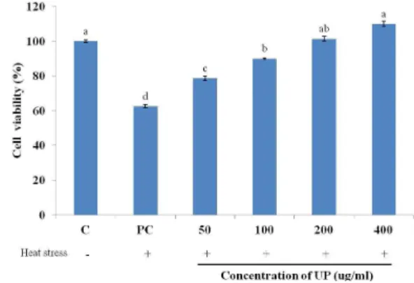
Fig. 1. The effect of UP treatment on the cell viability of heat stressed cells incubated for 24 hours. The results are expressed as mean + SD. Bars with different superscript are signi- ficantly different (P<0.05).
ATG ACC TCC TGG CAC TTG; HSP90 forward:
GTG TGC AAC AGC TGA AGG; HSP90 reverse: ACA GCA GCA CTG GTG TCA;
HSF1 forward: TGT TTG ACC AGG GCC AGT TT; HSF1 reverse: GTT CGA CTG CAC CAG TGA GA.
8. Western blot analysis
Protein was extracted by adding protein extraction solution (iNtRON Biotechnology). The lysates were clarified by centrifugation at 15,000 rpm for 15 min at 4ºC and the protein content of the supernatant was determined using a modified Bradford assay. Diluted 30 µg of the protein samples were separated by SDS- polyacrylamide gel electrophoresis and transferred to nitrocellulose transfer membranes. The membranes were blocked with 5% skimmed milk and hybridized with the following primary antibodies HSP70, HSP90, HSF1 and GAPDH (Abcam). Specific proteins were identified by further incubation of the corresponding membranes with horseradish peroxidase-conjugated secondary antibodies followed by a treatment with enhanced chemiluminescence (AB Frontier). The target proteins were exposed and detected to radiographic film.
9. Statistical analysis
All quantitative data are representative of at least three independent experiments and the results were expressed as means + S.D. Differences between means were evaluated using ANOVA test (one-way) followed by Duncan’s Multiple Range Test. Differences were considered signifi- cant at p<0.05. The statistical software package SAS v9.2 was used for the analysis.
RESULTS AND DISCUSSION
1. Effect on RAW264.7 cell viability
Exposure to heat stress significantly decreased the viability of RAW264.7 cells. As shown in Fig. 1, when cells are exposed to heat stress, the number of proliferating cells decreased significantly. However, upon treatment with increasing concentration of UP, the viability of cells significantly increase when compared to the heat stressed control and when treated with 400 ug/ml UP, the viability of cells have no significant difference with the cells that were not exposed to heat stress.
This shows that heat stress inhibits the proliferation of RAW264.7 cells when exposed to heat stress but the addition of different concentrations of UP negates this inhibitory effect in a dose dependent manner. This effect of UP may result from the stimulation of macrophage functions and/or amelioration of the heat stress condition.
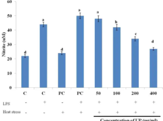
Fig. 2. The effect of UP on NO production in LPS-induced RAW264.7 macrophages incubated for 24 hours. The results are expressed as mean + SD. Bars with different superscript are signi- ficantly different (P<0.05).
2. Effect on nitric oxide (NO) production
NO assay was performed for better represent- ation of the protective effect of UP against heat stress. The effect of UP on NO production in LPS-induced RAW264.7 cells was presented in Fig. 2. NO production was down-regulated in a dose-dependent manner from 50 µg/mL to 400 µg/ml. Although exposure to heat stress did not drastically affected the production of NO, the treatment of UP in heat stressed RAW264.7 cells strongly inhibited the production of NO.
Macrophages play important roles in inflam- mation through the production of several pro- inflammatory molecules including NO. However, excessive production of NO has been associated with a wide range of inflammatory diseases including ischemic perfusion, hypertension, septic shock and arteriosclerosis (Terao, 2009).
In the present study, UP demonstrated that it can effectively inhibit the inflammatory process by suppressing the production of NO in LPS-
induced RAW264.7 macrophage cells. This result is in accordance with Van Morelle at al.
(2002), wherein induction of Hsp70 in vitro by heat shock response or through over expression reduced mortality in experimental models of septic shock and endotoxemia as well as down- regulate inflammatory gene expression.
3. Effect on phagocytic activity
Macrophages are the cells of the immune system, in particular of the innate immune system, which are responsible for the cell- mediated defense against invading foreign material through phagocytosis. Heat stress can negatively affect the animal’s immune competence to some bacterial or viral infection. If this characteristic function is lacking or disturbed then organisms are not able to protect themselves from external pathogens, such as bacteria and fungi, which are usually eliminated by phagocytosis. Thus, phagocytosis is an important indicator of macrophage effector activity. In this study, we evaluated the effect of heat stress on the phagocytic activity of RAW264.7 macrophage cells.
This is in agreement with previous studies that have shown that extracellular HSPs exert immunostimulatory effects (Johnson and Fleshner, 2006). Wang et al. (2006) have shown that extracellular HSP70 binds to the lipid raft microdomain on the plasma membrane of macrophages and enhances their phagocytic ability.
Fig. 3 showed that the exposure of RAW264.7 macrophage cells to heat stress significantly reduced the phagocytic activity of the cells.
However, treatment with UP significantly restored the phagocytic activity of heat stressed RAW264.7 cells in a dose dependent manner.
Fig. 3. The effect of UP on the phagocytic activity of heat stressed RAW264.7 incubated for 24 hours. The results are expressed as mean + SD. Bars with different superscript are signi- ficantly different (P<0.05).
Fig. 4. Effect of UP on the mRNA expres- sions of heat stress related genes in Raw264.7 macrophages.
The results demonstrated that UP can reverse the negative effect of heat stress on the phagocytic activity of macrophages and dose dependently ameliorates their function.
These data are clearly showing that the addition of UP was able to provide certain protection on heat stressed RAW264.7 cells.
To further investigate the mechanism of action behind the cytoprotective activity of UP, we analyzed the expressions of heat stress related genes and proteins.
4. RT-PCR for heat stress related genes
The Heat shock proteins (HSPs) are a group of highly conserved proteins that are induced in both prokaryotes and eukaryotes by elevated temperatures or a variety of cellular stresses (Ross et al., 2003). HSPs are traditionally classified by their molecular weight and the best understood are in the 27, 60, 70, 90 and 110kda classes (Prohaszka and Fust, 2004). The exact molecular targets for HSP protection from cellular stress remain unresolved, but cell membranes, DNA and proteins have all been
role that HSP have in cytoprotection during heat stress is shown by the fact that HSP overexpression protects against hyperthermia and cerebral ischemia during the heat stroke (Lee et al., 2006). The effect of UP on heat stressed Raw264.7 cells were determined further by the mRNA expressions of heat stress related genes with RT-PCR.
UP upregulated the biological activity of RAW264.7 macrophages such as proliferation, inhibition of NO production and phagocytic activity recovery under heat stress. But signals from the cells against heat stress should be revealed. We measured several heat shock protein expressions including Heat Shock Factor 1 (HSF1), Heat shock protein 70 (Hsp70) and Hsp90.
As seen in Fig. 4, heat stress induces HSF1, Hsp70 and Hsp90 significantly. After treatment with UP, the expression of HSF1 was not upregulated drastically but the gene expression was kept at an induced level. On the other hand, Hsp70 and Hsp90 expression are dose dependently upregulated by UP treatment.
5. Effects on heat stress related proteins As seen in Fig. 5, the same pattern was observed on the protein expression of heat stress related proteins as with the mRNA expression of heat stressed RAW264.7 cells treated with UP. HSF1 was even more induced
Fig. 5. Effect of UP on the protein expres- sions of heat stress related genes in Raw264.7 macrophages.
and dose dependently regulated by UP treatment.
Regulation of the transcription of heat shock protein genes is mediated by the interaction of heat shock factor (HSF) transcription factors with heat shock elements in the heat shock protein gene promoter regions (Voellmy, 1994).
In vertebrates, four HSFs have been identified, of which HSF1 and HSF2 are ubiquitously expressed but the main heat shock factor with a role in vertebrates’ response to physiological and environmental stress is HSF1 (Zuo et al., 1994). UP was able to induce and upregulate the mRNA and protein expression of HSF1 as seen in Fig. 4 and 5.
HSF1 is a central player controlling the heat stress response. Under heat shock conditions it upregulates several hundred genes including Hsp70 and Hsp90. Under normal condition, as a client protein, HSF1 is kept in an inactive monomeric form through the transient interaction with Hsp90 (Zou et al., 1998). Recent studies in plants and mammals revealed that Hsp90 is vital to stabilize Nod-like receptor proteins, which are conserved immune sensors to recognize pathogens (Mayor et al., 2007). It functions as part of a multichaperone complex via associ- ation with co-chaperone Hsp70.
The precise functions of the Hsp70 and Hsp90 protein have not been completely defined.
However, the high degree of conservation of
cell survival in various conditions, suggests that these HSPs are critical for both normal cellular function and survival after a stress.
Even though the exact mechanism of action for the cellular thermotolerance improvement through the increased levels of HSP is still unknown, it is enough to speculate that Hsp70 is involved in preventing protein denaturation and/or processing denatured proteins and protein fragments that are produced by stressors such as hyperthermia (Kregel, 2002).
CONCLUSIONS
In summary, many researchers have demon- strated that most HSPs have strong cytopro- tective effects, are involved in many regulatory pathways, and behave as molecular chaperones for other cellular proteins. The present study, suggests that the treatment of UP (up to 400 ug/ml) was able to enhance the biological activities of heat-stressed RAW264.7 macrophage cells and was able to induce the expression of heat stress related genes that can possibly account for the cytoprotective effect of UP on heat stressed RAW264.7 cells. Since the mechanisms for these cytoprotection are not yet fully understood, further research about the signaling pathway for the action of UP in ameliorating heat stress must be pursued.
ACKNOWLEDGEMENT
This work was carried out with the support of Cooperative Research Program for Agriculture Science & Technology Development (Project title: Study on development of livestock adaptation tool for climate change, Project No. PJ01007603) Rural Development Administration, Republic of Korea.
REFERENCES
1. Cheng, A.W., Wan, F.C., Wang, J.Q., Jin, Z.Y., Xu, X.M., 2008. Macrophage immunomodulatory activity of polysaccharides isolated from Glycyrrhiza uralensis fish.
International Immunopharmacology. 8:43-50.
2. Collier, R.J., Baumgard, L.H., Lock, A.L., Bauman, D.E., 2005. Physiological Limitations, Nutrient Partitioning. In Yield of Farmed Species. Constraints and Opportunities in the 21st Century. Nottingham University Press, Nottingham UK, pp.351-377.
3. Johnson, J.D., Fleshner, M., 2006. Releasing signals, secretory pathways, and immune function of endogenous extracellular heat shock protein 72. J. Leukoc. Biol. 79:425-434.
4. Kapila, N., Kishore, A., Sodhi, M., Sharma, A., Mohanty, A.K., Kumar, P., Mukesh, A., 2013. Temporal Changes In mRNA Expression Of Heat Shock Protein Genes In Mammary Epithelial Cells Of Riverine Buffalo In Response To Heat Stress In Vitro. I. J. Anim. Bio. 3:5-9.
5. Kregel, K.C., 2002. Molecular Biology of Thermoregulation Invited Review: Heat shock proteins: modifying factors in physiological stress responses and acquired thermotolerance.
J. Appl. Physiol. 92:2177-2186.
6. Lee, W.C., Wen, H.C., Chang, C.P., Chen, M.Y., Lin, M.T., 2006. Heat Shock Protein 72 Overexpression Protects Against Hyper- thermia, Circulatory Shock and Cerebral Ischemia During Heat Stroke. J. Appl.
Pysiol. 100:2073-2082.
7. Mayor, A., Martinon, F., De Smedt, T., Petrilli, V., Tschopp, J., 2007. A crucial function of SGT1 and HSP90 in inflammasome activity links mammalian and plant innate immune responses. Nat.
Immunol. 8:497-503.
8. Morange, F., 2006. HSFs in Development.
Handbook of Experimental Pharmacology.
9. Prohaszka, Z., Fust, G., 2004. Immunological Aspects of Heat-Shock Proteins - The Optimum Stress of Life. Molecular Immunology. 41:
29-44.
10. Ross, O.A., Curran, M.D., Crum, K.A., Rea, I.M., Barnett, Y.A., Middleton D., 2003. Increased Frequency of the 2437 T Allele of the Heat Shock Protein 70-Hom Gene in an Aged Irish Population. Experi- mental Gerontology. 38:561-565.
11. Terao, J., 2009. Dietary flavonoids as antioxidants. Forum Nutr. 61:87-94.
12. Wang, R., Kovalchin, J.T., Muhlenkamp, P., Chandawarkar, R.Y., 2006. Exogenous heat shock protein 70 binds macrophage lipid raft micro domain and stimulates phagocytosis, processing and MHC-II pre- sentation of antigens. Blood. 107:1636-1642.
13. Wang, D., Xia, M., Cui, Z., 2006. New triterpenoids isolated from the root bark of Ulmus pumila L. Chem. Pharm. Bull. 54:
775-778.
14. Van Morelle, W., Wielockx, B., Mahieu, T., Takada, M., Taniguchi, T., Sekikawa, K., Libert, C., 2002. HSP70 protects against TNF-induced lethal inflammatory shock.
Immunity 16:685-695.
15. Voellmy, R., 1994. Transduction of the stress signal and mechanisms of transcriptional regulation of heat shock/stress protein gene expression in higher eukaryotes. Crit. Rev.
Eukaryot. Gene Expr. 4:357-401.
16. Zuo, J., Baler, R., Dahl, G., Voellmy, R., 1994. Activation of the DNA-binding ability of human heat shock transcription factor 1 may involve the transition from an intramo- lecular triple-stranded coiled-coil structure.
Mol. Cell Biol. 14:7447-68.
17. Zou, J., Guo, Y., Guettouche, T., Smith, D.F., Voellmy, R., 1998. Repression of heat shock transcription factor HSF1 activation by HSP90 (HSP90 complex) that forms a stress-sensitive complex with HSF1.