Surgical Treatment for T4 Non-small Cell Lung Cancer Invading Mediastinal Structures
Background: Non-small cell lung cancer (NSCLC) with invasion of mediastinal structures is classified as stage IIIB, and has been considered surgically unresectable. However, in a selected group of these patients, better results after surgical resection compared to non-surgical group have been reported. The aim of this study is to evaluate the role of surgical resection in treatment of mediastinal T4 NSCLC. Material and Method: Among 1067 patients who underwent surgical intervention for non-small cell lung cancer from Aug 1987 to Dec 2001 in Korea cancer center hospital, 82 patients had an invasion of T4 mediastinal structures (7.7%). Resection was possible in 63 patients (63/82 resectability 76.8%). Their medical records in Data Base were reviewed, and they were followed up completely until Jun 2002. Surgical results and prognostic factors of NSCLC invading mediastinal structures were evaluated retrospectively. Result: Lung cancer was resected completely in 52 patients (63.4%, 52/82). Lung resec- tion was lobectomy (or more) in 14, pneumonectomy in 49. The mediastinal structures invaded by primary tumor were great vessel (61.9%), heart (19%), vagus nerve (9.5%), esophagus (7.9%), and vertebral body (7.9%). Nodal status was N0 in 11, N1 in 24, and N2 in 28 (44.4%). Neoadjuvant therapy was done in 6 (9.5%, 5 chemother- apy, 1 radiotherapy), and adjuvant therapy was added in 44 (69.8%, 15 chemotherapy, 29 radiotherapy) in resec- tion group (n=63). Complication was occurred in 23 (31.7%), and operative mortality was 9.5% in resection group.
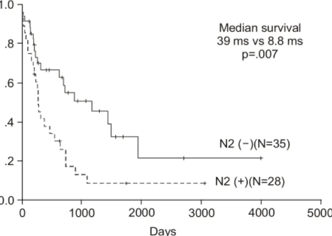
Median and 5 year overall survival including operative mortality was 18.1 months and 21.7% in resection group (n=63), 6.2months and 0 % in exploration only group (n=19, p=.001), 39 months and 32.9% in N2 (-) resection group (n=35), and 8.8 months and 8.6% in N2 (+) resection group (n=28, p=.007). The difference of overall survival by mediastinal structure was not significant. Conclusion: The operative risk of NSCLC invading mediastinal structures was high but acceptable, and long-term result of resection was favorable in selected group. Aggressive
종격동 구조물을 침범한 T4 비소세포폐암의 수술적 치료
resection is recommended in well selected pateints with good performace and especially N2 (-) NSCLC with mediastinal invasion.
(Korean J Thorac Cardiovasc Surg 2004;37:349-355) Key words: 1. Cancinoma, non-small cell, lung
2. Neoplasm metastasis
3. Mediastinal lymph nodes
4. Neoplasm staging
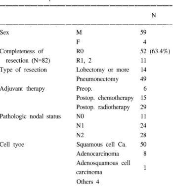
Table 1. Patient's profile Table 2. Invaded structures
Table 3. Procedures of resection
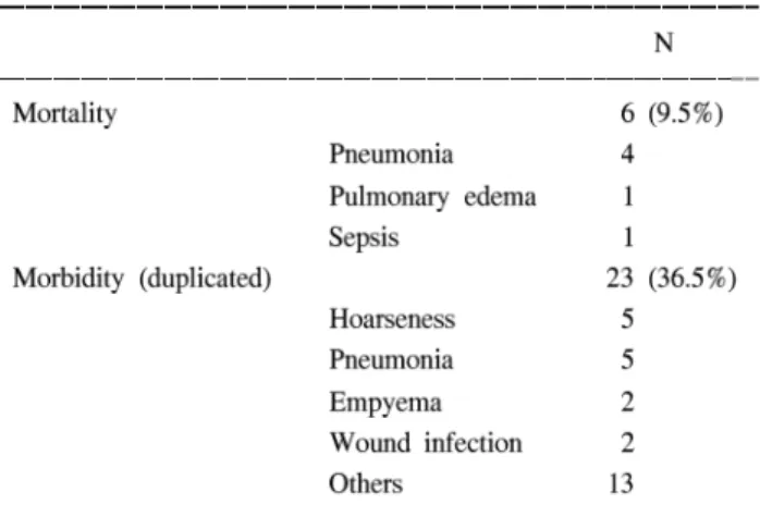
Table 4. Mortality and morbidity Table 5. Disease status
Table 6. Recurrence and treatment
Fig. 1. Postoperative overall survival curve after resection of Non-small cell lung cancer invading T4 mediastinal structures (5 YSR=21.7%).
0 1.0
140 Months
0.0
100 80 60 40 20 .8
.6
.4
.2
120
Fig. 2. Comparison of survival between resection and non- resection group (include Op. mortality)(p=.001).
0 1.0
5000 Days
N2 (+)(N=28) N2 ( )(N=35)- Median survival 39 ms vs 8.8 ms
p=.007
0.0
2000 3000 1000
.8
.6
.4
.2
4000
Fig. 3. Comparison of survival between N2 (+) and N2 (-) group (include op. mortality)(p=.007).
0 1.0
5000 Days
Non-resection (N=19)
Resection (N=63) Median survival 18.1 ms vs 6.2 ms
p=.001
0.0
3000 2000
1000 .8
.6
.4
.2
4000
Fig. 4. Comparison of effect on survival after postop. adjuvant therapy (include op mortality)(p .001).
0 1.0
12 Yrs
Adjuvant Tx (N=43)
No adjuvant Tx (N=20)
0.0 2 4 6
.8
.6
.4
.2
8 10
=국문 초록=
배경:
대상 및 방법:
결과:
결론:
중심 단어