전통 장류로부터 Exopolysaccharide 생성 유산균의 분리 및 특성
윤혜주1,2, 이유정1, 여수환1, 박혜영1, 박희동2, 백성열1
*
1농촌진흥청국립농업과학원농식품자원부발효식품과
2경북대학교농업생명과학대학식품공학과
Received : February 14, 2013 / Revised : April 29, 2013 / Accepted : April 30, 2013
서 론
유산균은자연계에널리분포하고탄수화물을혐기적으로 이용하여젖산을생성하는미생물로서유제품
,
육류,
채소등 의다양한발효식품가공에종균으로사용하고있으며,
식품 의보존성향상뿐만아니라관능적특성및영양적가치향 상에기여하고있다.
또한일부유산균속은다당류를생합성하여제품의조직과점성향상에도기여하고있다
[25].
미생물생성다당류에관한연구는
1942
년Leuconostoc mesenteroides
가생산하는dextran [12]
이혈장증강제로개 발된이래pullulan [7], xanthan gum [8]
등을비롯한여러 가지다당류에대한기초및응용연구가진행되어왔다.
미 생물이생성하는다당류는상업적으로이용되는식물다당류에비해물성이다양하고독특하며
,
부가가치가매우높아 각종산업의기능성신소재로서의잠재력이매우크다[32].
미생물 유래 다당류는크게 세포벽의일부로 존재하는
intracellular polysaccharide,
세포벽구조성분인structural polysaccharide,
세포벽 외부에 존재하는extracellular polysaccharide
등으로 나눌 수 있다.
특히extracellular polysaccharide
는 세포와의 구조적 관계에 따라slime, capsular, microcapsular
의세가지형태로분류할수있으 며이들을총칭하여exopolysaccharide (EPS)
라한다[30].
EPS
는세포벽의일부로서세포벽주위에협막을형성하거나세포벽외부에점질형태로서발효중에축적되는미 생물다당류로
, 1
차또는2
차대사산물이다[14]. EPS
는에 너지원으로이용되는것은아니고건조,
식균작용,
항생제,
독성화합물,
삼투압스트레스등과같은외부환경으로부터자신을보호하는작용을한다
[13]. EPS
는미생물이가장많이생성하는다당류로
,
배양액으로부터회수가쉽고정제비 용이적어상업적인잠재력이가장높은다당류이다[31].
최Isolation and Characterization of Exopolysaccharide Producing Lactic Acid Bacteria from Korean Soy Sauce and Soybean Paste. Yun, Hye Ju
1,2, You Jung Lee
1, Soo-Hwan Yeo
1, Hye Young Park
1, Heui-Dong Park
2, and Seong Yeol Baek
1*.
1Fermented Food Division, Department of Agro-food Resource, NAAS, RDA, Suwon 441-853, Korea,
2School of Food Science & Biotechnology, Kyungpook National University, Daegu 702-701, Korea
Three slime-forming lactic acid bacteria were isolated from traditional Korean fermented soy sauce and soybean paste and shown to produce exopolysaccharides (EPS) in sucrose media. By isolating the strains, examining their morphological charac- teristics and determining their 16S rDNA sequences, N58-5 and K6-7 were identified as Leuconostoc mesenteroides and N45- 10 as Leuconostoc citreum. The acid and bile tolerances of these three strains were investigated. Amongst the three lactic acid bacteria, Leuc. citreum N45-10 exhibited the highest viability (10
5-10
6CFU/ml) in 0.05 M sodium phosphate buffer (pH 0.3) for 2 h, in artificial gastric juice for 2 h and in 0.3%, 0.5% oxgall for 24h. Leuc. mesenteroides K6-7, N58-5 and Leuc. citreum N45- 10 were grown in sucrose liquid medium and 8.16 g/L, 3.65 g/L, 16.17 g/L of EPS was collected, respectively. The hydrolyzed EPS was analyzed by HPLC in order to determine the sugar composition of EPS. Leuc. mesenteroides K6-7 and N58-5 showed two peaks indicating glucose and fructose, thus they were determined to be hetero-type polysaccharides. Leuc. cit- reum N45-10 showed only the glucose polymer, indicating it to be a homo-type polysaccharide. In addition, all three lactic acid bacterial hemolysis did not demonstrate a clear zone in blood agar in the area surrounding a lactic acid bacteria colony.
Keywords: Lactic acid bacteria, exopolysaccharide, soy sauce, Leuconostoc sp.
*Corresponding author
Tel: +82-31-299-0581, Fax: +82-31-299-0554 E-mail: [email protected]
© 2013, The Korean Society for Microbiology and Biotechnology
근에유산균이생성하는
EPS
가식용다당류로서제품의점 도를높이고안정제,
유화제,
겔화및수분결합물질 등다 양한용도로사용이가능하므로EPS
를생산하는유산균에대한관심이점차높아지고있다
[33].
유산균이생성하는
EPS
는크게homo-polysaccharide
와hetero-polysaccharide
로나뉘어진다. Homo-polysaccharide
는 주로프락토오스와글루코오스와같은한가지형태의당으 로만 구성되어있는다당류로, Streptococcus salivarius
와Str. mutans
가생산하는fructan type
의levan
과inulin
이 있으며, glucan type
으로는Leuconostoc mesenteroides subsp.
mesenteroides
와Leuc. mesenteroides subsp. dextranicum
가 생산하는dextran
과Leuc. mesenteroides
가 생산하는alternan, Str. mutans
와Str. sobrinus
가생산하는mutan
등이있다[5, 25]. Hetero-polysaccharide
는2
종류이상의단 당,
주로glucose, galactose, fructose, rhamnose
가서로다 른비율로구성되어있으며,
경우에따라N-acetyl aminosugar, phosphate, acetyl, glycerol
과같은당이아닌물질들이존재 하기도 한다.
주로Lactococcus lactis subsp. lactis, Lac.
casei, Lac. sake
등의중온성유산균들과Lac. acidophilus, Lac. delbrueckii subsp. bulgalicus, Str. thermophilus
등 의 고온성 유산균들에서 생산된다.
일반적으로hetero- polysaccharide
는homo-polysaccharide
에비해생산성이낮 으며,
일시적으로생산된다는특성이있다.
이러한현상은미생물에서
EPS
생산에관여하는코딩유전자의불안정성에따른손실또는재배열에기인하며
, EPS
생산의유전적불안정성은산업적이용성에심각한문제가된다
[6, 25].
최근
EPS
가과거의단순물성기능소재가아닌생리기능 소재로 주목을 받고 있다.
유산균 중Bifidobacteria
유래EPS
와Lactobacillus delbrueckii subsp. bulgalicus, Lac.
helverticus var. jugurti
로부터유래한EPS
에항암효과가있다고보고되었다
[17].
지금까지유산균에서생성된EPS
에관한많은연구가이루어졌으나
,
대부분요구르트와치즈등 과같은발효유제품에서분리한유산균에국한되고우리나 라전통발효식품에서분리한유산균에관한연구는미흡한 실정이다[19].
전통장류는예로부터전승된우리나라의대표적인대두 발효식품으로서곡류단백질에서부족되기쉬운필수아미 노산
,
지방산,
유기산,
미네랄및비타민류등의영양소를보 충해줌으로써영양학적으로중요한기능을가진다[3].
최근,
전통발효식품에대한새로운인식과관심이높아짐에따라 이에대한연구가활발히이루어지고있으며[3, 21],
특히장 류에서생리활성물질과항암효과에대한많은연구결과로장류에대한이미지가새롭게변화하고있다
[21].
발효과정중미생물이생산하는
2
차대사산물의혈전용해능,
항산화 능,
항암활성,
면역증강,
혈압강하및항균효과등다양한생리활성이보고됨에따라기능성식품으로서전통장류에대 한관심이증가하고있는추세이다
[2, 22].
이와같이간장및 된장과같은전통장류의기능성에관한연구가활발하게진 행되면서전통장류에서분리된유산균의활용에대한인식 이재평가되고있다.
따라서본연구에서는 전통장류에서분리한유산균중
EPS
생산유산균을선별하였고,
내산성,
인공위액및인공 담즙저항성시험을통하여선발유산균의장내생존가능 성을조사하였으며, EPS
를분리,
정제하여그특성에대하 여연구하였다.
재료 및 방법
시료
본실험에서사용한재래식간장과된장은경기도양주시 와남양주시지역의재래시장에서구입하거나가정에서직 접담근장류를수집하였으며
,
표준균주Leuc. mesenteroides
KACC12312
는농촌진흥청한국농업미생물보존센터에서분양받아사용하였다
.
분석에사용한시약은Sigma Aldrich (ST. Louis, USA)
에서구입하여사용하였다.
균주 배양 및 선발
수집한장류시료
10 g
을90 ml
의0.85% NaCl
로현탁하 여MRS
고체배지에100
µl
도말하여37
oC
에서48
시간배 양하였다.
배양된미생물의형태적차이를이용하여1
차선 별하고,
유산균으로 확인된 균주를 슈크로오스 배지(1%
tryptone, 0.5% yeast extract, 0.5% dipotassium phosphate, 0.5% diammonium citrate, 5% sucrose, pH 7.0)
에도말하 여37
oC
에서48
시간배양하여mucoid colony
를나타내는점 질균을선별하였다.
이균주를슈크로오스액체배지에배양 하여점성물질을분비하는것을재확인한다음EPS
생성균 주로선발하여실험에사용하였다.
EPS 생산 균주의 동정
DNA
유전자 염기서열 분석은Solutions for Generic Technologies (Solgent)
사에의뢰하여분석하였으며, primer
로는Universal PCR primer 27F(5'-AGAGTTTGATCC TGGCTCAG-3')
와1492R(5'-GGTTACCTTGTTACGACTT- 3')
을사용하였다.
계통수작성은Lasergene
사의DNASTAR pro software (SeqMan Pro)
와The National Center for Biotechnology Information (NCBI, http://www.ncbi.nlm.
nih.gov/)
에서제공하는Advanced blast search
프로그램을통하여
GenBank
에보고된유사균주와의염기서열을비교,
계통분류학적유연관계를분석하였으며
, MEGA v4.0
을이 용하여Tamura-Nei distance model
과neighbor-joining
method [26]
에의해계통수를작성하였다.
내산성 및 인공위액 저항성분리균주의산저항성은단순산성
pH
에대한내성실험 과체내소화관조건과유사한환경에서측정하기위하여인 공위액내성실험을실시하여확인하였다.
산성pH
에대한 내성은0.05 M sodium phosphate
용액(pH 3.0)
을사용하여 측정하였다[24].
인공위액내성실험은Kobayashi
등[18]
의 방법을변형하여1 N HCl
로pH 3.0
으로조정한MRS
액체배지에펩신을
1,000 unit/ml
가되도록첨가하여인공위액을조제
,
실험하였다. EPS
생성을위해분리균주를슈크로 오스액체배지에1%(v/v)
접종하여37
oC
에서48
시간배양한 후,
원심분리(10,000
×g, 5 min, 4
oC)
하여균체를회수하였 다.
제거된상징액과동량의0.05 M sodium phosphate
용액(pH 3.0)
과인공위액을각각첨가하여37
oC
에서2
시간배양 한다음생균수를측정하여내산성과인공위액에대한저항 성을평가하였다.
대조군은MRS
배지에서배양하여(37
oC,
48 h), EPS
를생성하지않은조건에서실험구와동일하게처리하였다
.
인공 담즙 저항성
인공 담즙은
MRS
액체배지에0.45
µm filter
로여과한oxgall
용액을0.3%
첨가하여제조하였다[28].
담즙에대한 저항성은슈크로오스액체배지에균주를1%(v/v)
접종하여37
oC
에서48
시간배양한다음인공위액에2
시간처리하여 후인공담즙저항성을실험하였다.
인공위액처리를거친 유산균배양액을원심분리(10,000
×g, 5 min, 4
oC)
하여상징 액을제거,
균체를회수하였으며회수된균체에조제된인공 담즙액을제거한상징액과동량으로첨가하여현탁시킨후37
oC
에서24
시간배양하였고,
생균수를측정하여인공담즙 에대한저항성을조사하였다.
대조군은MRS
배지에서배 양하여(37
oC, 48 h), EPS
를생성하지않은조건에서실험구 와동일하게처리하였다.
EPS 분리, 정제 및 정량
분리균주를 슈크로오스 배지
(1% tryptone, 0.5% yeast extract, 0.5% dipotassium phosphate, 0.5% diammonium citrate, 5% sucrose, pH 7.0)
에접종하여37
oC
에서48
시간 배양하여EPS
를생성시켰다.
이배양액을4
oC
에서원심분 리(10,000
×g, 25 min, 4
oC)
하여균체를제거하고,
회수된상 징액에2
배냉각된95%
에탄올을첨가하여4
oC
에서15
시간 침전시켰다.
이침전물을다시원심분리(10,000
×g, 25 min,
4
oC)
하여회수하고,
남은에탄올을건조시킨다음동결건조하여이를
crude EPS
로하였다[29].
crude EPS
를정제하기위해배양액에trichloloacetic acid
를최종농도
4%(w/v)
가되도록첨가하고4
oC
에서2
시간처 리한후원심분리(10,000
×g, 25 min, 4
oC)
하여균체와침전 된단백질을제거하였다.
상징액은회수한후0.2
µm filter
로여과하여남은단백질을제거하고,
회수된상징액에2
배 량의95%
에탄올을첨가하여4
oC
에서15
시간침전시켜분 리하였다.
침전물은원심분리(10,000
×g, 25 min, 4
oC)
하여 회수하였고,
남은에탄올을건조시킨후3
차증류수에용해 하여dialysis sack
에넣고4
oC
에서24
시간동안투석한다 음,
동결건조하였다[34].
배양액에대한EPS
의생성량은g/L
로나타내었다.
EPS 구성 당 분석
EPS
의구성당은EPS
가수분해물을HPLC (Waters Co., USA)
로분석하였다. EPS
를2 N
황산으로100
oC
에서6
시간 동안 가수분해하고, 1 N NaOH
로 중화한 다음0.45
µm filter
로여과하여EPS
가수분해물시료로사용하였다.
분석 컬럼은탄수화물분석용컬럼(carbohydrate analysis column) (3.9 mm
×300 mm, Waters Co., USA)
을사용하였으며,
이 동상으로는75% (v/v)
아세토니트릴(acetonitrile)
을사용하 였다.
이동상의유속은1.5 ml/min
으로 하였으며,
시료는auto-sampler
를 이용하여20
µl
주입하여RI (Refractive Index)
검출기로검출하였다.
용혈성 및 젤라틴 액화시험
혈액한천배지
(tryptic soy agar
에5% sheep blood
첨가)
에분리한유산균을도말하고, 37
oC
에서48
시간배양한다 음균체주위에투명환의생성여부로용혈성을판단하였다[20].
젤라틴 영양 배지(beef extract 0.3%, peptone 0.5%, gelatin 12%)
에분리균주를접종한다음37
oC
에서48
시간 배양한후젤라틴영양배지를4
oC
에약4
시간동안냉장방 치하여배지의응고여부를확인하였다.
배지가응고되지않 으면액화반응양성으로판정하였다[27].
결과 및 고찰
EPS 생성 유산균의 선발 및 동정
전통장류시료에서미생물의형태적차이를분석
, 1
차선별하여약
1,000
여균주를선발하였고,
유산균으로확인된약
200
균주를슈크로오스배지에배양하여점성물질을분비 하는균주중높은활성을보이는N45-10, K6-7
및N58-5
의3
균주를선발하였다.
생성된EPS
는투명하면서물엿정도의 퍼짐을나타냈으며N45-10
균주가가장많은EPS
를생성하 였다.
선발한
3
종류유산균의16S rDNA
의염기서열을분석한결과
, N45-10, K6-7, N58-5
는Leuconostoc sp.
와높은상동성을 나타냈다
.
그리고 균주의 계통학적 유연관계를16S rDNA
유전자염기서열을기초로Leuconostoc
속의표준종 들과의유사도를조사한결과, N45-10
균주는Leuc. citreum
과K6-7, N58-5
균주는Leuc. mesenteroides
와100%
상동 성을보여기존에알려진유산균으로동정되었다.
내산성 및 인공 위액 저항성
유산균이살아있는상태로장내에도달하기위해서는염 산과각종효소가존재하는위를통과하여야하며
,
이때위 의pH
는상당히큰가변성을지니고있고음식물의섭취여 부에따라pH 2-8
의범위를나타낸다[10].
따라서위에도달 하는대부분의미생물은사멸하거나원래갖고있던활성이 저하된다.
전통장류에서선발된EPS
생성유산균N45-10, K6-7
및N58-5
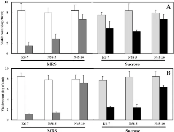
의산저항성에대한결과를Fig. 1
에나타내 었다.
슈크로오스액체배지에서EPS
생성을유도한균주를0.05 M sodium phosphate
용액(pH 3.0)
이나인공위액에서2
시간처리했을때생균수는EPS
를생성하지않은균주보다높게나타났다
. 3
균주중에서N45-10
은EPS
생성유무 와상관없이높은생균수(10
5-10
6CFU/ml)
를유지했을뿐만아니라높은내산성을가지고있어위장에서장으로이동할 수있는가능성이시사되었다
.
이결과는김치에서김등[15]
이 분리한
Leuc. kimchii GJ2, Leuc. citreum C3, Leuc.
mesenteroides C11
균주및이등[20]
이분리한Lac. plantarum NO.1
균주가EPS
를생성하였을때0.05 M sodium phosphate
용액(pH 3.0)
이나인공위액에서2
시간이경과한후에도사 멸하지않고초기균수를유지하여높은내산성을나타내었 다는결과와유사하였다.
반면K6-7, N58-5
균주는N45-10
균주보다비교적낮은생균수(10
1CFU/ml)
를나타내었다.
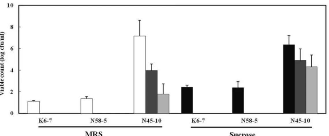
인공 담즙 저항성
유산균이장내에서정상적인기능을수행하려면장내담 즙의농도
(0.6 g/L)
보다훨씬많은oxgall
이함유된배지에서성장할수있는내성을가져야한다
[23].
섭취된유산균은실제로위를통과하여장으로이동되는것을고려하여선발된
3
균주를 인공 위액에서2
시간 처리한 후, 0.3%
및0.5%
oxgall
을함유한인공담즙액에처리하였다.
그결과Leuc.
mesenteroides K6-7
과N58-5
는 거의 사멸하였으나, Leuc.
citreum N45-10
은비교적높은생균수(10
3-10
4CFU/ml)
를Fig. 1. Acid tolerance of lactic acid bacteria isolated from korean fermented soy sauce and soybean paste in 0.05 M sodium phosphate (A) and artificial gastric juice (B).
Cells were precultured in MRS medium at 37
oC for 48 h ( ) and sucrose medium at 37
oC for 48 h ( ), subsequently treated in 0.05 M
sodium phosphate (pH 3.0) ( ) and artificial gastric juice (pH 3.0) ( ) for 2 h, respectively.
유지하는것으로보아담즙액저항성이가장우수하였다
(Fig.
2).
또한N45-10
균주는김등[15]
이보고한김치에서분리 한Leuc. kimchii GJ2, Leuc. citreum C3, Leuc. mesenteroides
C11
이높은내산성의기능을지니면서동시에담즙에대한높은저항성을나타낸다는결과와유사하였다
.
유산균의EPS
생성유무에따른담즙저항성을비교한결과
, MRS
배지보다슈크로오스배지에배양하였을때
,
생균수가더높게나타났다
.
이것은EPS
생성이유산균의세포벽주위에보호막으로작용한결과로생각된다
.
EPS 분리, 정제 및 정량
선발된유산균의탄소원으로슈크로오스를이용하여
EPS
생성능을 조사하였다. Leuc. citreum N45-10
과Leuc.
mesenteroides K6-7, N58-5
를슈크로오스액체배지에배양 한 후,
에탄올 침전법을 이용하여 각각16.173, 8.167
및3.652 g/L
의crude EPS
를 회수하였다(Table 1). Leuc.
citreum N45-10
은Leuc. mesenteroides K6-7, N58-5
보다2-5
배(16.173 g/L)
이상높은EPS
생산량을나타내었다.
이는 김 등
[15]
이 김치에서 분리한Leuc. citreum C3
에서16.46 g/L
의EPS
가생산되었다는결과와유사하였다.
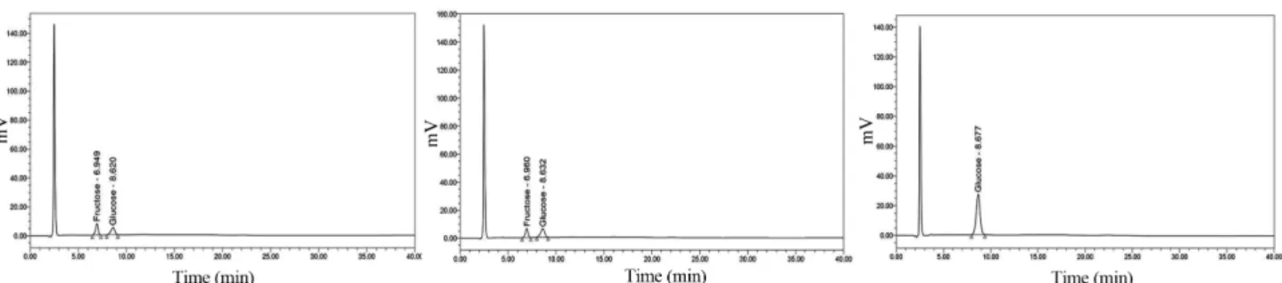
EPS 구성 당 분석
EPS
가수분해물을HPLC
로분석한결과, Leuc. citreum N45-10
는글루코오스, Leuc. mesenteroides K6-7, N58-5
는 프락토오스와글루코오스가주요구성당으로동정되어(Fig. 3) Leuc. citreum N45-10
이생산한EPS
는homo-polysaccharide
로, Leuc. mesenteroides K6-7, N58-5
이 생산한EPS
는hetero-polysaccharide
로구분되었다[5, 6, 25].
따라서Leuc.
citreum N45-10
과Leuc. mesenteroides K6-7, N58-5
의EPS
생산량은homo-polysaccharide
과hetero-polysaccharide
의 차이에기인한것으로나타났다.
유산균의 안전성
유산균은전통적으로식품에사용되어왔기때문에오랜 세월 동안 안전하다고 인지되어 왔다
(GRAS, generally recognized as safe).
이러한유산균은인간의건강에유용하 며안전하다고생각되기때문에안정성및독성연구가요구되 지않았다.
그러나여러종류의감염부위에서Lactobacillus, Leuconostoc, Pediococcus, Bifidobacterium
의 일부 종(Species)
이분리되어[1, 4, 9]
식품및의약품등에분리한새 로운유산균을상업적으로사용할때에는안전성확보가중 요하다고생각되어분리유산균의안전성실험을하였다.
용혈은적혈구가파괴되는정상적인작용과적혈구의유 전적인결함이나화학물질
,
뱀등의독액,
미생물이생성하 는독성물질에의해서형성되는비정상적인작용이다[16].
생체내용혈은주로비장
,
간장,
골수세포내피계세포에서Fig. 2. Bile tolerance of lactic acid bacteria from korean fermented soy sauce and soybean paste against 0.3% and 0.5% oxgall.
Cells were precultured in MRS medium at 37
oC for 48 h, subsequently treated in artificial gastric juice for 2 h ( ) and sucrose medium at 37
oC for 48 h, subsequently treated in artificial gastric juice for 2 h ( ) subsequently and than in 0.3% oxgall ( ) and 0.5% oxgall ( ) for 24 h.
Table 1. Production of exopolysaccharides by the lactic acid bacteria isolated from korean fermented soy sauce and soybean paste.
Strains EPS (g/L)
Leuc. mesenteroides KACC 12312 1.973 ± 0.229 Leuc. mesenteroides K6-7 8.167 ± 0.332 Leuc. mesenteroides N58-5 3.652 ± 0.670
Leuc. citreum N45-10 16.173 ± 0.521
Values are means ± SD (n = 3).
일어나며
,
세포내에서용혈현상이일어나면적혈구의산소 운반기능이없어져생체내에치명적인결과를가져온다.
선 발된유산균의안전성을증명하기위해용혈성유무를평가 하여Fig. 4
와같은결과를얻었다.
본연구에사용한3
종류 의유산균모두용혈성검사에서균체주위에적혈구가파괴 되어생기는투명환이나타나지않아안전성이확인되었다.
세균의병원성은세포의침입능력에좌우되는경우가많 으며,
세포침입에는단백질분해력이필요하다.
젤라틴액화 시험은젤라틴을가수분해하는능력을판정하는시험으로단백질분해능을조사하는방법중하나이다
[11].
선발유산균의단백질분해능을조사한결과를
Fig. 5
에나타내었다.
3
종류유산균모두젤라틴액화능을나타내지않았으며,
이러한결과는세포침입을위한단백질분해능력이없음을의 미하는것으로
Leuc. citreum N45-10
과Leuc. mesenteroides K6-7, N58-5
의안전성이입증되었다.
요 약
전통장류에서점성물질을분비하는유산균을분리하고 높은활성을보이는
N45-10, K6-7
및N58-5
의3
균주를선 발하였다.
선발한3
균주의16S rDNA
의염기서열을분석한 결과N45-10
은Leuc. citreum, K6-7, N58-5
는Leuc.
mesenteroides
로동정되었다.
산저항성과인공위액저항성 은3
균주중에서Leuc. citreum N45-10
이높은생균수(10
5- 10
6CFU/ml)
를나타내어산저항성과인공위액저항성이가 장우수하게나타났으며,
인공담즙저항성은Leuc. citreum N45-10
은높은생균수(10
3-10
4CFU/ml)
를유지하여담즙액 저항성이우수하게나타났다. 3
균주들중에서Leuc. citreum N45-10
은EPS
생성량이가장많았고, MRS
배지에서배양 하여EPS
가생성되지않았을때보다슈크로오스배지에서 배양하여EPS
를생성하였을때생균수가더높게나타났Fig. 3. HPLC chromatograms of acid hydrolyte of exopolysaccharides produced by the lactic acid bacteria isolated from korean fermented soy sauce and soybean paste.
(A) Leuc. mesenteroides K6-7, (B) Leuc. mesenteroides N58-5, (C) Leuc. citreum N45-10.
Fig. 4. Hemolysis test of the lactic acid bacteria isolated from korean fermented soy sauce and soybean paste.
(A) Leuc. mesenteroides K6-7, (B) Leuc. mesenteroides N58-5, (C) Leuc. citreum N45-10, (D) Leuc. mesenteroides KACC 12312.
Fig. 5. Gelatin liquefaction test of the lactic acid bacteria isolated from korean fermented soy sauce and soybean paste.
(A) Leuc. mesenteroides K6-7, (B) Leuc. mesenteroides N58-5,
(C) Leuc. citreum N45-10, (D) Leuc. mesenteroides KACC 12312,
(E) Positive control.
다
.
이것은EPS
생성이유산균의세포벽주위에보호막으로 작용한결과로생각된다.
선발유산균3
주의EPS
생산량은 슈크로오스액체배지에서각각16.173, 8.167, 3.652 g/L
였 으며, Leuc. citreum N45-10
은 글루코오스로만 이루어진homo-polysaccharide, Leuc. mesenteroides K6-7
과N58-5
는프락토오스,
글루코오스로구성된hetero-polysaccharides
로동정되었다.
선발유산균3
주모두는용혈음성반응을나 타내고,
젤라틴액화능을나타내지않아안전성이확인되었다.
Acknowledgement
This work was supported of Cooperative Research Program for Agriculture Science & Technology Development (Project No.
PJ006764) Rural Development Administration, Korea.
References
1. Aguirre, M. and M. D. Collins. 1993. Lactic acid bacteria and human clinical infection. J. Appl. Bacteriol. 75: 95-107.
2. Ahn, Y. S., Y. S. Kim, and D. H. Shin. 2006. Isolation, identifi- cation and fermentation characteristics of Bacillus sp. with high protease activity from traditional cheonggukjang. Korean J. Food Sci. Technol. 38: 82-87.
3. Bernard, F. G., Z. Alexandre, M. Robert, and M. Catherine.
2004. Production and characterization of bioactive peptides from soy hydrolysate and soy-fermented food. Food Res. Int.
37: 123-131.
4. Brook, I. 1996. Isolation of non-sporing anaerobic rods from infections in children. J. Med. Microbiol. 45: 21-6.
5. Chou, L. S. and B. Weimer. 1999. Isolation and chraracteriza- tion of acid- and bile-tolerant isolates from strains of Lactoba- cillus acidophilus. J. Dairy Sci. 82: 23-31.
6. Duboc, P. and B. Mollet. 2001. Applications of expolysaccha- rides in the dairy industry. Int. Dairy J. 11: 759-768.
7. Dufresne, R., J. Thibault, A. Leduy, and R. Lencki. 1990. The effects of pressure on the growth of Aureobasidium pullulans and the synthesis of pullulan. Appl. Microbiol. Biotech. 32:
526-532.
8. Fu, J. F. and Y. H. Tseng. 1990. Construction of lactose-utiliz- ing Xanthomonas campestris and production of xanthan gum from whey. Appl. Environ. Microbiol. 56: 919-923.
9. Gasser, F. 1994. Safety of lactic acid bacteria and their occur- rence in human clinical infections. Bull. Inst. Pasteur. 92: 45- 67.
10. Hood, S. K. and E. A. Zittola. 1998. Effect of low pH on the ability of Lactobacillus acidophilus to survive and adhere to human intestinal cells. J. Food Sci. 53: 1514-1516.
11. Isenberg, H. D. 1992. Gelatin liquefaction test. Clincal Microbi- ology Procedures Handbook. Vol. 1, pp. 1.19.42-1.19.43.
American Socity of microbiology, Washington D.D., USA.
12. Jeanes, A., D. A. Wilham, H. M. Tsuchiya, and W. C. Haynes.
1957. Properties of dextrans isolated from whole cultures at various stages of incubation. Arch. Biochem. Biophys. 71:
293-302.
13. Kang, H. J., S. C. Baick, and J. H. Yu. 2005. Studies on the properties of the stirred yogurt manufactured by exopolysac- charide producing lactic acid bacteria. Korean J. Food Sci.
Ani. Resour. 25: 84-91.
14. Kim, D. J. and S. Y. Lee. 2001. Isolation of exopolysaccharide producing Enterobacter sp. and physicochemical properties of the polysaccharide produced by this strain. Korean J. Biotech- nol. Bioeng. 16: 370-375.
15. Kim, H. J. and H. C. Chang. 2006. Isolation and characteriza- tion of exopolysaccharide producing lactic acid bacteria from kimchi. Korean J. Microbiol. Biotechnol. 34: 196-203.
16. Kim, Y. M., U. K. Park, J. S. Mok, and D. S. Chang. 1995.
Physiological characteristics of Listeria monocytogenes YM-7.
J. Korean Fish Soc. 28: 443-450.
17. Kimmel, S. A., R. F. Roberts, and G. R. Ziegler. 1998. Optimi- zation of exopolysaccharide production by Lactobacillus del- brueckii subsp. bulgaricus RR grown in a semidefined medium. Appl. Environ. Microbiol. 64: 659-664.
18. Kobayashi, Y., K. Tohyama, and T. Terashima. 1974. Biologi- cal characteristics of Lactobacillus. II. Tolerance of the multi- ple antibiotic resistance strain, L. casei PSR3002, to artificial digestive fluids. Jpn. J. Microbiol. 29: 691-697.
19. Lee, J. J., Y. M. Lee, H. C. Chang, and M. Y. Lee. 2007. Acute toxicity of Leuconostoc kimchii GJ2, an exopolysaccharide- producing lactic acid bacteria isolated from kimchi, in mice. J.
Life Sci. 17: 561-567.
20. Lee, Y. and H. C. Chang. 2008. Isolation and characterization of kimchi lactic acid bacteria showing anti-Helicobacter pylori activity. Korean J. Microbiol. Biotechnol. 36: 106-114.
21. Lee, Y. N., M. J. Sin, and B. N. Kim. 1991. A study on the present state of traditional food. Korean J. Dietary. Culture. 6:
71-81.
22. Oh, J. H., B. J. Lee, H. R. Paik, S. C. Jung, K. S. Baik, and S.
K. Choi. 2009. Isolation of bacteria from chunggukjang pre- pared by rice straw and identification of protease secreted. J.
Life Sci. 19: 397-402.
23. Paik, H. D., M. Y. Jung, H. Y. Jung, W. S. Kim, and K. T. Kim.
2002. Characterization of Bacillus polyfermenticus SCD for oral bacteriotherapy of gastrointestinal disorders. Korean J.
Food Sci. Technol. 34: 73-78.
24. Park, J. G., S. Y. Yun, S. Oh, J. G. Shin, and Y. J. Baek. 2003.
Probiotic characteristics of Lactobacillus acidophilus KY1909 isolated from Korean breast-fed infant. Korean J. Food Sci.
Technol. 35: 1244-1247.
25. Patricia, R. M., J. Hugenholtz, and P. Zoon. 2002. An over- view of the functionality of exopolysaccharides produced by lactic acid bacteria. Int. Dairy J. 12: 163-171.
26. Saitou, N. and M. Nei. 1987. The neighbor joining-methods: a new method for reconstructig phylogenetic trees. Mol. Biol.
Evol. 4: 406-425.
27. Shin, H. J. 2004. Studies on the probiotic characteristics of L.
paracasei LA44 isolated from Korean feces. Department of Food Biotechnology Graduate School Sungkyunkwan Univer- sity.
28. Shin, M. S., J. J. Lee, S. H. Na, H. S. Bae, C. S. Huh, and Y. J.
Baek. 1998. Characteristics of Bifidobacterium spp. Isolated from korean faces for probiotics. Korean J. Dairy Sci. 20: 273- 282.
29. Smitinont, T., C. Tansakul, S. Tanasupawat, S. Keeratipibul, L.
Navarini, M. Bosco, and P. Cescutti. 1999. Exopolysaccha- ride-producing lactid acid bacteria streins from traditional thai fermented foods: isolation, identification and exopolysaccha- ride characterization. Int. J. Food Microbiol. 51: 105-111.
30. Sutherland, I. W. 1972. Bacterial exopolysaccharides. Adv.
Microbiol. Physiol. 8: 143-213.
31. Sutherland, I. W. 1998. Novel and established applications of
microbial polysaccharides. Trends in Biotechnol. 16: 41-46.
32. Sutherland, I. W. 1996. Extracellular polysaccharides. pp.
613-657, In H. J. Rehm and G. Reed (eds.), Biotechnology: A Multi-Volume Comprehensive Treatise. 2nd, completely revised ed., vol. 6, VCH, Verlagsgesellschaft, Weinheim, Ger- many.
33. Van den Berg, D. J. C., G. W. Robijn, A. C. Janssen, M. L. F.
Giuseppin, R. Vreeker, J. P. Kamerling, J. F. G. Vliegenthart, A. M. Ledeboer, and C. T. Verrips. 1995. Production of a novel extracellular polysaccharide by Lactobacillus sake 0-1 and characterization of the polysaccharide. Appl. Environ. Micro- biol. 61: 2840-2844.
34. Yang, Z., E. Huttunen, M. Staaf, G. Widmalm, and H. Tenhu.
1999. Separation, purification and characterization of extracel- lular polysaccharides produced by slime-forming Lactococcus lactis ssp. cremoris strains. Int. Dairy J. 9: 631-638.