Detection and Typing of Human Papillomavirus in Cutaneous Common Warts by Multiplex Polymerase Chain Reaction
Soon Yong Choi
2, Jong Ho Lim
1, Eun Jung Kim
1, Hei Sung Kim
1, Beom Joon Kim
3, Hoon Kang
1and Young Min Park
1*
1
Department of Dermatology, College of Medicine, The Catholic University of Korea, Seoul 137-701, Korea
2
Department of Biotechnology, College of Life Science & Nanotechnology, Hannam University, Daejeon 306-791, Korea
3
Department of Dermatology, College of Medicine, Chung-Ang University, Seoul 156-756, Korea
Received May 9, 2011 /Revised June 21, 2011 /Accepted June 30, 2011A number of epidemiological studies have identified human papillomavirus (HPV) types 1, 2, 3, 4, 7, 10, 27, 57, and 65 in cutaneous common warts. However, identification of the HPV subtype by con- ventional polymerase chain reaction (PCR) is time consuming with its multi-step laboratory process.
In this study, we aim to develop a specific one-step multiplex polymerase chain reaction method which capably identifies six different HPV genotypes related to common warts. By HPV DNA se- quence analysis, 6 pairs of specific primers were designed from the intergenic regions of genes L1 to E6, and from genes E2 to L2. DNA sequence analysis with the L1 gene sequence of the sample was performed to measure the specificity of multiplex PCR. HPV-1, -2, -3, -4, -27, and -57 were identi- fied without cross amplification in 109 out of 129 samples. The sensitivity and specificity of our set of primers in detecting HPV were 85% and 99.5%, respectively. For the 20 samples where HPV type was not identifiable by our batch of primer sets, multiplex PCR with an additional set of HPV primers was done, where 7 were found positive for HPV-7 or -65. Our results demonstrate that the newly designed multiplex PCR can rapidly detect the specific HPV subtype involved in common warts with high accuracy.
Key words : Common wart, human papillomavirus (HPV), multiplex-PCR, PCR, wart
*Corresponding author
*Tel:+82-2-2258-6223, Fax:+82-2-594-3255
*E-mail : [email protected]
Introduction
Human papillomaviruses (HPVs) are small dou- ble-stranded DNA viruses with a genome size of approx- imately 8000 base pairs that infect epithelial cells causing benign proliferation of skin warts or malignant mucosal tu- mors [2,6]. From DNA sequence analysis [20], HPV is known to comprise a significantly heterogeneous family of more than 130 types [1,5]. The HPVs have been classified into cuta- neous, mucosal and epidermodysplasia verruciformis (EV) types according to their location and clinical context [4].
‘High-risk’ HPV types have been implicated in the develop- ment of intraepithelial lesions and cervical cancer [17]. The high-risk types include HPV-16, -18, -31, -33, and -45, while the low-risk types are HPV-6 and HPV-11 [14].
Epidemiological studies also revealed that cutaneous HPV types such as HPV-1,-2,-3,-4,-7, -10, -27, -57, and -65 cause common warts on the skin (verrucae vulgaris) [10], and pre- vail dominantly in the European population [11] as well as
Chinese [13]. They are often limited to the hands or the feet sparing other body sites.
The overwhelming number of HPV genotypes identified during the past decade has posed a challenge to the develop- ment of a simplified HPV DNA detection kit. The polymer- ase chain reaction (PCR) is proved to be the most sensitive method for identifying the presence of HPV infection in clin- ical samples [16]. The HPV genotype can be determined by analyzing the PCR product. However, the sequence diversity among various HPV types limits the number of viruses that can be detected by a single PCR primer set. Among the pri- mer combinations that can amplify DNA fragments from various regions of the HPV genome, MY09/MY11 and GP5/GP6 for the conserved capsid protein L1 region have been most widely used in clinical practice, allowing de- tection of a broad range of mucosal HPV types [15,19].
In this study, we aimed to develop a specific one-step
multiplex PCR method that can identify 6 different HPV
genotypes closely related to cutaneous common warts. The
multiplex PCR with specific primers were performed in the
129 wart tissue samples.
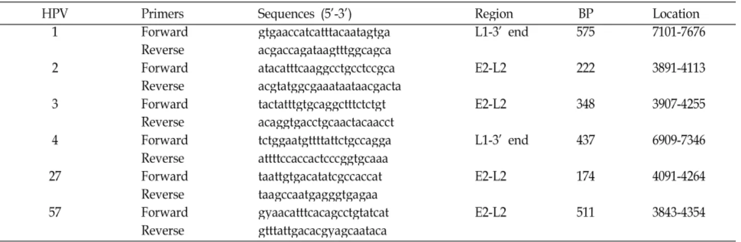
Table 1. Multiplex PCR primers used in this study
HPV Primers Sequences (5’-3’) Region BP Location
1 Forward gtgaaccatcatttacaatagtga L1-3’ end 575 7101-7676
Reverse acgaccagataagtttggcagca
2 Forward atacatttcaaggcctgcctccgca E2-L2 222 3891-4113
Reverse acgtatggcgaaataataacgacta
3 Forward tactatttgtgcaggctttctctgt E2-L2 348 3907-4255
Reverse acaggtgacctgcaactacaacct
4 Forward tctggaatgttttattctgccagga L1-3’ end 437 6909-7346
Reverse attttccaccactcccggtgcaaa
27 Forward taattgtgacatatcgccaccat E2-L2 174 4091-4264
Reverse taagccaatgagggtgagaa
57 Forward gyaacatttcacagcctgtatcat E2-L2 511 3843-4354
Reverse gtttattgacacgyagcaataca HPV: Human papilloma virus; BP: base pairs
Clinical samples
A total of 129 tissue specimens were obtained from 109 patients with cutaneous warts on the hands and feet. Seoul St. Mary’s Hospital and Chung-Ang University Yongsan Hospital participated in this study. Peeling followed by cry- otherapy with liquid nitrogen is the routine treatment proto- col for cutaneous common warts. The warty tissue was ob- tained by the peeling process. An informed consent was ob- tained from all patients who participated in the study.
Primer design
The HPV genome is composed of six early (E1, E2, E3, E4, E6 and E7) and two late (L1 and L2) proteins. Aligned sequences from the intergenic region between E2 to L2 and L1 to 3’-end of the genomes in the Genbank Database were manually compared to identify regions with the least degree of nucleotide sequence match. The nucleotide sequences of the six specific primers are described in Table 1. The HPV type 57-specific primer contains a single Y (T or C) base in its sequence. Primers MY09 (5’-CGTCCMARRGGAWA CTGATC-3’) and MY11 (5’-GCMCAGGGWCATAATAAT GG-3’) were used as degenerate PCR primers for HPV typ- ing [15]. To demonstrate that DNA samples are positive tem- plates for the PCR, primers PC03 (5’-ACACAACTGTGT TCACTAGC-3’) and PC04 (5’-CAACTTCATCCACGTTCA CC-3’) were used to amplify the endogenous β-globin region of human genome.
Preparation of sample DNA
The frozen tissues were digested with lysis buffer (10 mM
200 μg/ml of Proteinase K) at 37℃ overnight. After remov- ing protein debris twice by phenol-chloroform extraction, the DNA was precipitated with ethanol and re-suspended in water.
Multiplex PCR
The PCR was performed over a solution with a total vol- ume of 30 μl, containing six primer pairs (10 μM each), DNA extract from a frozen sample, and a 1× Premix solution (Solgent, Korea). 30 cycles of amplification was carried out with each cycle running in the following parameter; denatu- ration at 94℃ for 30 sec, annealing at 56℃ for 30 sec, and extension at 72℃ for 1 min.
Results Design of multiplex PCR primers
HPV-1, -2, -3, -4, -7, -27, -57 and -65 are well known to
cause common warts. Especially, HPV-2, -27, and -57 are
closely related and associated with skin warts. Among them,
we chose common warts-related 6 HPV subtypes for identi-
fication by the multiplex PCR [3,9]. To set up a multiplex
PCR for rapid identification of HPV-1, -2, -3, -4, 27, and -57,
type-specific primers were designed from the intergenic re-
gion of each viral genome (Fig. 1). By comparing the HPV
DNA sequences, we found that most variable regions were
located between E2 and L2 open reading frame (ORF) or
L1 and 3’ end of the genome. Based on the aligned se-
quences, pairs of primers with a length of 20 bp and without
homology were designed, amplifying distinguishable sizes
of 575, 222, 348, 437, 174 and 511 bp, respectively (Table 1).
Fig. 1. Schematic diagram of HPV genome. : Target region for the amplification of HPV-2, -3, -27 and -57; : target region for HPV-1 and -4.
Table 2. Distribution of HPV types identified by multiplex PCR in common warts samples
Type 1 2 3 4 7 27 57 65 Mixed Negative Total
Number
(%) 12
(9.3) 42
(32.6) 2 (1.5) 9
(7) 3
(2.3) 23
(17.8) 17
(13.2) 4
(3.1) 4
(3.1) 13
(10) 129
HPV: Human papilloma virus
Preparation of the standard HPV type DNA
With HPV-1, -2, -3, -4, -27 and -57 type-specific primers, specific sequences flanking the target region were obtained.
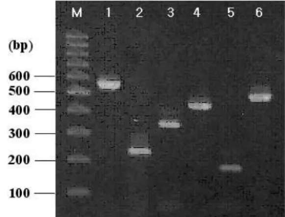
The HPV types were confirmed by DNA sequencing and Blast DNA database analysis. Each DNA from the 6 positive PCR products was then cloned into pHNT-T vector and the resultant recombinant plasmids were used as standard HPV type DNA [12]. PCRs of standard HPVs were performed us- ing its specific primer to test whether DNA of the expected size was produced. From PCR amplification, the bands of 575, 222, 348, 437, 174 and 511 bp of HPV-1, -2, -3, -4, -27 and -57 type were detected, respectively (Fig. 2).
Genotyping of HPVs of common warts by multiplex PCR amplification
All HPV-1, -2, -3, -4, -27 and -57 types of different sizes were amplified simultaneously from the combined 6 stand- ard HPV DNA mixed with their primer sets (Fig. 3, lane 1). The difference in size of the amplification products on agarose gel allowed rapid and specific discrimination be- tween HPV type 1, 2, 3, 4, 27 and 57.
After confirming that the multiplex amplification works properly, a total of 129 tissue specimens of common warts from Korean patients were subjected to multiplex PCR.
Figure 3 shows some results. By comparing the size of DNA on 2% agarose gel by standard multiplex PCR, we were able to identify their genotypes. The testing of 129 samples by multiplex PCR showed that 85% could be identified by their HPV types. The prevalence of HPV-1, 2, 3, 4, 27 and 57 in skin warts was 9.3%, 32.6%, 1.5%, 7%, 17.8%, and 13.2%, re- spectively (Table 2). Additional positive warts detected by the multiplex PCR system comprised of 4 samples co-in- fected with HPV types 2 and 27, or 2 and 57 (data not
Fig. 2. Independent PCR reaction of standard HPV type DNA with its specific primer pairs. Lane 1: HPV-1; lane 2:
HPV-2; lane 3: HPV-3; lane 4: HPV-4; lane 5: HPV-27;
lane 6: HPV-57.
shown). All samples negative with multiplex PCR primers were positive for β-globin amplification.
Specificity and sensitivity
During PCR, there can be an experimental risk of cross-contamination between the reactions, leading to mis- typing of the wart samples. To make sure the HPV typing results are correct, the 109 HPV positive samples were am- plified with the degenerate primers corresponding to the L1 ORF region. DNA sequencing and BLAST analysis of L1 PCR DNA fragment showed that HPV types determined by multiplex PCR were 99% correct, indicating high specificity of the specific primer sets in multiplex PCR (Table 3).
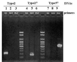
Among the HPV types causing cutaneous warts, type 2, 27
and 57 are closely related and show more DNA homology
to each other. In HPV identified cases, the specificity of the
primer pairs for HPV-2, -27, or -57 type was analyzed by
3 independent PCR. As illustrated in Fig. 4, each PCR primer
Specific primer set (multiplex) MY09/11primers (consensus) Confirmed by sequencing
Detection positive 109/129a (85%) 34/129 (27%) 108/109b (99%)
Detection negative 20/129 (15%) 95/129 (73%)
HPV typing was confirmed by DNA sequencing of each L1 region of positive samples.
a Total sample number
b Positive sample number of six HPV types
Fig. 3. Multiplex PCR with the wart samples. Lane 1: multiplex PCR with mixed standard HPV-1. -2. -3, -4, -27 and -57 DNAs; lane 2: sample 1; lane 3: sample 2; lane 4: sample 3; lane 5: sample 4; lane 6: sample 5; lane 7: sample 6.
Numbers denoted under the bands indicates HPV type.
was specific to the corresponding HPV type because there was no cross reaction with the other two sets of primer. The result demonstrates that the type-specific primer set can dis- criminate even closely related HPV types with high DNA homologies.
To evaluate the sensitivity of the multiplex PCR, the de- generate primer pairs MY09/11 were applied in PCR re- action of the same 129 samples tested with multiplex PCR.
Out of 129 wart samples, PCR amplified bands were ob- served in 39 cases (26.4%) with MY09/11 primers, while the multiplex PCR products were detected from 109 samples (85%) with 99% specificity (Table 3). This clearly shows that our specific primer sets designed for multiplex PCR have high specificity and sensitivity.
Discussion
PCR with primers of matching nucleotide identity and conserved sequences in the L1 and E1 open reading frame has been successfully used to detect a wide range of genital HPV types in mucosal warts or epidermodysplasia verruci-
Fig. 4. Specificity of multiplex PCR primers. HPV-2 samples (lanes 1, 2, 3), HPV-27 samples (lanes 4, 5, 6) and HPV-57 samples (lanes 7, 8, 9) were amplified independently with HPV type 2-specific primers (lanes 1, 4, 7), type 27-specific primers (lanes 2, 5, 8), and type 57 primers (lanes 3, 6, 9), respectively.
formis [7,19]. However, the identification of HPV types with degenerated primers require nested PCR, RFLP, or hybridization. In addition, compared to the large number of molecular epidemiological studies performed on HPV in- fection in genital warts, HPV infections in common warts have not been widely analyzed.
In this study, we present a specific one-step multiplex
PCR method which permits identification of 6 different HPV
genotypes related to common warts. Using a com-
puter-mediated HPV DNA sequence analysis, 6 pairs of spe-
cific primers were designed from the intergenic regions be-
tween genes L1 and 3’-end of genome, or between genes
E2 and L2. The detection of each HPV type depends on the
identification of 6 amplification products of different sizes
on agarose gel following multiplex PCR. We ran the multi-
plex PCR with HPV DNA isolated from 129 samples of com-
mon warts. Out of the HPV positive 109 samples, HPV-1,
-2, -3, -4, -27, and -57 were identified without any cross
amplification. The specificity of HPV-positive samples by the
multiplex PCR assay was confirmed by DNA sequence anal-
ysis with the L1 gene sequence. The clinical specificity of the primer sets was 85%, which was significantly higher than those from PCR with the consensus sequence primers MY09/11 [8]. In this study, the most frequently observed HPV type in common warts was HPV type 2. HPV-27, -57 and -1 were also prevalent, showing similar results with the German [11] and Chinese study [13]. However, Hagiwara et al. [9] reported that the most frequent HPV types found in common warts among Japanese patient were HPV-1, 4, and 65. For the 20 samples with unidentified HPV, primers for HPV-7, 10 and 65 were added for trial. Of the 20 cases, 3 were positive to HPV type 7, and 4 to HPV type 65 (Table 2). Since the remaining samples negative with multiplex PCR were positive with β-globin PCR, these seem to contain oth- er types of HPV. Therefore, we feel that there is a need to add a number of primers for different HPV types.
In conclusion, our data demonstrate that multiplex PCR with specific primers is a rapid and efficient method to iden- tify the HPV types involved in common warts. The tissue samples required for the multiplex PCR is safe and easy to obtain- we can simply use the peeled- off products preceding cryotherapy. We hope our multiplex-PCR method encour- ages many to participate in studies with cutaneous common warts, of which the importance has been under-estimated despite its prevalence in the past.
Acknowledgements
This work was supported by the Korean Research Foundation Grant funded by the Korean Government (KRF-2008-314-E00097), in which main calculations were performed by using the supercomputing resource of the Korean Institute of Science and Technology Information (KISTI).
References
1. Bernard, H. U., I. E. Calleja-Macias, and S. T. Dunn. 2006.
Genome variation of human papillomavirus types:
Phylogenetic and medical implications.
Int. J. Cancer
118, 1071-1076.2. Burhart, C. G. 2004. The endogenous, exogenous, and latent infections with human papillomavirus.
Int. J. Dermatol.
43, 548-549.3. Chen, S. L., Y. P. Tsao, J. W. Lee, W. C. Sheu, and Y. T.
Liu. 1993. Characterization and analysis of human papil- lomaviruses of skin warts.
Arch. Dermatol. Res.
285, 460-465.4. de Villiers, E. M. 1994. Human pathogenic papillomavirus
types: an update.
Curr. Top. Microbiol. Immunol.
186, 1-12.5. de Villiers, E. M., C. Fauquet, T. R. Broker, H. U. Bernard, and H. zur Hausen. 2004. Classification of papillomaviruses.
Virology
324, 17-27.6. Doorbar, J. 2006. Molecular biology of human papil- lomavirus infection and cervical cancer.
Clin. Sci.
110, 525-541.7. Forslund, O., A. Antonsson, P. Nordin, B. Stenquist, and B. G. Hansson. 1999. A broad range of human papil- lomavirus types detected with a general PCR method suit- able for analysis of cutaneous tumors and normal skin.
J.
Gen. Virol.
80, 2437-2443.8. Gravitt, P. E., C. L. Pryton, T. Q. Alessi, C. M. Wheeler, F. Coutlée, A. Hildesheim, M. H. Schiffman, D. R. Scott, and R. J. Apple. 2000. Improved amplification of genital hu- man papillomavirus.
J. Clin. Microbiol.
38, 357-361.9. Hagiwara, K., H. Uezato, H. Arakaki, S. Nonaka, K. Nonaka, H. Nonaka, T. Asato, M. Oshiro, K. Kariya, and A. Hattori.
2005. A genotype distribution of human papillomaviruses detected by polymerase chain reaction and direct sequenc- ing analysis in a large sample of common warts in Japan.
J. Med. Virol.
77, 107-112.10. Handisurya, A., C. Schellenbacher, and R. Kirnbauer. 2009.
Diseases caused by human papillomaviruses (HPV).
J.
German Soc. Dermatol.
7, 453-466.11. Iftner, A., S. J. Klug, C. Garbe, A. Blum, A. Stancu, S. P.
Wilczynski, and T. Iftner. 2003. The prevalence of human papillomavirus genotypes in nonmelanoma skin cancers of nonimmunosuppressed individuals identifies high-risk gen- ital types as possible risk factors.
Cancer Res.
63, 7515-7519.12. Kwon, J., K. S. Park, S. W. Park, and S. Y. Choi. 1998.
Construction of PCR cloning T-vector for the direct selection with green fluorescent protein.
BioTechniques
25, 192-196.13. Lei, Y. J., C. Gao, Q. Shi, J. M. Chen, Y. K. Yuan, C. Wang, J. Han, and X. P. Dong. 2008. Development of a multiplex PCR method for detecting and typing human papil- lomaviruses in verrucae vulgaris.
J. Virol. Methods
147, 72-77.14. Longworth, M. S. and L. A. Laimins. 2004. Pathogenesis of human papillomaviruses in differentiating epithelia.
Microbiol. Mol. Biol. Rev.
68, 362-372.15. Munoz, N., F. X. Bosch, S. de Sanjose, R. Herrero, X.
Castellsagué, K. V. Shah, P. J. Snijders, and C. J. Meijer.
International Agency for Research on Cancer Multicenter Cervical Cancer Study Group. 2003. Epidermiologic classi- fication of human papillomavirus types associated with cer- vical cancer.
N. Engl. J. Med.
348, 518-527.16. Qu, W., G. Jiang, Y. Cruz, C. J. Chang, G. Y. Ho, R. S. Klein, and R. D. Burk. 1997. PCR detection of human papil- lomavirus: comparison between MY09/MY1 and GP5+/GP6+ primer systems.
J. Clin. Microbiol.
36, 1304-1310.17. Sanclemente, G. and D. K. Gill. 2002. Human papillomavirus molecular biology and pathogenesis.
J. Eur. Acad. Dermatol.
Venereol.
16, 231-240.18. Snijders, P. J., A. J. van den Brule, M. V. Jacobs, R. P. Pol, and C. J. Meijer. 2005. HPV DNA detection and typing in cervical scrapes.
Methods Mol. Med.
119, 101-114.초록:Multiplex PCR 기법을 이용한 보통사마귀 내 인유두종바이러스 검출 및 분류 최순용
2· 임종호
1․김은정
1․김혜성
1․김범준
3․강훈
1․박영민
1*
(
1가톨릭대학교 의과대학 피부과학교실,
2한남대학교 생명·나노과학대학 생명공학과,
3중앙대학교 의과대학
피부과학교실)
현재까지 다수의 역학연구를 통해 피부에 발생한 보통사마귀에서 제 1, 2, 3, 4, 7, 10, 27, 57 및 65형의 인유두종바이러스가 검출되었다. 그러나 기존의 중합효소연쇄반응(conventional polymerase chain reaction, PCR)을 이용하는 경우 절차가 복잡하여 시간이 오래 걸리는 단점이 있었다. 이번 연구를 통해 저자들은 보통사 마귀에서 가장 흔히 검출되는 6가지 유전자형의 인유두종바이러스를 한번에 확인 가능한 간편한 muliplex PCR 의 개발을 목표로 하였다. 인유두종바이러스의 염기서열분석을 통해, L1에서 E6, 그리고 E2에서 L2 사이의 유전 자간영역(intergenic region)으로 부터 6쌍의 primer를 고안하였으며, L1 유전자서열 분석을 통해 multiplex PCR 의 특이성을 확인하였다. 총 129개의 조직표본 중 109개에서 제 1, 2, 3, 4, 27, 57형의 인유두종바이러스를 확인하 였다. 이번 연구의 primer를 이용한 인유두종바이러스 검출의 민감도와 특이도는 각각 85%와 99.5%였다. 이러한 primer 세트로 인유두종바이러스가 검출되지 않은 20개의 조직표본의 경우, 또 다른 HPV primer를 사용한 추가 적인 multiplex PCR을 시행하여 7개 표본에서 제 7형 및 65형의 인유두종바이러스가 검출되었다. 이상의 결과는 본 연구를 통해 새롭게 고안된 multiplex PCR 기법을 통해 보통사마귀에서의 인유두종바이러스를 보다 정확하 고 빠르게 검출할 수 있다는 것을 보여 준다.
G. Snow, C. J. Meijer, and J. M. Walboomers. 1990. The use of general primers in the polymerase chain reaction permits the detection of broad spectrum of human papillomavirus genotypes.
J. Gen. Virol.
71, 173-181.20. Surentheran, T., C. A. Harwood, P. J. Spink, A. L. Sinclair,
1998. Detection and typing of human papillomaviruses in mucosal and cutaneous biopsies from immunosuppressed and immunocompetent patients and patients with epi- dermodysplasia verruciformis: a uified diagnostic approach.