대한치과재료학회지 42(3) : 229-238, 2015 ISSN:2384-4434 (Print); 2384-3268 (Online) Available online at http://www.kadm.org http://dx.doi.org/10.14815/kjdm.2015.42.3.229
치면열구전색재와 충전재료에 대한 우식유발 세균의 부착특성 비교
강재민1), 임상욱1), 조화영1), 마재경2), 김정숙3), 김교한4), 송근배1)*, 최연희1)
경북대학교 치의학전문대학원 예방치과학교실1), 닥터홍치과의원2), 대구보건대학 치기공과3),
경북대학교 치의학전문대학원 치과생체재료학교실4)
<Abstract>
Adhesive characteristics of Mutans Streptococci on the surface of filling materials and sealant
Jae-Min Kang
1), Sang-Uk Im
1), Hwa-Young Jo
1), Jae-Kyung Ma
2), Jeong-Sook Kim
3), Kyo-Han Kim
4), Keun-Bae Song
1)*, Youn-Hee Choi
1)Department of Preventive Dentistry, School of Dentistry, Kyungpook National University, Daegu1), Doctor Hong Dental Clinic, Daegu2), Department of Dental Technology, Daegu Health college3), Department of Dental Biomaterials, School of Dentistry, Kyungpook National University, Daegu4)
Streptococcus mutans (S. mutans) is most commonly founded Mutans Streptococci group in human oral cavity. This study aimed to compare the adhesion ability of Xylitol-sensitive (Xs) and Xylitol-resistant (Xr) on three filling materials which are currently used by dentists.
Hydroxyapatite (HA) disk (control group, n=36), sealant (n=36), resin (n=36), glass-ionomer (n=36) disks were made by mold (10.2 mm Ø, 3 mm hight). Xs and Xr of S. mutans KCTC3065 and S. mutans UA159 were formed and cultured in TYE media. After culture, 500 ㎕ of 0.01% crystal violet was added to the plate, dyeing was performing, and mixed 1 ㎖ solution of ethanol:acetone (4:1 w/w) for reaction termination. The absorbance of the extracted solution was measured by using ELISA reader and the extracted solution on disk was observed with SEM. The adhesion ability was calculated by ELISA reader.
Adhesion ability of Xs and Xr from S. mutans of resin was higher than those of HA disk and the adhesion ability of sealant was lower compared to those of HA disk.
Sealant had the lowest adhesion ability of dental caries-inducing bacteria among three filling materials when viewed in terms of relevance of the material and decay-inducing bacteria in oral cavity.
Key words: Adhesion characteristics, Dental caries, Filling materials
* Correspondence: Keun-Bae Song
Department of Preventive Dentistry, School of Dentistry, Kyungpook National University, 2177 Dalgubeol-daero, Jung-gu, Daegu, 700-412, Korea
Tel: +82-53-660-6875, Fax: +82-53-423-2947 E-mail: [email protected]
Ⅰ. INTRODUCTION
치아우식증은 구강 내 세균에 의한 대표적인 감염성 질환 으로, 세균성 요인, 식이성 요인, 숙주 요인들이 유기적으로 결합하여 발생한다(van Houte J, 1994). 치아우식증 발생과정 의 초기에서는 치아의 내산성을 증대시키거나 재석회화를 촉 진시키는 방법과 산을 생성하는 세균의 성장을 억제시키는
방법 등을 이용하면 치아우식증을 예방할 수 있다(Song G, 2006).
사람의 구강 내에는 매우 다양한 종의 세균이 존재하는데 치아우식증과 밀접한 관련이 있는 것으로 알려진 대표적인 세균은 Mutans streptococci (MS)로(Oho T, 2000) 치아우식 은 이른 시기에 구강 내로 MS 집락이 형성될수록 발생할 가 능성이 높다(Kishi M, 2009). Mutans streptococci 중에서도 Streptococcus mutans (S. mutans)가 인간의 구강 내에서 가 장 많이 발견되고, 치아우식증과 깊은 관련성을 갖고 우식부 위의 치면세균막 내에 나타나는 것으로 알려져 있다 (Loesche WJ, 1986). 가장 주목을 받고 있는 치아우식 예방 물질은 자일리톨이다(Hilderbrandt GH와 Sparks BS, 2006;
Burt BA, 2006). 치아우식증을 예방하기 위해 설탕을 대체하 는 감미료로 사용되고 있는 자일리톨은 치면세균막의 형성과 우식증의 발생률을 감소시킨다고 보고되었다(Alanen P 등, 1994; Machiulskiene V 등, 2001; Autio JT, 2002; Twetman S와 Stecksen-Blicks C, 2003; Thorild I 등, 2004). 최근에 자일리톨을 장기간 섭취한 환자의 구강 내에서 자일리톨에 저항성을 가지는 균주가 발견되었다(Gauthier L 등, 1984).
Trahan 등(1992)은 세균이 자일리톨에 장기간 노출되면 감 수성이 점차 둔화되어 자일리톨 내성균주가 더 많았으 며, 자일리톨 섭취를 자제한 후에도 내성이 유지됨을 확인하 였다. 자일리톨에 의해 성장이 억제되는 균주를 감성균주 (Xylitol-sensitive; Xs)라고 하며, 자일리톨이 포함된 배지에 지속적으로 배양되어 자일리톨에 의해 더 이상 성장 억제가 나타나지 않는 균주를 자일리톨 내성균주(Xylitol-resistant;
Xr)라 한다(Trahan L 등, 1996).
현재 치과에서 치아우식으로 이환된 치아의 수복에 사용되 는 수복재료는 매우 다양하다. 충전을 위한 충전 및 수복재료 에는 레진(Resin-based composite)(Ferracane JL와 Mitchem JC, 1998), 글라스아이오노머(Glass-ionomer; G.I.)(Hembree JH, 1986), 치면열구전색재(Sealant)등(Ripa LW, 1985)이 사 용된다. sealant는 치아우식 발생초기에 교합면 우식증을 예 방 또는 억제할 수 있다(Hatibovic-Kofman S 등 1998). 우리 나라에서는 정부적 차원의 치아우식예방사업으로 치과충전 재료 중 sealant의 사용이 점점 많아지고 있다. 소아치과영역 에서 글라스아이오노머 시멘트와 더불어 가장 많이 사용되는
심미재료인 레진은 1960년대 중반 전치용 수복재료로서 개발 되었다(Kim JC와 Kim CW, 1990). 글라스아이오노머는 Wilson과 Kent에(1972) 의해 처음 소개되었고, 치아우식 충 전 시 아말감 와동형성시보다 치질의 삭제량이 크게 감소하였 고(Hicks MJ 등, 1986) 우수한 심미성(Maldonado, 1978) 때 문에 어린이의 치아우식 충전재로 선호되었다.
따라서 이번 연구에서는 현재 치과에서 사용되고 있는 sealant와 수복재료인 레진, 글라스아이오노머를 우식유발 세 균인 자일리톨 감성균주와 내성균주를 이용한 부착특성을 비 교하고자 하였다.
Ⅱ. MATERIALS AND METHODS
1. 연구재료
1.1 sealant와 충전재료의 시편 제작
sealant와 충전재료의 시편을 제작하기 위해 Putty 타입 의 부가중합형 폴리비닐실리콘 인상재(CharmFlexⓇ Putty, Dentkist, Korea)를 사용하여 직경 10.2 mm, 높이 3 mm의 균일한 원형 Hydroxyapatite (HA, control group) 디스크의 mold를 제작하였다. 완성된 mold를 이용하여 현재 치과 에서 대중적으로 사용되는 우식예방재료인 치면열구전색재 (ClinproTM Sealant, 3M Espe, USA)와 충전재료인 복합레진 (DenfilTM Flow Flowable Light-cured Composite Resin, Vericom, Korea)은 약간 넘치게 채운 후 광중합기(D1, DXM, Korea)를 이용하여 표면에 1mm 상방에서 수직방향으로 제 조사의 지시에 따라 1,500 mW/cm2의 강도로 20초간 중합 시킨 후 4분간 상온에서 방치한 후 증류수에 담아 보관하였 으며 글라스아이오노머(Fuji IX GP, GC, Japan)도 역시 약 간 넘치게 채운 후 자가중합시켰다. 중합된 시편은 24 시간 후 시료 표면의 표준화를 위하여 시편 전 처리장비 LaboPol (Laboforce-3, Struers, Denmark) 자동연마기를 이용하여 600 grit 연마지(CC-600Cw , DAESUNG, Korea)를 사용하여 직경 10.2 mm, 높이 3 mm가 되도록 균일하게 연마하였다.
이때 연마된 한쪽 면에서 남아있을 가능성이 높은 중합도가 낮은 층의 존재여부는 확인하지 못하였고, 다만 시료의 물성
적인 표준화만 고려하였다. 이렇게 제작된 시편들은(n=36) 밀폐된 용기에 시편이 충분히 잠길 정도로 distilled H2O (dH2O)를 담아 37 ℃ incubator에 24시간 보관하였다. 24시 간이 지난 후 Ethylene Oxide gas (EO gas) 멸균기를 이용하 여 멸균하였다.
2. 연구방법
2.1. 자일리톨 내성균주 형성 및 배양
자일리톨에 내성을 가지는 균주(Xr)를 형성하기 위해 S.
mutans KCTC3065와 S. mutans UA159를 0.5% 포도당 (Sigma, MO, USA)이 포함된 5 ㎖의 TYE 액체배지(1.7%
Bacto Tryptone: Difco Laboratories, MD, USA; 0.3% Bacto Yeast Extract: Difco Laboratories, MD, USA; 0.5% Sodium Chloride: Sigma, MO, USA; 0.25% Potassium Phosphate:
Sigma, MO, USA)에 1% (66 mM)(Kim JH 등, 2010) 자일리 톨을 첨가하여 37 ℃, 5% CO2 조건에서 24시간 간격으로 계대 배양하여 이를 30일간 반복하였다. 자일리톨 감성균주 (Xs)의 경우 자일리톨을 첨가하지 않고 동일한 조건으로 배 양하였다. 실험에 사용한 S. mutans KCTC3065는 한국생명 공학연구원 생명자원센터로부터 분양받았고, S. mutans UA159 (#700610) 는 ATCC (Manassas, VA, USA)로부터 분 양받았다.
2.2. 부착정도 측정
sealant와 충전재료의 종류에 따른 Xs와 Xr 균주의 biofilm 형성 정도에 따른 부착정도 변화를 알아보기 위해서 microtiter plate assay 방법을 약간 수정하여 사용하였다.
24시간 배양된 S. mutans를 OD600이 0.05가 되도록 희석하여 시편과 자일리톨 유무에 의해 구분된 24-well polystyrene microtiter plate에 700 ㎕씩을 접종하고 37 ℃, 5% CO2에서 총 24시간 동안 배양하였다. 배양이 끝나면 핀셋으로 시편을 꺼내어 dH2O를 이용하여 남아있는 부유세균을 제거하기 위 해 총 2회 세척하였다. 종이타올로 물기를 제거하고 공기 중에 15분 동안 건조시킨 다음 0.01% crystal violet 500 ㎕를 plate에 첨가하여 염색을 실시하였다. 15분간 실온에서 보관 한 다음 dH2O로 시편을 2회 씻어주고 Ethanol:Acetone=4:1
로 섞인 용액을 1 ㎖을 넣어 37 ℃ incubator에서 30분 동안 방치하여 염색된 dye가 추출되게 하였다. 추출된 dye의 흡 광도를 부착정도로 측정했다. dye를 96-well plate에 well 당 200 ㎕씩 옮겨 ELISA reader (ASYS, Austria)를 사용하여 570 nm에서 solution의 흡광도를 3-well 씩 3번 반복 측정 하였다.
2.3. 주사전자현미경(Scanning Electron Microscope;
SEM)을 이용한 sealant와 충전재료에 따른 S. mutans의 형태학적 변화
부착정도 측정을 위해 준비한 방법과 동일하게, 24-well polystyrene microtiter plate에 OD600이 0.05가 되도록 접종 하고 37 ℃, 5% CO2에서 총 24시간 동안 배양한 후 핀셋으로 시편을 꺼내어 dH2O를 이용하여 남아있는 부유세균을 제거하기 위해 총 2회 세척하였다. 그런 후 ethanol 30%, 50%, 70%, 90% 순으로 탈수시킨 후 완전히 알코올이 건조되도록 24시간 방치한 후 주사전자현미경을 촬영하기 전까지 실리카젤(Silicagel, Duksan, Korea)이 들어있는 밀폐 된 용기에 보관하였다. 준비된 시료는 주사전자현미경을 촬영을 위해 백금코팅을 한 후 주사전자현미경(SU8820, HITACHI, Japan)을 사용하여 5 KV의 전압에서 10,000배의 배율로 관찰하였다.
2.4. 통계분석
sealant와 충전재료의 종류에 따른 우식유발 세균인 자일리 톨 감성균주와 내성균주를 이용한 부착특성을 비교를 확인하 기 위하여 Kruskal-Wallis 검정을 수행하였다. 각각의 군을 Mann-Whitney 검정을 이용하여 사후분석을 시행하였다. 모 든 통계 분석은 통계분석용 소프트웨어인 SPSS 22.0 (SPSS Inc, Chicago, IL, USA)를 이용하여 수행하였으며, 유의성 판 정을 위한 유의수준은 5%로 설정하였다.
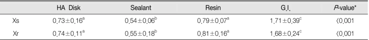
Table 1. The mean values of eluted dye between Xs and Xr of S. mutans KCTC3065 for filling materials and sealant
HA Disk Sealant Resin G.I. P-value*
Xs 0.73±0.16a 0.54±0.06b 0.79±0.07a 1.71±0.39c <0.001
Xr 0.74±0.11a 0.55±0.18b 0.81±0.16a 1.68±0.24c <0.001
Values are Mean±S.D.
*Kruskal-Wallis test.
a,b,c; Same letter denotes there is no significant difference among groups by Mann-Whitney test.
Table 2. The mean values of eluted dye between Xs and Xr of S. mutans UA159 for filling materials and sealant.
HA Disk Sealant Resin G.I. P-value*
Xs 0.87±0.11a 0.49±0.23b 1.05±0.10c 1.86±0.40d <0.001
Xr 0.91±0.12a 0.72±0.14b 0.96±0.17a 1.94±0.24c <0.001
Values are Mean±S.D.
*Kruskal-Wallis test.
a,b,c,d; Same letter denotes there is no significant difference among groups by Mann-Whitney test.
Ⅲ. RESULTS
1. sealant와 충전재료에 따른 S. mutans KCTC3065와 S. mutans UA159의 Xs와 Xr의 부착정도
Table 1은 sealant와 충전재료에 따른 S. mutans KCTC 3065의 Xs와 Xr의 부착정도 비교 값을 나타낸 것이다. 각 균 주에서 sealant와 충전재료에 따른 부착정도를 비교하였을 때, Xs와 Xr 모두에서 4가지 재료에 대해 유의한 차이가 나타났다 (P<0.001). Xs와 Xr에서 각각 G.I.가 가장 큰 부착정도를 보 였고, 다음으로 Resin, HA Disk로 나타났다. Xs와 Xr 모두 Sealant에서 가장 낮은 부착정도를 보였다. sealant와 충전재 료들 간의 차이가 있는지 알아본 결과 HA Disk와 Resin 사이 에서는 통계적으로 유의한 차이를 보이지 않았으며, 나머지 모든 재료 사이에서는 유의한 차이가 나타났다(P<0.008).
Table 2는 sealant와 충전재료에 따른 S. mutans UA159의 Xs와 Xr의 부착정도 비교 값을 나타낸 것이다. 각 균주에서 sealant와 충전재료에 따른 부착정도를 비교하였을 때, Xs와 Xr 모두에서 4가지 재료에 대해 유의한 차이가 나타났다 (P<0.001). Xs와 Xr에서 각각 G.I.가 가장 큰 부착정도를 보 였고, 다음으로 Resin, HA Disk로 나타났다. S. mutans
KCTC3065와 마찬가지로 S. mutans UA159의 Xs와 Xr 모두 Sealant에서 가장 낮은 부착정도를 보였다. sealant와 충전재 료들 간의 차이가 있는지 알아본 결과 S. mutans UA159의 Xs에서는 모든 재료 사이에 유의한 차이가 나타났지만 (P<0.008), S. mutans UA159의 Xr에서는 HA Disk와 Resin 사이에서 통계적으로 유의한 차이를 보이지 않았으며, 나머지 모든 재료 사이에서는 유의한 차이가 나타났다(P<0.008).
2. 주사전자현미경을 이용한 S. mutans의 형태학적 관찰
sealant와 충전재료 별 S. mutans에 의해 형성되는 세균집 락의 부착정도 차이를 확인하기 위하여 주사전자현미경을 통해 관찰하였다. Figure 1은 sealant와 충전재료에 따른 S. mutans KCTC3065 Xs, Xr을 관찰한 결과이며, Figure 2는 S. mutans UA159 Xs, Xr을 관찰한 결과이다. S. mutans KCTC3065 Xs, Xr과 S. mutans UA159 Xs, Xr 모두 자일리톨 에 내성이 있는 균주와 없는 균주 간 부착정도의 차이는 없었 다(Figure 1, 2). G.I.에서 형성되는 세균집락의 부착정도가 가장 많았고 다음으로 Resin과 HA Disk 순으로 세균집락의 부착정도가 많았다. Sealant의 경우 세균집락의 부착정도가 가장 적었다.
Figure 1. SEM images of Xs and Xr of S. mutans KCTC3065 for filling materials and sealant. A: Xs for HA Disk; B: Xs for Sealant; C: Xs for Resin; D: Xs for G.I.; E: Xr for HA Disk; F: Xr for Sealant; G: Xr for Resin; H: Xr for G.I.
Figure 2. SEM images of Xs and Xr of S. mutans UA159 for filling materials. A: Xs for HA Disk; B: Xs for Sealant; C: Xs for Resin; D: Xs for G.I.; E: Xr for HA Disk; F: Xr for Sealant; G: Xr for Resin;
H: Xr for G.I.
IV. DISCUSSION
S. mutans는 사람에게서 일어나는 가장 흔한 감염성 질병 중 하나인 치아우식증을 일으키는 주요 원인균이다(Oho T 등, 2000; Hamada S와 Slade HD, 1980). 전체 치과치료에서 가장 빈도가 높은 치료이며, 보존치료에 의하여 치아의 기능 과 심미성을 회복하고 주위조직들과 조화를 이루도록 하는 치아우식증 치료는 구강건강은 물론 정신건강의 증진까지를 최종 목표로 하고 있다.
본 연구에서는 sealant와 충전재료 별 시편을 만들어 치아 우식유발 세균인 S. mutans의 자일리톨 감성균주(Xs)와 내성 균주(Xr)의 부착정도를 비교하였다. S. mutans KCTC3065, S.
mutans UA159의 Xs와 Xr의 부착정도를 비교한 결과, 모든 재료에 유의한 차이가 나타났다. 부착정도는 S. mutans KCTC3065, S. mutans UA159 모두에서 G.I., Resin, HA Disk, Sealant 순으로 낮아졌음을 알 수 있었다. G.I.와 Resin에 치 아우식 유발 세균이 더 잘 부착하는 것은 물성의 특징에 의한 것으로 추정된다. Nicholson JW 등(1992)은 G.I.는 친수성 성분의 존재로 인해 수분에의 노출이 물리적 성질을 변화시킨 다고 하였다. Resin은 중합 후 분해되어 방출되거나 잔존하는 자유 모노머의 독성에 대해 많은 연구가 있었는데, 자유 모노 머가 치아우식을 야기할 수 있는 미생물의 증식을 일으키며 치수의 변성과 치은경계 퇴축, 일부 환자에게 알레르기 반응 을 유발한다고 알려졌다(Emmler J 등, 2008; Gwinnett AJ와 Tay F, 1999; Schweikl H 등, 2006; Singh J 등, 2009). 스웨덴 치과의사들은 교합면 충전재로 Resin을 선호하였고, 덴마크 치과의사들은 아말감을, 노르웨이의 치과의사들은 Resin과 G.I.를 선호하였다고 Espelid I와 Tveit AB(2001)는 보고하였 다. 본 연구의 결과를 바탕으로 치아우식유발 세균에 가장 낮은 부착정도를 보인 Sealant가 우식을 예방하는데 효과적이 라고 판단된다.
이제까지 치과용 교정 와이어(Lee HJ 등, 2011; Lee SP 등, 2009), 치과용 심미수복재료(Kim DH, 2013)의 우식유발 세균 의 부착에 관한 연구는 있었지만 Sealant와 충전재료인 Resin, G.I.에 따른 치아우식유발 세균인 S. mutans의 부착정도 비 교, 또한 Sealant, Resin, G.I. 재료에 따른 자일리톨 Xs와 Xr의 부착정도를 동일한 방법으로 비교한 연구는 국내에서 첫 시도
라고 할 수 있다. 그러나 본 연구에는 몇 가지 제한점이 있다.
먼저, 현재 치과에서 대중적으로 많이 사용되는 sealant와 충 전재료 별 Resin, G.I.를 한 가지씩만 사용하였다. 이번 연구 의 결과가 단순한 재료 별 제품 특징의 차이인지 Sealant, Resin, G.I.의 그룹 간의 차이인지 확인하지 못하였으므로, 우 식을 유발시키는 세균에 대해 Sealant와 충전재료의 부착정도 를 비교한 결과가 과대평가 되었을 수도 있겠다. 다음으로는 G.I.의 물성의 차이로 인해 시편 제작 시 표면에 균열이 발생 하였다. 이것으로 인해 우식유발 세균의 부착정도에 미친 영 향을 배제할 수는 없다. 그럼에도 불구하고, 본 연구는 sealant 와 충전재료 별 치아우식유발 세균인 S. mutans의 자일리톨 Xs와 Xr의 부착정도를 비교한 첫 연구였다는 점과 Sealant와 충전재료를 시편형태로 만들고 그 위에 세균을 배양하여 dye 가 용출되는 양을 확인함으로써 부착정도를 비교하였다는 점 에서 의의가 있다고 하겠다. 따라서 향후 이러한 제한점들을 보완하여 포괄적이고 추가적인 실험방법의 개발이 진행되어 우식유발 방지에 좋은 우식예방재료를 선택할 수 있도록 기여 하겠다.
V. CONCLUSIONS
본 연구는 Sealant와 충전재료인 Resin, G.I.에 대한 S.
mutans 자일리톨 감성균주(Xs)와 내성균주(Xr)의 부착정도를 비교하여 다음과 같은 결론을 얻었다.
1. Sealant와 충전재료에 따른 S. mutans KCTC3065의 Xs 와 Xr의 부착정도를 비교하였을 때, Xs와 Xr 모두에서 4가지 재료에 대해 유의한 차이가 나타났으며(P<0.001), 글라스아이오노머, 레진, HA Disk, sealant 순으로 낮아 졌다. Sealant와 충전재료들 간의 차이가 있는지 알아본 결과 HA Disk와 Resin 사이에서는 통계적으로 유의한 차이를 보이지 않았으며, 나머지 모든 재료 사이에서는 유의한 차이가 나타났다(P<0.008).
2. Sealant와 충전재료에 따른 S. mutans UA159의 Xs와 Xr의 부착정도를 비교한 결과도 Xs와 Xr 모두에서 4가 지 재료에 대해 유의한 차이가 나타났다(P<0.001).
Sealant와 충전재료들 간의 차이가 있는지 알아본 결과 S. mutans UA159의 Xs에서는 모든 재료 사이에 유의한 차이가 나타났지만(P<0.008), S. mutans UA159의 Xr에 서는 HA Disk와 Resin 사이에서 통계적으로 유의한 차 이를 보이지 않았으며, 나머지 모든 재료 사이에서는 유 의한 차이가 나타났다(P<0.008).
3. Sealant와 충전재료 별 S. mutans에 의해 형성되는 세균 집락의 부착정도 차이를 확인하기 위하여 주사전자현미 경을 통해 관찰한 결과, 모두 자일리톨에 내성이 있는 균주와 없는 균주 간 부착정도의 차이는 없었다. G.I.에 서 형성되는 세균집락의 부착정도가 가장 많았고 다음 으로 Resin과 HA Disk 순으로 세균집락의 부착정도가 많았다. Sealant의 경우 세균집락의 부착 밀도가 가장 낮게 나타났다.
이상의 결과들을 종합해 보았을 때, 치아우식유발 세균의 부착정도가 가장 낮게 나타난 Sealant가 재료와 구강 내 우식 을 예방하는데 효과적이라고 판단된다.
REFERENCES
Alanen P, Hurskainen K, Isokangas P (1994). Clinician’s ability to identify caries risk subjects. Community Dent Oral Epidemiol 22(2):86-89.
Autio JT (2002). Effect of xylitol chewing gum on salivary Streptococcus mutans in preschool children. ASDC J Dent Child 69(1):81-86.
Burt BA (2006). The use of sorbitol-and xylitol- sweetened chewing gum in caries control. J Am Dent Assoc 137(2):190-196.
Emmler J, Seiss M, Kreppel H, Reichl FX, Hickel R, Kehe K (2008). Cytotoxicity of the dental composite component TEGDMA and selected metabolic by-products in human pulmonary cells. Dent Mater 24(12):1670-1675.
Espelid I, Tveit AB (2001). Restorative treatment decisions
on occlusal caries in Scandinavia. Acta Odontol Scand 59(1):21-27.
Ferracane JL, Mitchem JC (1998). Fluoride penetration into the hybrid layer from a dentin adhesive. Am J Dent 11(1):23-28.
Gauthier L, Vadeboncoeur C, Mayrand D (1984). Loss of sensitivity to xylitol by Streptococcus mutans LG-1.
Caries Res 18(4):289-295.
Gwinnett AJ, Tay F (1999). Early and intermediate time response of the dental pulp to an acid etch technique in vivo. Elsevier 82(5):524.
Hamada S, Slade HD (1980). Biology, immunology and cariogenicity of Streptococcus mutans. Microbiol Rev 44(2):331-384.
Hatibovic-Kofman S, Wright GZ, Braverman I (1998).
Microleakage of sealants after conventional, bur, and air-abrasion preparation of pits and fissures. Pediatr Dent 20(3):173-176.
Hembree JH (1986). In vitro microleakage of a new dental adhesive system. J Prosthet Dent 55(4):442-445.
Hilderbrandt GH, Sparks BS (2006). Maintaining mutans Streptococci suppression with xylitol chewing gum. J Am Dent Assoc 131(7):909-916.
Hicks MJ, Flaitz CM, Silverstone LM (1986). Secondary caries formation in vitro around glass ionomer restorations. Quintessence Int 17(9):527-532.
Kim DH (2013). Adherence of Streptococcus mutans to tooth-colored restorative materials. [박사학위논문]. 경북 대학교 대학원.
Kim JC, Kim CW (1990). Comparative studies on the charateristics of dental composite resins. Kor J Dent Mater 17(2):179-195.
Kim JH, Lee YE, Chung SY, Ahn SH, Choi YH, Song KB (2010). The effect of xylitol on the use of various carbohydrates by Streptococcus mutans. J Korean Acad Oral Health 34(1):1-8.
Kishi M, Abe A, Kishi K, Ohara-Nemoto Y, Kimura S,
Yonemitsu M (2009). Relationship of quantitative salivary levels of Streptococcus mutans and S. sobrinus in mothers to caries status and colonization of mutans streptococci in plaque in their 2.5-year-old children.
Community Dent Oral Epidemiol 37(3):241-249.
Lee HJ, Park HS, Kim KH, Kwon TY, Hong SH (2011).
Effect of garlic on bacterial biofilm formation on orthodontic wire. Angle Orthod 81(5):895-900.
Lee SP, Lee SJ, Lim BS, Ahn SJ (2009). Surface characteristics of orthodontic materials and their effects on adhesion of mutans streptococci. Angle Orthod 79(2):353-360.
Loesche WJ (1986). Role of Streptococcus mutans in human dental decay. Microbiol Rev 50(4):353-380.
Machiulskiene V, Nyvad B, Baelum V (2001). Caries preventive effect of sugar-substituted chewing gum.
Community Dent Oral Epidemiol 29(4):278-288.
Maldonado (1978). An in vitro study certain properties of a glass ionomer cement. J Am Dent Assoc 96(5):785-791.
Nicholson JW, Anstice HM, McLean JW (1992). A preliminary report on the effect of storage in water on the properties of commercial light-cured glass-ionomer cements. Br Dent J 173(3):98-101.
Oho T, Yamashita Y, Shimazaki Y, Kushiyama M, Koga T (2000). Simple and rapid detection of Streptococcus mutans and Streptococcus sobrinus in human saliva by polymerase chain reaction. Oral Microbiol Immunol 15(4):258-262.
Ripa LW (1985). The current status of pit and fissure sealants. A review. J Can Dent Assoc 51(5):367-380.
Schweikl H, Spagnuolo G, Schmalz G (2006). Genetic and cellular toxicology of dental resin monomers. J Dent Res 85(10):870-877.
Singh J, Khalichi P, Cvitkovitch DG (2009). Composite resin degradation products from BisGMA monomer modulate the expression of genes associated with biofilm formation and other virulence factors in Streptococcus mutans. J Bio Mat 88(2):551-560.
Song G, Luo Q, Qin J, Wang L, Shi Y, Sun C (2006). Effects of oxymatrine on proliferation and apoptosis in human hepatoma cell. Colloids Surf B Biointerfaces 48(1):1-5.
Thorild I, Lindau B, Twetman S (2004). Salivary mutans streptococci and dental caries in three-year-old children after maternal exposure to chewing gums containing combinations of xylitol, sorbitol, chlorhexidine, and fluoride. Acta Odontol Scand 62(5):245-250.
Trahan L, Soderling E, Drean MF, Chevrier MC, Isokangas P (1992). Effect of xylitol consumption on the plaque-saliva distribution of mutans streptococci and the occurrence and long-term survival of xylitol-resistant strains. J Dent Res 71(11):1785-1791.
Trahan L, Bourgeau G, Breton R (1996). Emergence of multiple xylitol- resistant (fructose PTS-) mutants from human isolates of mutans streptococci during growth on dietary sugars in the presence of xylitol. J Dent Res 75(11):1892-1900.
Twetman S, Stecksen-Blicks C (2003). Effect of xylitol-containing chewing gums on lactic acid production in dental plaque from caries active pre-school children. Oral Health Prev Dent 1(3):
195-199.
van Houte J (1994). Role of micro-organisms in caries etiology. J Dent Res 73(3):672-681.
Wilson AD, Kent BE (1972). A new translucent cement for dentistry. The glass ionomer cement. Br Dent J 15(4):133-135.