Leptin as a Key between Obesity and Cardiovascular Disease
Ki-Woon Kang1, Minho Ok2, Seong-Kyu Lee3,4,*
1Division of Cardiology, Department of Internal Medicine, Eulji University School of Medicine, Daejeon; 2Department of Cardiovascular Pharmacology, Mokpo National University, Mokpo; 3Division of Endocrinology, Department of Internal Medicine and 4Department of Biochemistry-Molecular Biology, Eulji University School of Medicine, Daejeon, Korea
Obesity increases the risk of cardiovascular disease through various influencing factors. Leptin, which is predom- inantly secreted by adipose tissue, regulates satiety homeostasis and energy balance, and influences cardiovas- cular functions directly and indirectly. Leptin appears to play a role in heart protection in leptin-deficient and leptin-receptor-deficient rodent model experiments. Hyperleptinemia or leptin resistance in human obesity in- fluences the vascular endothelium, cardiovascular structure and functions, inflammation, and sympathetic ac- tivity, which may lead to cardiovascular disease. Leptin is involved in many processes, including signal transduc- tion, vascular endothelial function, and cardiac structural remodeling. However, the dual (positive and negative) regulator effect of leptin and its receptor on cardiovascular disease has not been completely understood. The protective role of leptin signaling in cardiovascular disease could be a promising target for cardiovascular dis- ease prevention in obese patients.
Key words: Obesity, Leptin, Cardiovascular disease
Received November 19, 2020 Reviewed December 3, 2020 Accepted December 13, 2020
* Corresponding author Seong-Kyu Lee
https://orcid.org/0000-0002-5999-7656 Division of Endocrinology, Department of Internal Medicine, Eulji University Hospital and Department of Biochemistry-Molecular Biology, Eulji University School of Medicine, 95 Dunsanseo-ro, Seo-gu, Daejeon 35233, Korea Tel: +82-42-259-1642 Fax: +82-42-259-1539 E-mail: lskendo@hanmail.net
Copyright © 2020 Korean Society for the Study of Obesity
This is an Open Access article distributed under the terms of the Creative Commons Attribution Non-Commercial License (https://creativecommons.org/licenses/by-nc/4.0/) which permits unrestricted non-commercial use, distribution, and reproduction in any medium, provided the original work is properly cited.
2017-03-16 https://crossmark-cdn.crossref.org/widget/v2.0/logos/CROSSMARK_Color_square.svg
INTRODUCTION
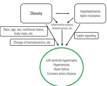
Obesity increases the risk of cardiovascular disease through vari- ous influencing factors,1 such as hemodynamic changes, cardiac structure and cardiac function, inflammation, neurohumoral chang- es, and cellular remodeling (Fig. 1).2 The presence of large emerg- ing adipocytes may be directly associated with production of leptin, angiotensin, proinflammatory cytokines, and reactive oxygen spe- cies.3 Moreover, progressive inflammation processes, oxidative stress, and hyperleptinemia in obesity is significantly correlated with de- veloping cardiovascular diseases4 and hypertension (Fig. 1).5-7 Adi- pocyte-derived leptin exhibits pleiotropic effects. In obese people, hyperleptinemia is not sufficient to prevent energy balance dysreg-
ulation, indicating that obese individuals are leptin resistant. Al- though most obese cases are associated with hypothalamic leptin resistance, the peripheral effects of leptin signaling or leptin resis- tance in obesity are not fully elucidated. Moreover, the net effects of hyperleptinemia or leptin resistance on cardiovascular disease in obese people are complex and not completely understood. In this review, we discuss leptin as a key between obesity and cardiovascu- lar disease (Fig. 1).
RELATIONSHIP BETWEEN OBESITY AND CARDIOVASCULAR DISEASE
Obesity can lead to cardiac structural remodeling, causing left
Leptin as a Key between Obesity and Cardiovascular Disease
Ki-Woon Kang1, Minho Ok2, Seong-Kyu Lee3,4,*
1Division of Cardiology, Department of Internal Medicine, Eulji University School of Medicine, Daejeon; 2Department of Cardiovascular Pharmacology, Mokpo National University, Mokpo; 3Division of Endocrinology, Department of Internal Medicine and 4Department of Biochemistry-Molecular Biology, Eulji University School of Medicine, Daejeon, Korea
Obesity increases the risk of cardiovascular disease through various influencing factors. Leptin, which is predom- inantly secreted by adipose tissue, regulates satiety homeostasis and energy balance, and influences cardiovas- cular functions directly and indirectly. Leptin appears to play a role in heart protection in leptin-deficient and leptin-receptor-deficient rodent model experiments. Hyperleptinemia or leptin resistance in human obesity in- fluences the vascular endothelium, cardiovascular structure and functions, inflammation, and sympathetic ac- tivity, which may lead to cardiovascular disease. Leptin is involved in many processes, including signal transduc- tion, vascular endothelial function, and cardiac structural remodeling. However, the dual (positive and negative) regulator effect of leptin and its receptor on cardiovascular disease has not been completely understood. The protective role of leptin signaling in cardiovascular disease could be a promising target for cardiovascular dis- ease prevention in obese patients.
Key words: Obesity, Leptin, Cardiovascular disease
Received November 19, 2020 Reviewed December 3, 2020 Accepted December 13, 2020
* Corresponding author Seong-Kyu Lee
https://orcid.org/0000-0002-5999-7656 Division of Endocrinology, Department of Internal Medicine, Eulji University Hospital and Department of Biochemistry-Molecular Biology, Eulji University School of Medicine, 95 Dunsanseo-ro, Seo-gu, Daejeon 35233, Korea Tel: +82-42-259-1642 Fax: +82-42-259-1539 E-mail: lskendo@hanmail.net
ventricular (LV) hypertrophy.8,9 When body mass index (BMI) in- creases by 1 kg/m2, the risk of LV hypertrophy increases by 5.1%, and when waist circumference increases by 1 cm, the risk of LV hy- pertrophy increases by 2.6%.10 Furthermore, obesity is associated with vascular injuries, such as increased arterial stiffness,11 coronary artery calcification,12 increased carotid intima-media thickness, and higher incidence of carotid stenosis,13 all of which contributes to early vascular aging.14 Obesity has been recognized as an indepen- dent predictor of coronary artery disease15,16 and carries an approxi- mately two-fold higher risk of developing heart failure than a healthy weight.17,18 Studies have also shown correlations between stroke and BMI and waist-hip ratio,19,20 and population-based co- hort studies have reported a 49% higher risk of arteriosclerosis in obese patients compared with non-obese patients.21
In contrast, among patients with heart failure, those who were overweight or obese (BMI > 27.8 kg/m2) were clinically shown to have a higher survival rate.22 A follow-up analysis of 7,767 patients with chronic heart failure reported lower hazard ratios (HRs) in overweight or obese subjects (HR, 0.81; 95% confidence interval [CI], 0.72–0.92) than in normal-weight subjects (HR, 0.88; 95%
CI, 0.80–0.96).23 Similar results were observed in patients who ex- perienced sudden cardiac arrest, wherein a higher BMI was associ- ated with reduced mortality.24 Higher BMI was also associated with
lower mortality in cases of coronary artery disease, heart failure, and diabetes.25 In general, obese patients have higher mortality;
however, paradoxically, higher mortality is often observed in nor- mal-weight patients compared with obese patients.26 This phenom- enon, in which survival rates are higher among obese patients, in contrast to conventional expectations, is known as the “obesity par- adox,” and is most commonly observed in patients with coronary artery disease or heart failure.27
THE ROLE OF LEPTIN
Leptin is an important hormone involved in weight regulation and energy homeostasis.28-31 It is a 16-kDa protein with 167 amino acids.32 Leptin is predominantly secreted by adipose tissue and is also secreted from other tissues, including the heart, via autocrine or paracrine effects.33,34 Leptin regulates appetite by controlling sa- tiety signals to the central nervous system (CNS)35 and it influenc- es cardiovascular functions either directly or indirectly via second- ary responses mediated by the vasculature (such as hypertension, endothelial function, atherosclerosis, and thrombopoiesis) or the CNS.36
The ob (obesity) gene mutation and leptin receptor (LepR) mu- tants—ob/ob and db/db mouse models, respectively—and fa/fa Zucker rat models were developed as obesity animal models.37 Heart failure is common to these animals,38 suggesting that leptin is linked to cardiovascular disease. In research using leptin- or LepR- deficient rodent models, leptin appears to play a role in heart pro- tection.
Despite varying interpretations of the results of a meta-analysis of the effects of leptin on coronary artery disease,39 in the Multi- ethnic Study of Atherosclerosis study that included 1,910 patients with atherosclerosis only, leptin was not significantly correlated with cardiovascular disease.40 However, contradictory results have been reported in other studies that found a correlation between leptin and cardiovascular disease. In contrast, hyperleptinemia was found to be correlated with a positive prognosis for cardiovascular disease through coronary artery vasodilation, endothelial nitric ox- ide synthase (eNOS) activation, endothelial precursor cell activa- tion, and reduced lipid accumulation.41
Figure 1. Overview of leptin as a key between obesity and cardiovascular disease.
Obesity increases the risk of cardiovascular disease through the various factors:
obesity-induced changes in hemodynamics, cardiac structure and cardiac function, inflammation, neurohumoral changes, and cellular remodeling, as well as hyper- leptinemia and leptin resistance.
Obesity Hyperleptinemia
leptin resistance
Left ventricle hypertrophy Hypertension
Heart failure Coronary artery disease Race, age, sex, nutritional status,
body mass, etc.
Change of hemodynamics, etc.
Leptin signaling Inflammation process,
oxidative stress, etc.
LEPTIN RECEPTOR AND SIGNAL TRANSDUCTION
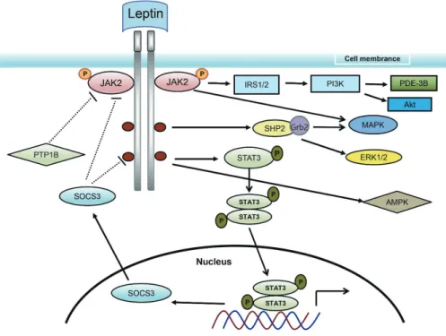
LepR is simultaneously expressed in adipose, heart, muscle, lung, small intestine, and liver tissue, as well as the CNS, particularly the hypothalamus. LepR is a type-I cytokine receptor, from which six subtypes (LepRa to LepRf) are generated by selective splicing.42 Leptin signaling relies on LepR autophosphorylation, which trig- gers the pathways for Janus kinase (JAK), signal transducer and ac- tivator of transcription (STAT), insulin receptor substrate (IRS)/
phosphatidylinositol 3 kinase (PI3K), mitogen-activated protein kinase (MAPK), extracellular signal-regulated protein kinase (ERK), and 5΄-adenosine monophosphate-activated protein kinase (AMPK) (Fig. 2). Leptin may activate JAK2, IRS1, and ERK via LepRa; how- ever, LepRb, with its long intracellular tail, appears to be the only subtype capable of mediating JAK/STAT signaling (Fig. 2).43,44
JAK/STAT signaling is triggered by JAK2 phosphorylation, fol- lowed by STAT3 phosphorylation and a conformational change due to binding. STAT3 forms a dimer that enters the nucleus to
regulate the expression of genes governing food ingestion.44 Such signaling pathways include negative feedback—suppressor of cyto- kine signaling 3 (SOC3) negatively regulates the JAK/STAT path- way by interfering with the phosphorylation of the tyrosine residue of LepR (Fig. 2). Moreover, STAT3 repressor hinders STAT3 bind- ing to DNA, and protein tyrosine phosphatase-1B interrupts phos- phorylation of JAK2 and STAT3 and negatively regulates leptin signaling (Fig. 2).44
Leptin also mediates MAPK and ERK signaling (Fig. 2). Binding of leptin results in SH2-containing protein tyrosine phosphatase 2 (SHP2) phosphorylation, and growth factor receptor-bound pro- tein 2 (Grb2) activates ERK. Moreover, irrespective of LepRb phosphorylation, JAK2 activates downstream signaling via Grb2 and SHP2.45 PI3K is a dimer and has a component that regulates signaling by acting as a catalyst. LepR activation facilitates the inter- action and complex formation of JAK2/IRS1 to downregulate tar- gets such as protein kinase B (Akt).44
Elevated leptin levels are generally observed in obese people, which led to a hypothesis that elevated leptin levels may be linked
Figure 2. Signal transduction process of leptin. Leptin signaling relies on leptin receptor autophosphorylation, which triggers the JAK2, STAT3, IRS/PI3K, MAPK, ERK, and AMPK pathways. JAK, Janus kinase; IRS, insulin receptor substrate; PI3K, phosphatidylinositol 3 kinase; PDE, phosphodiesterase; Akt, protein kinase B; SHP2, SH2-con- taining protein tyrosine phosphatase 2; Grb2, growth factor receptor-bound protein 2; MAPK, mitogen-activated protein kinase; PTP, protein tyrosine phosphatase; STAT, signal transducer and activator of transcription; ERK, extracellular signal-regulated protein kinase; SOCS3, suppressor of cytokine signaling 3; AMPK, 5’-adenosine mono- phosphate-activated protein kinase.
to leptin resistance.35,46 If leptin resistance causes changes in leptin signaling (such as changes in LepR tyrosine residues or the leptin- binding site), additional studies are required to understand this phenomena. Whether leptin resistance is just inhibition of leptin signaling that affects the heart or whether other adipokines also af- fect the heart needs to be investigated.
RELATIONSHIP BETWEEN LEPTIN AND CARDIAC HYPERTROPHY
The increased blood volume in obese patients increases cardiac output and stimulates biomechanical stress and structural remodel- ing that may result in cardiac hypertrophy.8,9,47 A correlation be- tween plasma leptin levels and cardiac hypertrophy has been clini- cally demonstrated.48 Numerous studies observed that leptin di- rectly induces cardiac hypertrophy in humans and mice.33,49-55 Leptin induces cardiac hypertrophy through diverse pathways such as the mammalian target of rapamycin signaling,56 calcineurin acti- vation and nuclear factor of activated T cells transportation into the nucleus,57 peroxisome proliferator-activated receptor-α activation,58 MAPK 14 (p38) activation and transportation into the nucleus,50,52 activation of Rho and actin dynamics,49 and increases in intracellu- lar reactive oxygen species.51,59
LV hypertrophy is often observed in ob/ob and db/db mice morbidly predisposed to obesity.60-63 However, when adequate leptin levels are provided, the LV returns to its normal thickness, regardless of the mouse’s weight.62 Hyperleptinemia is observed in the heart in a diet-induced obese mouse model, and LepR con- stantly responds to elevated plasma or cardiac leptin levels. This appears to provide protection against cardiac hypertrophy via STAT3 activation and its downstream pathways and by affecting p38 and p42/44 MAPK as well as Akt, in comparison to LepR mu- tants or db/db mouse models.60
Despite the studies that conclude that leptin causes cardiac hy- pertrophy, research by Leifheit-Nestler et al.60 showed protective effects of leptin on cardiac hypertrophy. They investigated LepR in relation to STAT3, but other effects of leptin also require investiga- tion. Influential factors that require consideration are the mouse model species, leptin resistance in obesity, age, sex, and patient nu- tritional status. Without this knowledge, it is unclear whether obe-
sity-induced cardiac hypertrophy is the result of leptin-driven car- diac hypertrophy or resistance to the cardio-protective effects of leptin.60
RELATIONSHIP BETWEEN LEPTIN AND CARDIAC FUNCTION
Ca2+ influx into the heart acts as a multifunctional signal that causes the heart muscles to contract, controls the period of action potential maintenance, and regulates gene expression.64 During the period of excitation-contraction coupling in the heart, β-adrenergic signals activate the Na+/Ca2+ exchange channels via protein kinase A (PKA) signaling while causing depolarization of the sarcolem- ma.65 Depolarization of the sarcolemma leads to the opening of high-voltage-activated L-type calcium channels and allows Ca2+ en- try into the cytoplasm, which, in turn, causes Ca2+ release through ryanodine receptor channels into the sarcoplasmic reticulum (SR) for muscle contraction initiation.65 Sarcoplasmic/endoplasmic re- ticulum calcium ATPase 2 (SERCA2) and phospholamban (PLN) play crucial roles in transporting Ca2+ from myocytes and the cyto- plasm.66 Abnormal Ca2+ circulation in the SR characterizes cardiac diseases such as heart failure and arteriosclerosis and contributes to the pathophysiology of disease progression.66,67
Decreased activation of SERCA2 and Na+/Ca2+ exchange chan- nels is observed in leptin-deficient mice,68 whereas leptin treatment to the myocytes of ob/ob mice improved β-adrenergic signaling and increased Gsα expression, PKA activation, and PLN phos- phorylation.69 These results suggest a definite correlation between leptin and cardiac function.70,71 Moreover, leptin treatment to car- diomyocytes of adult mice also resulted in suppressed contraction via different pathways (e.g., endothelin-1 receptor- nicotinamide adenine dinucleotide phosphate H oxidase pathway,70,71 nitric ox- ide,72 JAK/STAT pathway,73 interleukin-1β signaling,74 autophagy induction75).
RELATIONSHIP BETWEEN LEPTIN AND CARDIOMYOCYTE APOPTOSIS
Apoptosis, through strict regulation of specific signaling steps, is the main cause of cell loss in the heart.76 Cardiomyocyte apoptosis
plays a key role in the progression of heart failure and is especially important for compensatory remodeling in heart failure.77 Cells undergoing apoptosis experience structural changes including shrinking, plasma membrane blebbing, nuclear condensation, and DNA and nuclear fragmentation.78 Once cells become apoptotic bodies, they are removed by macrophages.76 Apoptosis is mediated via two different pathways: the first is the extrinsic or death recep- tor pathway activated by death-domain-containing receptors in the plasma membrane, and the second is the intrinsic or mitochondrial pathway activated by intracellular stress from growth factors, low oxygen concentration, oxidative stress, and DNA damage.76 Apop- totic signals instantly activate caspases and disable the mitochon- dria, leading to cell death.
Zucker rats showed increased apoptosis via both the extrinsic and intrinsic pathways.79,80 In ob/ob and db/db mice and fa/fa rats, impaired leptin signaling caused a rise in triglyceride levels, which led to lipid accumulation and induction of cardiomyocyte apopto- sis. However, when normal leptin levels were reached, excessive lip- id accumulation was prevented and cardiac function was re- stored,61,81 indicating that increased apoptosis in obese mouse models was not due to aging or cellular damage but directly associ- ated with impaired leptin signaling. Moreover, in knock-out mice models with cardiomyocyte-specific deletions of LepR, impaired leptin signaling directly led to increased cardiac hypertrophy, apop- tosis, deterioration of cardiac structure and function, and impair- ment of energy, glucose, and fatty acid metabolism, which further accelerated heart damage due to myocardial infarction.82
Apoptosis can be induced by a mechanism where the opening of the mitochondrial permeability transition pore (mPTP) releases cytochrome C into the cytoplasm.83 Leptin prevents the opening of mPTP in mouse cardiomyocytes84-86 and can also protect cardio- myocytes from apoptosis induced by H2O2 or hypoxia-reoxygen- ation conditions.87,88
Tumor necrosis factor (TNF)-α at high concentrations binds to TNFR-1 to cause cardiomyocyte apoptosis, leading to various car- diovascular diseases.89-91 Leptin treatment in mouse cardiomyo- cytes interfered with the caspase-3 fragmentation and the intrinsic mitochondrial pathway, thereby preventing TNF-α-induced apop- tosis.87 Despite reports that leptin treatment causes apoptosis,92,93 it seems clear that leptin plays a protective role against apoptosis pro-
gression under stressful physiological conditions. Furthermore, it should be noted that leptin treatment could prevent apoptosis by blocking TNF-α–induced pathways (e.g., caspase-3 fragmentation, poly adenosine diphosphate ribose polymerase segmentation, p38 MAPK/nuclear factor kappa B phosphorylation, Bax transport).94 If these downstream pathways could be regulated by leptin, cardio- myocyte apoptosis could be controlled and cardiovascular disease progression could be halted.
RELATIONSHIP BETWEEN LEPTIN AND ENDOTHELIAL VASCULAR FUNCTION
Leptin was initially thought to stimulate the sympathetic nerve,95 raising blood pressure. However, leptin was not found to exert sig- nificant effects on blood pressure on its own.96-98 A number of ex- periments demonstrated that leptin is involved in endothelium-de- pendent vasodilation via nitric oxide,99-101 which was further ob- served in leptin-deficient ob/ob mice, where arterial vessel contrac- tion mediated by noradrenaline or phenylephrine increased while vasodilation by acetylcholine weakened. These anomalies disap- peared when leptin levels were restored.102
Leptin induces Akt phosphorylation103 and phosphorylated Akt then induces phosphorylation of the eNOS serine residue, which heightens its activity. eNOS produces nitric oxide, which activates soluble guanylyl cyclase. This stimulates cyclic guanosine mono- phosphate synthesis in smooth muscle cells, leading to vasodila- tion. Although eNOS can also be activated by insulin, leptin is ca- pable of activating the PI3K-independent Akt/eNOS pathway.104 Leptin also seems to mediate vasodilation via endothelium-derived hyperpolarizing factor (EDHF).105
Leptin resistance is observed in obesity and metabolic syn- drome,35,46 where the effect of leptin on nitric-oxide-induced vaso- dilation becomes less significant. During the early stages, a com- pensatory response may originate from EDHF, but even this be- comes inadequate as leptin resistance duration increases. Conse- quently, lack of dilation and continued stimulation of the sympa- thetic nerve causes hypertension and atherosclerosis.106 Nonethe- less, it is anticipated that elucidating the signaling pathway between leptin and endothelial cells and uncovering the mechanism of the role of leptin resistance in blood vessels would lead to a reliable
treatment strategy for obesity-associated vascular diseases.
Leptin causes atherosclerosis through diverse mechanisms in- cluding the entry of monocytes into blood vessels, transformation of macrophages into foam cells, proliferation of vascular smooth muscle cells, and secretion of atherosclerotic cytokines, which sug- gests that leptin indirectly causes atherosclerosis.107 A recent study reported a protective role for leptin against atherosclerosis in a con- centration-dependent manner in low-density lipoprotein recep- tor–/–ob/ob mice.108 Leptin exerts protective effects by reducing liv- er cholesterol or lowering its synthase mRNA expression.109-111 Fur- thermore, adiposity and related inflammation are independent prognostic factors alongside fibrosis in the progression and detec- tion of atherosclerosis, and leptin relieves local inflammation in the liver by down-regulating inflammatory cytokines such as mono- cyte chemoattractant protein-1, TNF-α, and fibrosis marker trans- forming growth factor-β.112,113 Based on these findings, it seems im- portant to elucidate the mode of liver cholesterol metabolism un- der physiological conditions and in leptin deficiency for ameliorat- ing atherosclerosis progression. Moreover, the facilitated release of nitric oxide by leptin and the association of nitric oxide and sympa- thetic nerve activation may be another mechanism that prevents atherosclerosis.114 Such results, however, do not contradict the role of leptin in causing atherosclerosis when its levels are higher than normal. As previously mentioned, leptin causes atherosclerosis in- directly. Leptin and its receptors are expressed in atherosclerotic plaques in mice as well as humans,115,116 and high levels of leptin are known to elevate cardiovascular risk factors such as plasma fibrino- gens, which suggest that high levels of leptin may contribute to ath- erosclerosis.117
RELATIONSHIP BETWEEN LEPTIN AND SYMPATHETIC STIMULATION
Sympathetic nerve activation is observed in obese patients118 as well as in animal models of obesity.119 In several tissue types, leptin seems to promote sympathetic nerve activation involved in cardio- vascular regulation, which raises arterial pressure.120 Furthermore, although leptin, when administered, was ineffective in regulating energy homeostasis in obesity, its influence on cardiovascular sym- pathetic nerve hyperactivity and blood pressure was maintained.121
This indicates a selective influence of leptin resistance. This is sup- ported by the finding that blood pressure decreased when sympa- thetic nerve activity was inhibited and central leptin signaling was blocked in obese rabbits.122
It is broadly agreed that hypertension is caused by sympathetic nerve activation that affects peripheral resistance or blood flow in the kidneys, and that leptin levels are elevated in obese individu- als.123 However, because leptin treatment in healthy individuals does not affect hypertension, it is worth considering whether hy- pertension is caused by elevated leptin levels or leptin signaling de- ficiency due to leptin resistance or by the burden of cardiac hyper- trophy due to obesity. Additionally, demographic factors besides leptin (e.g., race, age, sex) should be considered. If it is the elevated leptin or leptin signaling deficiency due to leptin resistance in obe- sity that causes hypertension, then the condition might be con- trolled by regulating the downstream pathways (STATs, PI3K, ERK1/2, SOC3, etc.) (Fig. 2).124
CONCLUSION
Leptin exerts its influence on the cardiovascular system in a num- ber of ways. Obesity and leptin cannot be simply said to have either a negative or a protective role in cardiovascular disease. Nevertheless, a number of studies have shown that leptin has protective effects on cardiovascular disease. Future investigations should confirm wheth- er elevated leptin levels themselves are responsible for cardiovascular disease or whether leptin resistance is responsible for cardiovascular disease in human obesity. If leptin plays different roles under differ- ent conditions, other factors including race, age, sex, nutritional sta- tus, BMI, and leptin resistance should be considered. The protective role of leptin signaling in cardiovascular disease could be a promis- ing target to prevent cardiovascular disease in obese patients.
CONFLICTS OF INTEREST
The authors declare no conflict of interest.
AUTHOR CONTRIBUTIONS
Study concept and design: KWK and SKL; analysis and interpre-
tation of data: KWK and MO; drafting of the manuscript: KWK and MO; critical revision of the manuscript: SKL; and study su- pervision: SKL.
REFERENCES
1. Sharma AM. Adipose tissue: a mediator of cardiovascular risk. Int J Obes Relat Metab Disord 2002;26 Suppl 4:S5-7.
2. Lavie CJ, Milani RV, Ventura HO. Obesity and cardiovascu- lar disease: risk factor, paradox, and impact of weight loss. J Am Coll Cardiol 2009;53:1925-32.
3. Pausova Z. From big fat cells to high blood pressure: a path- way to obesity-associated hypertension. Curr Opin Nephrol Hypertens 2006;15:173-8.
4. Ridker PM. High-sensitivity C-reactive protein: potential adjunct for global risk assessment in the primary prevention of cardiovascular disease. Circulation 2001;103:1813-8.
5. Harrison DG, Guzik TJ, Lob HE, Madhur MS, Marvar PJ, Thabet SR, et al. Inflammation, immunity, and hyperten- sion. Hypertension 2011;57:132-40.
6. Mathieu P, Lemieux I, Després JP. Obesity, inflammation, and cardiovascular risk. Clin Pharmacol Ther 2010;87:407-16.
7. Pou KM, Massaro JM, Hoffmann U, Vasan RS, Maurovich- Horvat P, Larson MG, et al. Visceral and subcutaneous adi- pose tissue volumes are cross-sectionally related to markers of inflammation and oxidative stress: the Framingham Heart Study. Circulation 2007;116:1234-41.
8. Kuch B, Muscholl M, Luchner A, Döring A, Riegger GA, Schunkert H, et al. Gender specific differences in left ven- tricular adaptation to obesity and hypertension. J Hum Hy- pertens 1998;12:685-91.
9. Kuperstein R, Hanly P, Niroumand M, Sasson Z. The im- portance of age and obesity on the relation between diabe- tes and left ventricular mass. J Am Coll Cardiol 2001;37:
1957-62.
10. Bombelli M, Facchetti R, Sega R, Carugo S, Fodri D, Bram- billa G, et al. Impact of body mass index and waist circum- ference on the long-term risk of diabetes mellitus, hyperten- sion, and cardiac organ damage. Hypertension 2011;58:
1029-35.
11. Zebekakis PE, Nawrot T, Thijs L, Balkestein EJ, van der Heijden-Spek J, Van Bortel LM, et al. Obesity is associated with increased arterial stiffness from adolescence until old age. J Hypertens 2005;23:1839-46.
12. Cassidy AE, Bielak LF, Zhou Y, Sheedy PF 2nd, Turner ST, Breen JF, et al. Progression of subclinical coronary athero- sclerosis: does obesity make a difference? Circulation 2005;
111:1877-82.
13. Ingelsson E, Sullivan LM, Fox CS, Murabito JM, Benjamin EJ, Polak JF, et al. Burden and prognostic importance of subclinical cardiovascular disease in overweight and obese individuals. Circulation 2007;116:375-84.
14. Nilsson PM, Lurbe E, Laurent S. The early life origins of vascular ageing and cardiovascular risk: the EVA syndrome.
J Hypertens 2008;26:1049-57.
15. Hubert HB, Feinleib M, McNamara PM, Castelli WP. Obe- sity as an independent risk factor for cardiovascular disease:
a 26-year follow-up of participants in the Framingham Heart Study. Circulation 1983;67:968-77.
16. Manson JE, Colditz GA, Stampfer MJ, Willett WC, Rosner B, Monson RR, et al. A prospective study of obesity and risk of coronary heart disease in women. N Engl J Med 1990;322:
882-9.
17. Kenchaiah S, Evans JC, Levy D, Wilson PW, Benjamin EJ, Larson MG, et al. Obesity and the risk of heart failure. N Engl J Med 2002;347:305-13.
18. Lee DS, Massaro JM, Wang TJ, Kannel WB, Benjamin EJ, Kenchaiah S, et al. Antecedent blood pressure, body mass index, and the risk of incident heart failure in later life. Hy- pertension 2007;50:869-76.
19. Rexrode KM, Hennekens CH, Willett WC, Colditz GA, Stampfer MJ, Rich-Edwards JW, et al. A prospective study of body mass index, weight change, and risk of stroke in wom- en. JAMA 1997;277:1539-45.
20. Walker SP, Rimm EB, Ascherio A, Kawachi I, Stampfer MJ, Willett WC. Body size and fat distribution as predictors of stroke among US men. Am J Epidemiol 1996;144:1143-50.
21. Wanahita N, Messerli FH, Bangalore S, Gami AS, Somers VK, Steinberg JS. Atrial fibrillation and obesity--results of a meta-analysis. Am Heart J 2008;155:310-5.
22. Horwich TB, Fonarow GC, Hamilton MA, MacLellan WR, Woo MA, Tillisch JH. The relationship between obesity and mortality in patients with heart failure. J Am Coll Cardiol 2001;38:789-95.
23. Bozkurt B, Deswal A. Obesity as a prognostic factor in chron- ic symptomatic heart failure. Am Heart J 2005;150:1233-9.
24. Matinrazm S, Ladejobi A, Pasupula DK, Javed A, Durrani A, Ahmad S, et al. Effect of body mass index on survival after sudden cardiac arrest. Clin Cardiol 2018;41:46-50.
25. Goyal A, Nimmakayala KR, Zonszein J. Is there a paradox in obesity? Cardiol Rev 2014;22:163-70.
26. Braun N, Gomes F, Schütz P. “The obesity paradox” in dis- ease--is the protective effect of obesity true? Swiss Med Wkly 2015;145:w14265.
27. Wannamethee SG, Shaper AG, Whincup PH, Lennon L, Papacosta O, Sattar N. The obesity paradox in men with coronary heart disease and heart failure: the role of muscle mass and leptin. Int J Cardiol 2014;171:49-55.
28. Friedman JM. Leptin, leptin receptors, and the control of body weight. Nutr Rev 1998;56(2 Pt 2):s38-46.
29. Park HK, Ahima RS. Leptin signaling. F1000Prime Rep 2014;
6:73.
30. Pelleymounter MA, Cullen MJ, Baker MB, Hecht R, Win- ters D, Boone T, et al. Effects of the obese gene product on body weight regulation in ob/ob mice. Science 1995;269:
540-3.
31. Rosenbaum M, Leibel RL. 20 years of leptin: role of leptin in energy homeostasis in humans. J Endocrinol 2014;223:
T83-96.
32. Faggioni R, Feingold KR, Grunfeld C. Leptin regulation of the immune response and the immunodeficiency of malnu- trition. FASEB J 2001;15:2565-71.
33. Rajapurohitam V, Javadov S, Purdham DM, Kirshenbaum LA, Karmazyn M. An autocrine role for leptin in mediating the cardiomyocyte hypertrophic effects of angiotensin II and endothelin-1. J Mol Cell Cardiol 2006;41:265-74.
34. Purdham DM, Zou MX, Rajapurohitam V, Karmazyn M.
Rat heart is a site of leptin production and action. Am J Physiol Heart Circ Physiol 2004;287:H2877-84.
35. Ahima RS, Flier JS. Leptin. Annu Rev Physiol 2000;62:
413-37.
36. Koh KK, Park SM, Quon MJ. Leptin and cardiovascular dis- ease: response to therapeutic interventions. Circulation 2008;
117:3238-49.
37. Lutz TA, Woods SC. Overview of animal models of obesity.
Curr Protoc Pharmacol 2012;Chapter 5:Unit5.61.
38. Ren J, Dong F, Cai GJ, Zhao P, Nunn JM, Wold LE, et al.
Interaction between age and obesity on cardiomyocyte con- tractile function: role of leptin and stress signaling. PLoS One 2010;5:e10085.
39. Sweeney G. Cardiovascular effects of leptin. Nat Rev Cardiol 2010;7:22-9.
40. Martin SS, Blaha MJ, Muse ED, Qasim AN, Reilly MP, Blu- menthal RS, et al. Leptin and incident cardiovascular dis- ease: the Multi-ethnic Study of Atherosclerosis (MESA).
Atherosclerosis 2015;239:67-72.
41. Wolk R, Bertolet M, Singh P, Brooks MM, Pratley RE, Frye RL, et al. Prognostic value of adipokines in predicting car- diovascular outcome: explaining the obesity paradox. Mayo Clin Proc 2016;91:858-66.
42. Mercer JG, Hoggard N, Williams LM, Lawrence CB, Han- nah LT, Trayhurn P. Localization of leptin receptor mRNA and the long form splice variant (Ob-Rb) in mouse hypo- thalamus and adjacent brain regions by in situ hybridization.
FEBS Lett 1996;387:113-6.
43. Tartaglia LA, Dembski M, Weng X, Deng N, Culpepper J, Devos R, et al. Identification and expression cloning of a leptin receptor, OB-R. Cell 1995;83:1263-71.
44. Wada N, Hirako S, Takenoya F, Kageyama H, Okabe M, Sh- ioda S. Leptin and its receptors. J Chem Neuroanat 2014;61- 62:191-9.
45. Myers MG Jr. Leptin receptor signaling and the regulation of mammalian physiology. Recent Prog Horm Res 2004;59:
287-304.
46. Martin SS, Qasim A, Reilly MP. Leptin resistance: a possible interface of inflammation and metabolism in obesity-related cardiovascular disease. J Am Coll Cardiol 2008;52:1201-10.
47. Carreño JE, Apablaza F, Ocaranza MP, Jalil JE. Cardiac hy- pertrophy: molecular and cellular events. Rev Esp Cardiol 2006;59:473-86.
48. Paolisso G, Tagliamonte MR, Galderisi M, Zito GA, Petro- celli A, Carella C, et al. Plasma leptin level is associated with myocardial wall thickness in hypertensive insulin-resistant men. Hypertension 1999;34:1047-52.
49. Zeidan A, Javadov S, Karmazyn M. Essential role of Rho/
ROCK-dependent processes and actin dynamics in mediat- ing leptin-induced hypertrophy in rat neonatal ventricular myocytes. Cardiovasc Res 2006;72:101-11.
50. Zeidan A, Javadov S, Chakrabarti S, Karmazyn M. Leptin- induced cardiomyocyte hypertrophy involves selective cave- olae and RhoA/ROCK-dependent p38 MAPK translocation to nuclei. Cardiovasc Res 2008;77:64-72.
51. Xu FP, Chen MS, Wang YZ, Yi Q, Lin SB, Chen AF, et al.
Leptin induces hypertrophy via endothelin-1-reactive oxy- gen species pathway in cultured neonatal rat cardiomyocytes.
Circulation 2004;110:1269-75.
52. Rajapurohitam V, Gan XT, Kirshenbaum LA, Karmazyn M.
The obesity-associated peptide leptin induces hypertrophy in neonatal rat ventricular myocytes. Circ Res 2003;93:
277-9.
53. Majumdar P, Chen S, George B, Sen S, Karmazyn M, Chakrab- arti S. Leptin and endothelin-1 mediated increased extracel- lular matrix protein production and cardiomyocyte hyper- trophy in diabetic heart disease. Diabetes Metab Res Rev 2009;25:452-63.
54. Madani S, De Girolamo S, Muñoz DM, Li RK, Sweeney G.
Direct effects of leptin on size and extracellular matrix com- ponents of human pediatric ventricular myocytes. Cardiovasc Res 2006;69:716-25.
55. Abe Y, Ono K, Kawamura T, Wada H, Kita T, Shimatsu A, et al. Leptin induces elongation of cardiac myocytes and causes eccentric left ventricular dilatation with compensation.
Am J Physiol Heart Circ Physiol 2007;292:H2387-96.
56. Zeidan A, Hunter JC, Javadov S, Karmazyn M. mTOR me- diates RhoA-dependent leptin-induced cardiomyocyte hy- pertrophy. Mol Cell Biochem 2011;352:99-108.
57. Rajapurohitam V, Izaddoustdar F, Martinez-Abundis E, Karma- zyn M. Leptin-induced cardiomyocyte hypertrophy reveals both calcium-dependent and calcium-independent/RhoA- dependent calcineurin activation and NFAT nuclear translo-
cation. Cell Signal 2012;24:2283-90.
58. Hou N, Luo MS, Liu SM, Zhang HN, Xiao Q, Sun P, et al.
Leptin induces hypertrophy through activating the peroxi- some proliferator-activated receptor α pathway in cultured neonatal rat cardiomyocytes. Clin Exp Pharmacol Physiol 2010;37:1087-95.
59. Hu TP, Xu FP, Li YJ, Luo JD. Simvastatin inhibits leptin-in- duced hypertrophy in cultured neonatal rat cardiomyocytes.
Acta Pharmacol Sin 2006;27:419-22.
60. Leifheit-Nestler M, Wagner NM, Gogiraju R, Didié M, Konstantinides S, Hasenfuss G, et al. Importance of leptin signaling and signal transducer and activator of transcrip- tion-3 activation in mediating the cardiac hypertrophy asso- ciated with obesity. J Transl Med 2013;11:170.
61. Barouch LA, Gao D, Chen L, Miller KL, Xu W, Phan AC, et al. Cardiac myocyte apoptosis is associated with increased DNA damage and decreased survival in murine models of obesity. Circ Res 2006;98:119-24.
62. Barouch LA, Berkowitz DE, Harrison RW, O’Donnell CP, Hare JM. Disruption of leptin signaling contributes to cardi- ac hypertrophy independently of body weight in mice. Cir- culation 2003;108:754-9.
63. Raju SV, Zheng M, Schuleri KH, Phan AC, Bedja D, Saraiva RM, et al. Activation of the cardiac ciliary neurotrophic fac- tor receptor reverses left ventricular hypertrophy in leptin-de- ficient and leptin-resistant obesity. Proc Natl Acad Sci USA 2006;103:4222-7.
64. Shaw RM, Colecraft HM. L-type calcium channel targeting and local signalling in cardiac myocytes. Cardiovasc Res 2013;98:177-86.
65. Bers DM, Shannon TR. Calcium movements inside the sar- coplasmic reticulum of cardiac myocytes. J Mol Cell Cardiol 2013;58:59-66.
66. Kho C, Lee A, Hajjar RJ. Altered sarcoplasmic reticulum calcium cycling: targets for heart failure therapy. Nat Rev Cardiol 2012;9:717-33.
67. Ibrahim M, Gorelik J, Yacoub MH, Terracciano CM. The structure and function of cardiac t-tubules in health and dis- ease. Proc Biol Sci 2011;278:2714-23.
68. Dong F, Yang X, Sreejayan N, Ren J. Chromium (D-phenyl-
alanine)3 improves obesity-induced cardiac contractile de- fect in ob/ob mice. Obesity (Silver Spring) 2007;15:2699- 711.
69. Minhas KM, Khan SA, Raju SV, Phan AC, Gonzalez DR, Skaf MW, et al. Leptin repletion restores depressed β‐adren- ergic contractility in ob/ob mice independently of cardiac hypertrophy. J Physiol 2005;565(Pt 2):463-74.
70. Dong F, Zhang X, Yang X, Esberg LB, Yang H, Zhang Z, et al. Impaired cardiac contractile function in ventricular myo- cytes from leptin-deficient ob/ob obese mice. J Endocrinol 2006;188:25-36.
71. Dong F, Zhang X, Ren J. Leptin regulates cardiomyocyte contractile function through endothelin-1 receptor-NADPH oxidase pathway. Hypertension 2006;47:222-9.
72. Nickola MW, Wold LE, Colligan PB, Wang GJ, Samson WK, Ren J. Leptin attenuates cardiac contraction in rat ventricular myocytes: role of NO. Hypertension 2000;36:501-5.
73. Wold LE, Relling DP, Duan J, Norby FL, Ren J. Abrogated leptin-induced cardiac contractile response in ventricular myocytes under spontaneous hypertension: role of Jak/
STAT pathway. Hypertension 2002;39:69-74.
74. Radin MJ, Holycross BJ, McCune SA, Altschuld RA. Cross- talk between lepti and interleukin-1β abrogates negative ino- tropic effects in a model of chronic hyperleptinemia. Exp Biol Med (Maywood) 2011;236:1263-73.
75. Kandadi MR, Roe ND, Ren J. Autophagy inhibition rescues against leptin- induced cardiac contractile dysfunction. Curr Pharm Des 2014;20:675-83.
76. Orogo AM, Gustafsson ÅB. Cell death in the myocardium:
my heart won’t go on. IUBMB Life 2013;65:651-6.
77. Lee Y, Gustafsson AB. Role of apoptosis in cardiovascular disease. Apoptosis 2009;14:536-48.
78. Zhang Y, Chen X, Gueydan C, Han J. Plasma membrane changes during programmed cell deaths. Cell Res 2018;28:
9-21.
79. Lee SD, Tzang BS, Kuo WW, Lin YM, Yang AL, Chen SH, et al. Cardiac fas receptor-dependent apoptotic pathway in obese Zucker rats. Obesity (Silver Spring) 2007;15:2407-15.
80. Lu MC, Tzang BS, Kuo WW, Wu FL, Chen YS, Tsai CH, et al. More activated cardiac mitochondrial-dependent apop-
totic pathway in obese Zucker rats. Obesity (Silver Spring) 2007;15:2634-42.
81. Hall ME, Maready MW, Hall JE, Stec DE. Rescue of cardiac leptin receptors in db/db mice prevents myocardial triglyc- eride accumulation. Am J Physiol Endocrinol Metab 2014;
307:E316-25.
82. McGaffin KR, Witham WG, Yester KA, Romano LC, O’Doherty RM, McTiernan CF, et al. Cardiac-specific leptin receptor deletion exacerbates ischaemic heart failure in mice. Cardio- vasc Res 2011;89:60-71.
83. Bernardi P, Di Lisa F. The mitochondrial permeability tran- sition pore: molecular nature and role as a target in cardio- protection. J Mol Cell Cardiol 2015;78:100-6.
84. Dixon RA, Davidson SM, Wynne AM, Yellon DM, Smith CC. The cardioprotective actions of leptin are lost in the Zucker obese (fa/fa) rat. J Cardiovasc Pharmacol 2009;53:
311-7.
85. Smith CC, Dixon RA, Wynne AM, Theodorou L, Ong SG, Subrayan S, et al. Leptin-induced cardioprotection involves JAK/STAT signaling that may be linked to the mitochondri- al permeability transition pore. Am J Physiol Heart Circ Physiol 2010;299:H1265-70.
86. Smith CC, Mocanu MM, Davidson SM, Wynne AM, Simp- kin JC, Yellon DM. Leptin, the obesity-associated hormone, exhibits direct cardioprotective effects. Br J Pharmacol 2006;
149:5-13.
87. Eguchi M, Liu Y, Shin EJ, Sweeney G. Leptin protects H9c2 rat cardiomyocytes from H2O2-induced apoptosis. FEBS J 2008;275:3136-44.
88. Shin EJ, Schram K, Zheng XL, Sweeney G. Leptin attenuates hypoxia/reoxygenation-induced activation of the intrinsic pathway of apoptosis in rat H9c2 cells. J Cell Physiol 2009;
221:490-7.
89. Engel D, Peshock R, Armstong RC, Sivasubramanian N, Mann DL. Cardiac myocyte apoptosis provokes adverse cardiac re- modeling in transgenic mice with targeted TNF overexpres- sion. Am J Physiol Heart Circ Physiol 2004;287:H1303-11.
90. Packer M. Is tumor necrosis factor an important neurohor- monal mechanism in chronic heart failure? Circulation 1995;
92:1379-82.
91. Micheau O, Tschopp J. Induction of TNF receptor I-mediat- ed apoptosis via two sequential signaling complexes. Cell 2003;114:181-90.
92. Martinez-Abundis E, Rajapurohitam V, Haist JV, Gan XT, Karmazyn M. The obesity-related peptide leptin sensitizes cardiac mitochondria to calcium-induced permeability tran- sition pore opening and apoptosis. PLoS One 2012;7:e41612.
93. Chen J, Mo H, Guo R, You Q, Huang R, Wu K. Inhibition of the leptin-induced activation of the p38 MAPK pathway contributes to the protective effects of naringin against high glucose-induced injury in H9c2 cardiac cells. Int J Mol Med 2014;33:605-12.
94. Yu L, Zhao Y, Xu S, Jin C, Wang M, Fu G. Leptin confers protection against TNF-α-induced apoptosis in rat cardio- myocytes. Biochem Biophys Res Commun 2014;455:126-32.
95. Haynes WG, Morgan DA, Walsh SA, Mark AL, Sivitz WI.
Receptor-mediated regional sympathetic nerve activation by leptin. J Clin Invest 1997;100:270-8.
96. Dunbar JC, Hu Y, Lu H. Intracerebroventricular leptin in- creases lumbar and renal sympathetic nerve activity and blood pressure in normal rats. Diabetes 1997;46:2040-3.
97. Casto RM, VanNess JM, Overton JM. Effects of central leptin administration on blood pressure in normotensive rats. Neurosci Lett 1998;246:29-32.
98. Gardiner SM, Kemp PA, March JE, Bennett T. Regional haemodynamic effects of recombinant murine or human leptin in conscious rats. Br J Pharmacol 2000;130:805-10.
99. Bełtowski J, Jochem J, Wójcicka G, Zwirska-Korczala K. In- fluence of intravenously administered leptin on nitric oxide production, renal hemodynamics and renal function in the rat. Regul Pept 2004;120:59-67.
100. Beltowski J, Wójcicka G, Borkowska E. Human leptin stimu- lates systemic nitric oxide production in the rat. Obes Res 2002;10:939-46.
101. Lembo G, Vecchione C, Fratta L, Marino G, Trimarco V, d’Amati G, et al. Leptin induces direct vasodilation through distinct endothelial mechanisms. Diabetes 2000;49:293-7.
102. Winters B, Mo Z, Brooks-Asplund E, Kim S, Shoukas A, Li D, et al. Reduction of obesity, as induced by leptin, reverses en- dothelial dysfunction in obese (Lepob) mice. J Appl Physiol
(1985) 2000;89:2382-90.
103. Procopio C, Andreozzi F, Laratta E, Cassese A, Beguinot F, Arturi F, et al. Leptin-stimulated endothelial nitric-oxide synthase via an adenosine 5’-monophosphate-activated pro- tein kinase/Akt signaling pathway is attenuated by interaction with C-reactive protein. Endocrinology 2009;150:3584-93.
104. Bełtowski J, Wójcicka G, Jamroz-Wiśniewska A, Borkowska E. Role of PI3K and PKB/Akt in acute natriuretic and NO- mimetic effects of leptin. Regul Pept 2007;140:168-77.
105. Jamroz-Wiśniewska A, Gertler A, Solomon G, Wood ME, Whiteman M, Bełtowski J. Leptin-induced endothelium-de- pendent vasorelaxation of peripheral arteries in lean and obese rats: role of nitric oxide and hydrogen sulfide. PLoS One 2014;9:e86744.
106. Bełtowski J. Leptin and the regulation of endothelial func- tion in physiological and pathological conditions. Clin Exp Pharmacol Physiol 2012;39:168-78.
107. Beltowski J. Leptin and atherosclerosis. Atherosclerosis 2006;
189:47-60.
108. Hoffmann A, Ebert T, Klöting N, Dokas J, Jeromin F, Jess- nitzer B, et al. Leptin dose-dependently decreases atheroscle- rosis by attenuation of hypercholesterolemia and induction of adiponectin. Biochim Biophys Acta 2016;1862:113-20.
109. Shimomura I, Hammer RE, Ikemoto S, Brown MS, Gold- stein JL. Leptin reverses insulin resistance and diabetes mel- litus in mice with congenital lipodystrophy. Nature 1999;
401:73-6.
110. Liang CP, Tall AR. Transcriptional profiling reveals global defects in energy metabolism, lipoprotein, and bile acid syn- thesis and transport with reversal by leptin treatment in ob/
ob mouse liver. J Biol Chem 2001;276:49066-76.
111. Singh A, Wirtz M, Parker N, Hogan M, Strahler J, Michaili- dis G, et al. Leptin-mediated changes in hepatic mitochon- drial metabolism, structure, and protein levels. Proc Natl Acad Sci U S A 2009;106:13100-5.
112. Targher G. Associations between liver histology and early carotid atherosclerosis in subjects with nonalcoholic fatty liver disease. Hepatology 2005;42:974-5.
113. Alkhouri N, Tamimi TA, Yerian L, Lopez R, Zein NN, Feld- stein AE. The inflamed liver and atherosclerosis: a link between
histologic severity of nonalcoholic fatty liver disease and in- creased cardiovascular risk. Dig Dis Sci 2010;55:2644-50.
114. Frühbeck G. Pivotal role of nitric oxide in the control of blood pressure after leptin administration. Diabetes 1999;
48:903-8.
115. Park HY, Kwon HM, Lim HJ, Hong BK, Lee JY, Park BE, et al. Potential role of leptin in angiogenesis: leptin induces en- dothelial cell proliferation and expression of matrix metallo- proteinases in vivo and in vitro. Exp Mol Med 2001;33:95- 102.
116. Parhami F, Tintut Y, Ballard A, Fogelman AM, Demer LL.
Leptin enhances the calcification of vascular cells: artery wall as a target of leptin. Circ Res 2001;88:954-60.
117. Gómez-Ambrosi J, Salvador J, Páramo JA, Orbe J, de Irala J, Diez-Caballero A, et al. Involvement of leptin in the associa- tion between percentage of body fat and cardiovascular risk factors. Clin Biochem 2002;35:315-20.
118. Esler M, Straznicky N, Eikelis N, Masuo K, Lambert G, Lambert E. Mechanisms of sympathetic activation in obesi- ty-related hypertension. Hypertension 2006;48:787-96.
119. Muntzel MS, Al-Naimi OA, Barclay A, Ajasin D. Cafeteria diet increases fat mass and chronically elevates lumbar sym- pathetic nerve activity in rats. Hypertension 2012;60:1498- 502.
120. Simonds SE, Pryor JT, Ravussin E, Greenway FL, Dileone R, Allen AM, et al. Leptin mediates the increase in blood pres- sure associated with obesity. Cell 2014;159:1404-16.
121. Simonds SE, Cowley MA, Enriori PJ. Leptin increasing sym- pathetic nerve outflow in obesity: a cure for obesity or a po- tential contributor to metabolic syndrome? Adipocyte 2012;
1:177-81.
122. Lim K, Burke SL, Head GA. Obesity-related hypertension and the role of insulin and leptin in high-fat-fed rabbits. Hy- pertension 2013;61:628-34.
123. Rahmouni K, Morgan DA, Morgan GM, Mark AL, Haynes WG. Role of selective leptin resistance in diet-induced obe- sity hypertension. Diabetes 2005;54:2012-8.
124. Bell BB, Rahmouni K. Leptin as a mediator of obesity-in- duced hypertension. Curr Obes Rep 2016;5:397-404.