Volume 08 | Number 01
JANUARY 2020
I. Who Are at Risk for COPD?
II. 조기 COPD 진단과 평가
III. Does Earlier Intervention Have Better Outcomes in COPD?
IV. Review on Non-inhaled Medications in COPD V. How Can the Lung Cancer Screening Program Be Applied in COPD?
VI. Air Pollution and Asthma
Ⅶ. Predictor of Severe Exacerbation in Asthma Ⅷ. 천식 환자는 ICS를 끊으면 안 된다 - Pro
IX. Should ICS Be Maintained for Whole Life in Mild Asthma?
X. 위생 가설(Hygiene Hypothesis)과 천식
Number 01 JANUARY 2020
I. Who Are at Risk for COPD? ··· 1
정지예 (연세대학교 의과대학 세브란스병원 호흡기내과)II. 조기 COPD 진단과 평가 ··· 7
엄수정 (동아대학교 의과대학 내과학교실)III. Does Earlier Intervention Have Better Outcomes in COPD? ··· 12
강지은, 이재승 (울산대학교 의과대학 서울아산병원 호흡기내과)IV. Review on Non-inhaled Medications in COPD ··· 17
이진국 (가톨릭대학교 의과대학 서울성모병원 호흡기내과)V. How Can the Lung Cancer Screening Program Be Applied in COPD? ··· 21
장승훈 (한림대학교 의과대학 내과학교실, 한림대학교성심병원 호흡기-알레르기내과)VI. Air Pollution and Asthma ··· 27
김정현 (국립중앙의료원 호흡기내과)
VII. Predictor of Severe Exacerbation in Asthma ··· 32
조용숙 (한림대학교 의과대학 강동성심병원 호흡기내과)VIII. 천식 환자는 ICS를 끊으면 안 된다 - Pro ··· 37
이창훈 (서울대학교병원 내과)IX. Should ICS Be Maintained for Whole Life in Mild Asthma? ··· 42
이세원 (울산대학교 의과대학 서울아산병원 호흡기내과)X. 위생 가설(Hygiene Hypothesis)과 천식 ··· 47
강성윤1, 박소영2(1가천대학교 의과대학 가천대학교길병원 호흡기알레르기내과,
2건국대학교 의과대학 건국대학교병원 호흡기알레르기내과)
I Who Are at Risk for COPD?
정지예
연세대학교 의과대학 세브란스병원 호흡기내과
Chronic obstructive pulmonary disease (COPD) is a chronic inflammatory lung disease that causes ob- structed airflow from the lungs. COPD is the result of complex interplay between clinical and molecular (ie, genetic) risk factors. It is caused by long-term exposure to irritating gases or particulate matter, most often from cigarette smoke. People with COPD are at increased risk of developing heart disease, lung cancer and a variety of other conditions. Besides cigarette smoke, genetic factors, age and sex, reduced growth and development of lung, exposure to particles, asthma and airway hyper-reactivity, chronic bronchitis, and infections are important risk factors for COPD. Identifying risk factors for COPD and better understanding their interactions may lead to strategies that reduce the prevalence of COPD.
Key Words: Chronic obstructive pulmonary disease, Risk factor Corresponding author: Ji Ye Jung, M.D., Ph.D.
Division of Pulmonology, Department of Internal Medicine, Severance Hospital, Yonsei University College of Medicine, 50-1, Yonsei-ro, Seodaemun-gu, Seoul 03722, Korea
Tel: +82-2-2228-1980, Fax: 82-2-393-6884, E-mail: [email protected]
1. 서론
흡연이 만성폐쇄성폐질환(COPD) 발생의 주요 원인 인자이지만, COPD 발병에는 다른 다양한 원인이 있다.
여러 역학 연구에 따르면 비흡연자의 상당수에서도 만성적인 기류 제한 소견이 관찰된다1. 많은 단면 연구를 통해서 살펴보았던 COPD 위험 인자들에 대한 분석은 인과관계가 아닌 상관관계에 대한 접근이다. 기류 제한이 있는 비흡연자는 COPD가 있는 흡연자에 비해서 증상의 정도가 덜 하고, 전신 염증반응 상태와 질환의 중증도가 낮다. 흥미롭게도 기류 제한이 있는 비흡연자는 기류 제한이 없는 비흡연자에 비해서 폐암과 심혈관계 동반 질환의 발병 위험이 높지 않다. 그러나 폐렴 발생 그리고 호흡부전으로 인한 사망 위험은 높다2.
COPD 발병의 다양한 원인들의 상호 작용에 대한 이해는 COPD 발생을 줄이기 위한 전략을 만드는 데 도움이 될 것이다. 따라서 저자는 COPD 발생의 다양한 위험 인자에 대해 살펴보고자 한다.
2. 본론
1) 유전적 요인
가장 잘 입증된 유전적 위험 인자로는 serine protease의 억제제인 alpha-1 antitrypsin 유전적 결핍이다3. Alpha-1 antitrypsin 결핍은 소수에서만 관련이 있지만 COPD가 발생하기까지 유전자와 환경 노출 간의 상호작 용을 잘 보여주는 질환이다.
흡연자이면서 형제 중 중증의 COPD가 있는 경우 가족성으로 기류 제한이 발생할 위험이 높다고 보고되었고, 이는 유전적 요인과 환경적 요인의 조합이 COPD 발생의 민감성을 형성한 것이다4. 단일 유전자로 matrix metal- loproteinase-12 (MMP-12)와 glutathione S-transferase는 폐기능 감소와 COPD 발병의 위험 인자로 알려져 있다5,6.
2) 나이와 성별
노화는 COPD의 위험 요인 중 하나로, 기도와 폐실질의 노화는 COPD와 관련된 여러 구조적인 변화들과 유사함을 보인다7. 과거 대부분의 연구는 여성보다 남성들에서 COPD의 유병률과 사망률이 높게 보고되었지만 최근 더 많은 데이터에서 특히 선진국들에서는 COPD 유병률이 남성과 여성에서 거의 동일하다고 보고하였는데 이러한 변화는 담배 흡연 패턴의 변화와 연관되어 있을 것이다8. 논란이 있기는 하지만 일부 연구들은 여성들이 남성들보다 담배연기의 영향에 더 민감하게 반응하여 동일한 양의 흡연에 노출이 되었을 때 중증도가 더 높은 질병 발생으로 이어질 수 있다고 하였다9-11. 이는 동물실험과 COPD 환자들의 검체 병리학적 소견에서도 흡연이 남성에 비해 여성의 소기도 질환의 더 큰 변화에 영향을 미쳤다12,13.
3) 폐 성장과 발달
폐기능은 20∼30대의 성인 초기에 최대 성장에 이르고, 이후 서서히 감소하기 시작한다. 그러나 이 시기에 최대 성장의 정상적인 폐기능을 보여주는 경우도 있지만, 감소된 폐기능으로 최대 성장이 이루어지는 경우도 있다. Lange 등이 제시한 FEV1의 변화 궤적(trajectory)을 보면 COPD 환자의 반은 정상 폐기능의 최대 성장을 보인 이들에서, 나머지 반은 감소된 폐기능의 최대 성장을 보인 이들에서 발생한다14. 이를 다시 보면, 정상 폐기능으로 최대 성장한 이들에서 7% 그리고 감소된 폐기능의 최대 성장을 보인 이들 중 26%에서 COPD가 발생한다. 따라서 감소된 폐기능의 최대 성장을 보이는 이들은 향후 COPD 발생의 고위험군에 해당된다.
감소된 폐기능의 최대 성장이 발생하는 원인은 산전과 산후 요인으로 나눠볼 수 있다. 산전 요인으로 산모의 흡연, 저체중아, 조산 등이 있으며, 산후 요인으로 호흡기 감염, 흡연, 대기 오염, 소아 천식 등이 있다.
유아기와 청소년기에 임신, 출생 및 노출 중에 발생하는 모든 요인들은 폐 성장에 영향을 미친다15,16. 성인 초기에 최대 도달 폐 기능이 감소되어 있을 경우 COPD 발생의 위험인자를 갖게 된다. 따라서 임신과 유년기 동안 폐 성장에 영향을 미치는 모든 요인은 COPD의 발병 위험을 증가시킬 수 있다17. 대규모 연구와 메타 분석에서 출생 시 체중과 성인기에 FEV1 사이의 양의 상관 관계를 확인하였고, 유아기 폐 감염 또한 영향을 미치는 것으로 보고되었다.
성인기에 폐기능을 예측하는 데 흡연만큼이나 중요한 요소가 유아기 시절의 “위험요소”라고 불리는 요인들이 다. 가정 내 과밀 인원과 유아 호흡기 감염은 흡연과 더불어 폐기능 저하에 시너지 상호작용을 보여주었다18.
4) 대기 오염 노출
흡연은 COPD의 가장 흔한 위험 인자이며, 다른 종류의 담배들(파이프 담배, 시가, 물담배)과 마리주아 또한 COPD의 위험 요인이다19-21. 간접 흡연을 포함한 환경 담배 연기(environmental tobacco smoke, ETS)는 폐 내 흡인되는 입자와 가스 양이 증가되면서 호흡기 증상을 증가시키고 COPD 발생에 영향을 미친다22. 임신 중 흡연은 태아에게 폐의 성장과 발달, 그리고 면역 체계 형성에 영향을 준다23.
유기 및 무기 분진, 화학 물질 및 가스를 포함한 직업상 노출 또한 COPD의 주요 위험 요인이다24. 단면적 관찰 연구에서는 작업장 먼지와 매연에 노출이 기도 기류 제한과 호흡기 증상과 관련이 있었고, 흉부컴퓨터단층 촬영 상 페기종과 공기 걸림(air trapping) 소견이 더 많이 관찰되었다25. 미국 국민건강영양조사 결과 COPD 발생의 작업장 노출은 기여분율이 19.2%였고, 비흡연자에서는 31.1%로 보고되었다26.
연료나 난로에서 연소되는 나무, 동물의 배설물, 농작물 잔류물, 석탄은 매우 높은 실내 공기 오염을 야기할 수 있다27. 요리하는 동안에 발생하는 실내 바이오매스에 노출되는 여성들은 COPD 발생에 취약해질 수 있다28,29.
높은 수준의 도시 대기 오염은 기저질환으로 심장질환이나 폐질환을 가진 사람들에게 더 큰 해가 될 수 있다. COPD의 위험요인으로서의 대기 오염의 역할은 분명하지 않지만 흡연에 비해 성인의 경우 상대적으로 적은 것으로 보인다30. 최근 중국에서 단면 연구 분석 결과, 미세먼지(PM2.5/10)와 COPD 유병과의 연관성을 보고하였다31. 또한 대기 오염이 폐의 성숙과 발달에 큰 영향을 미친다는 증거가 있다. 어린이들을 대상으로 한 연구에 따르면 대기 이산화질소(NO2)와 PM2.5가 가장 높은 지역 사회의 어린이들은 NO2와 PM2.5가 가장 낮은 지역사회의 어린이들에 비해 폐 기능이 5배 이상 감소되어 있었다32. 또한, 대기 NO2와 PM2.5 농도의 감소는 폐 성장 저하의 위험을 줄여주었다33. 그러나 단기간의 고농도 노출과 장기간의 저농도 노출의 상대적 영향은 아직 명확히 알려져 있지 않다.
5) 천식과 기도 과반응
천식은 만성 기류 제한과 COPD의 발달을 위한 위험인자일 수 있다. 기류 제한 질환에 대한 Tucson 역학 연구에서 천식이 있는 성인은 천식이 없는 성인에 비해 COPD 발생의 12배의 위험이 있다34. 다른 종방향 코호트 에 따르면 천식이 있는 환자들의 20%에서 비가역적인 기류 제한이 발생하였다35. 천식이 있는 소아들의 폐 성장 및 폐기능 감소 추이를 살펴보면 성인 초기에 11%에서 COPD에 합당한 기류 제한을 보인다36. 천식 비흡연 자와 비천식 흡연자들의 만성 기류 제한의 병리학적 소견을 살펴보면 비록 감소된 폐기능이라는 공통점이 있지 만 두 질환은 독립적 질환의 소견은 보이는데 임상에서 두 질환은 구별하는 것은 어렵다34,37,38.
기도 과민반응은 천식의 임상적 진단 없이 존재할 수 있으며 COPD의 독립적 예측인자이며 일반인들에서 호흡계 사망을 증가시키고, 경미한 COPD를 가진 환자의 급속한 폐기능 감소의 위험 요인이기도 하다39,40.
6) 만성 기관지염
Fletcher와 Peto는 만성 기관지염이 폐기능의 급속한 감소와 관련성이 없다고 보고하였지만, 이후 연구에서는 점액의 과다 분비는 FEV1의 감소와의 연관성이 관찰되었고, 담배를 피우는 젊은 성인에서 만성 기관지염이 있으면 COPD 발생 가능성이 더 증가되었다. 만성 기관지염은 급성악화의 중증도와 빈도를 높이기도 한다41-43.
7) 감염
소아기에 중증 호흡기 감염은 성인기에 감소된 폐기능과 호흡기 증상 발생과의 관련이 있다44. 감염의 취약함 은 COPD 급성악화 발생의 주요 요인이지만 COPD 발병 자체를 증가시킨다는 연관성은 명확하지 않다. 그러나 HIV 감염 환자는 HIV 비감염 환자에 비해 COPD 발병의 위험성이 높다고 보고되었다(11개의 연구에서 풀드 오즈비=1.14)45. 결핵 감염은 COPD 발병의 위험요인이다46.
3. 결론
COPD는 임상 및 분자(즉, 유전적) 위험인자 사이의 복잡한 상호작용에서 비롯된다. COPD에 대한 주요 위험 요인으로는 흡입 노출(예: 흡연), 기도 반응성 증가, 아토피 등이 있다. COPD에 대한 분자 위험요인에는 다양한 유전자 다형성, 항산화 관련 효소 기능장애, 금속단백질효소 이질조절장애, 과도한 엘라스타제를 일으키 는 이상 등이 포함된다.
COPD 발생과 연관된 일부 임상적 위험 요인들은 개선을 통해 폐 기능 감소를 예방할 수 있다. 금연은 흡연자 중에서 폐기능 저하와 COPD 발생 위험을 줄이는 데 가장 중요하다. 미세입자, 먼지, 가스, 증기 및 매연에 대한 환경적 노출을 방지하거나 줄이는 것도 COPD 예방에 도움이 될 것이다. 신체적 활동이 폐 기능 저하를 완화시킬 수 있지만 추가 연구가 필요하다.
References
1. Lamprecht B, McBurnie MA, Vollmer WM, Gudmundsson G, Welte T, Nizankowska-Mogilnicka E, et al. COPD in never smokers: results from the population-based burden of obstructive lung disease study. Chest 2011;139:
752-63.
2. Thomsen M, Nordestgaard BG, Vestbo J, Lange P. Characteristics and outcomes of chronic obstructive pulmo- nary disease in never smokers in Denmark: a prospective population study. Lancet Respir Med 2013;1:543-50.
3. Stoller JK, Aboussouan LS. Alpha1-antitrypsin deficiency. Lancet 2005;365:2225-36.
4. McCloskey SC, Patel BD, Hinchliffe SJ, Reid ED, Wareham NJ, Lomas DA. Siblings of patients with severe chronic obstructive pulmonary disease have a significant risk of airflow obstruction. Am J Respir Crit Care Med 2001;164:1419-24.
5. Hunninghake GM, Cho MH, Tesfaigzi Y, Soto-Quiros ME, Avila L, Lasky-Su J, et al. MMP12, lung function, and COPD in high-risk populations. N Engl J Med 2009;361:2599-608.
6. Ding Z, Wang K, Li J, Tan Q, Tan W, Guo G. Association between glutathione S-transferase gene M1 and T1 polymorphisms and chronic obstructive pulmonary disease risk: a meta-analysis. Clin Genet 2019;95:53-62.
7. Mercado N, Ito K, Barnes PJ. Accelerated ageing of the lung in COPD: new concepts. Thorax 2015;70:482-9.
8. Landis SH, Muellerova H, Mannino DM, Menezes AM, Han MK, van der Molen T, et al. Continuing to Confront COPD International Patient Survey: methods, COPD prevalence, and disease burden in 2012-2013. Int J Chron Obstruct Pulmon Dis 2014;9:597-611.
9. Foreman MG, Zhang L, Murphy J, Hansel NN, Make B, Hokanson JE, et al. Early-onset chronic obstructive pulmonary disease is associated with female sex, maternal factors, and African American race in the COPDGene Study. Am J Respir Crit Care Med 2011;184:414-20.
10. Lopez Varela MV, Montes de Oca M, Halbert RJ, Muiño A, Perez-Padilla R, Tálamo C, et al. Sex-related differ- ences in COPD in five Latin American cities: the PLATINO study. Eur Respir J 2010;36:1034-41.
11. Silverman EK, Weiss ST, Drazen JM, Chapman HA, Carey V, Campbell EJ, et al. Gender-related differences in severe, early-onset chronic obstructive pulmonary disease. Am J Respir Crit Care Med 2000;162:2152-8.
12. Martinez FJ, Curtis JL, Sciurba F, Mumford J, Giardino ND, Weinmann G, et al. Sex differences in severe pulmo- nary emphysema. Am J Respir Crit Care Med 2007;176:243-52.
13. Tam A, Churg A, Wright JL, Zhou S, Kirby M, Coxson HO, et al. Sex differences in airway remodeling in a mouse model of chronic obstructive pulmonary disease. Am J Respir Crit Care Med 2016;193:825-34.
14. Lange P, Celli B, Agustí A, Boje Jensen G, Divo M, Faner R, et al. Lung-function trajectories leading to chronic obstructive pulmonary disease. N Engl J Med 2015;373:111-22.
15. Barker DJ, Godfrey KM, Fall C, Osmond C, Winter PD, Shaheen SO. Relation of birth weight and childhood respiratory infection to adult lung function and death from chronic obstructive airways disease. BMJ 1991;303:
671-5.
16. Todisco T, de Benedictis FM, Iannacci L, Baglioni S, Eslami A, Todisco E, et al. Mild prematurity and respiratory functions. Eur J Pediatr 1993;152:55-8.
17. Lawlor DA, Ebrahim S, Davey Smith G. Association of birth weight with adult lung function: findings from the British Women's Heart and Health Study and a meta-analysis. Thorax 2005;60:851-8.
18. Allinson JP, Hardy R, Donaldson GC, Shaheen SO, Kuh D, Wedzicha JA. Combined impact of smoking and early-life exposures on adult lung function trajectories. Am J Respir Crit Care Med 2017;196:1021-30.
19. Kohansal R, Martinez-Camblor P, Agustí A, Buist AS, Mannino DM, Soriano JB. The natural history of chronic airflow obstruction revisited: an analysis of the Framingham offspring cohort. Am J Respir Crit Care Med 2009;
180:3-10.
20. Raad D, Gaddam S, Schunemann HJ, Irani J, Abou Jaoude P, Honeine R, et al. Effects of water-pipe smoking on lung function: a systematic review and meta-analysis. Chest 2011;139:764-74.
21. Tan WC, Lo C, Jong A, Xing L, Fitzgerald MJ, Vollmer WM, et al. Marijuana and chronic obstructive lung
disease: a population-based study. CMAJ 2009;180:814-20.
22. Yin P, Jiang CQ, Cheng KK, Lam TH, Lam KH, Miller MR, et al. Passive smoking exposure and risk of COPD among adults in China: the Guangzhou Biobank Cohort Study. Lancet 2007;370:751-7.
23. Tager IB, Ngo L, Hanrahan JP. Maternal smoking during pregnancy. Effects on lung function during the first 18 months of life. Am J Respir Crit Care Med 1995;152:977-83.
24. Paulin LM, Diette GB, Blanc PD, Putcha N, Eisner MD, Kanner RE, et al. Occupational exposures are associated with worse morbidity in patients with chronic obstructive pulmonary disease. Am J Respir Crit Care Med 2015;
191:557-65.
25. Marchetti N, Garshick E, Kinney GL, McKenzie A, Stinson D, Lutz SM, et al. Association between occupational exposure and lung function, respiratory symptoms, and high-resolution computed tomography imaging in COPDGene. Am J Respir Crit Care Med 2014;190:756-62.
26. Hnizdo E, Sullivan PA, Bang KM, Wagner G. Association between chronic obstructive pulmonary disease and employment by industry and occupation in the US population: a study of data from the Third National Health and Nutrition Examination Survey. Am J Epidemiol 2002;156:738-46.
27. Orozco-Levi M, Garcia-Aymerich J, Villar J, Ramírez-Sarmiento A, Antó JM, Gea J. Wood smoke exposure and risk of chronic obstructive pulmonary disease. Eur Respir J 2006;27:542-6.
28. Ezzati M. Indoor air pollution and health in developing countries. Lancet 2005;366:104-6.
29. Zhou Y, Zou Y, Li X, Chen S, Zhao Z, He F, et al. Lung function and incidence of chronic obstructive pulmo- nary disease after improved cooking fuels and kitchen ventilation: a 9-year prospective cohort study. PLoS Med 2014;11:e1001621.
30. Eisner MD, Anthonisen N, Coultas D, Kuenzli N, Perez-Padilla R, Postma D, et al. An official American Thoracic Society public policy statement: Novel risk factors and the global burden of chronic obstructive pulmonary disease. Am J Respir Crit Care Med 2010;182:693-718.
31. Liu S, Zhou Y, Liu S, Chen X, Zou W, Zhao D, et al. Association between exposure to ambient particulate matter and chronic obstructive pulmonary disease: results from a cross-sectional study in China. Thorax 2017;72:788-95.
32. Gauderman WJ, Avol E, Gilliland F, Vora H, Thomas D, Berhane K, et al. The effect of air pollution on lung development from 10 to 18 years of age. N Engl J Med 2004;351:1057-67.
33. Gauderman WJ, Urman R, Avol E, Berhane K, McConnell R, Rappaport E, et al. Association of improved air quality with lung development in children. N Engl J Med 2015;372:905-13.
34. Silva GE, Sherrill DL, Guerra S, Barbee RA. Asthma as a risk factor for COPD in a longitudinal study. Chest 2004;126:59-65.
35. Vonk JM, Jongepier H, Panhuysen CI, Schouten JP, Bleecker ER, Postma DS. Risk factors associated with the presence of irreversible airflow limitation and reduced transfer coefficient in patients with asthma after 26 years of follow up. Thorax 2003;58:322-7.
36. McGeachie MJ, Yates KP, Zhou X, Guo F, Sternberg AL, Van Natta ML, et al. Patterns of growth and decline in lung function in persistent childhood asthma. N Engl J Med 2016;374:1842-52.
37. Fabbri LM, Romagnoli M, Corbetta L, Casoni G, Busljetic K, Turato G, et al. Differences in airway inflammation in patients with fixed airflow obstruction due to asthma or chronic obstructive pulmonary disease. Am J Respir Crit Care Med 2003;167:418-24.
38. To T, Zhu J, Larsen K, Simatovic J, Feldman L, Ryckman K, et al. Progression from asthma to chronic obstructive pulmonary disease. Is air pollution a risk factor? Am J Respir Crit Care Med 2016;194:429-38.
39. Hospers JJ, Postma DS, Rijcken B, Weiss ST, Schouten JP. Histamine airway hyper-responsiveness and mortality from chronic obstructive pulmonary disease: a cohort study. Lancet 2000;356:1313-7.
40. Tashkin DP, Altose MD, Connett JE, Kanner RE, Lee WW, Wise RA. Methacholine reactivity predicts changes in lung function over time in smokers with early chronic obstructive pulmonary disease. The Lung Health Study Research Group. Am J Respir Crit Care Med 1996;153:1802-11.
41. Fletcher C, Peto R. The natural history of chronic airflow obstruction. Br Med J 1977;1:1645-8.
42. Guerra S, Sherrill DL, Venker C, Ceccato CM, Halonen M, Martinez FD. Chronic bronchitis before age 50 years predicts incident airflow limitation and mortality risk. Thorax 2009;64:894-900.
43. Kim V, Han MK, Vance GB, Make BJ, Newell JD, Hokanson JE, et al. The chronic bronchitic phenotype of COPD: an analysis of the COPDGene Study. Chest 2011;140:626-33.
44. de Marco R, Accordini S, Marcon A, Cerveri I, Antó JM, Gislason T, et al. Risk factors for chronic obstructive pulmonary disease in a European cohort of young adults. Am J Respir Crit Care Med 2011;183:891-7.
45. Bigna JJ, Kenne AM, Asangbeh SL, Sibetcheu AT. Prevalence of chronic obstructive pulmonary disease in the global population with HIV: a systematic review and meta-analysis. Lancet Glob Health 2018;6:e193-e202.
46. Menezes AM, Hallal PC, Perez-Padilla R, Jardim JR, Muiño A, Lopez MV, et al. Tuberculosis and airflow ob- struction: evidence from the PLATINO study in Latin America. Eur Respir J 2007;30:1180-5.
II 조기 COPD 진단과 평가
엄수정
동아대학교 의과대학 내과학교실
Chronic obstructive pulmonary disease is frequently undiagnosed. The U.S. Preventive Services Task Force recommends against routine screening of asymptomatic individuals with spirometry. However, case finding strategy is encouraged instead. Questionnaires to identify at risk individuals are feasible and mostly available method. In addition to questionnaire, simple instruments reflecting lung function such as peak flow or COPD-6 are commonly used. There is an attempt to identify a group of individuals who could progress to COPD so that better understand mechanisms of disease development and where disease-modifying inter- ventions are most likely to be successful. An operational definition of the group, early COPD, has recently been proposed. Assessment methods of early COPD are developing area. The symptom of chronic bronchitis in the absence of air flow limitation can be a marker of early disease. New parameters of spirometry and Forced Oscillation Technique / Impulse Oscillometry System are promising tool for early recognition of small airway disease in the absence of conventional air flow limitation. Computed tomography is good assessment tool for detecting early changes of emphysema and small airway disease.
Key Words: Early COPD, Case finding, Airflow limitation, Spirometry, Chest computed tomography Corresponding author: Soo-Jung Um, M.D.
Division of Pulmonology, Department of Internal Medicine, Dong-A University Hospital, 26, Daesingongwon- ro, Seo-gu, Busan 49201, Korea
Tel: +82-51-240-2769, Fax: +82-51-240-5852, E-mail: [email protected]
1. 서론
COPD는 치료가 되지 않는 질병으로 알려져 왔으나 최근에는 치료 가능하고 예방 가능한 질환으로 개념이 변화하였다. 특히 경증 질환자 즉 GOLD 1, 2 단계(GOLD stage 1, 2) 환자들에 대한 대규모 무작위 대조군 연구결과 폐기능 향상 효과와 악화 예방 효과를 입증함으로써 COPD를 조기에 진단하고 치료하면 기류제한의 진행을 막을 수 있을 것이라고 생각된다1. 따라서 조기 COPD 라는 개념에 대한 관심이 증가하고 있다. 최근 연구 결과를 통해 조기 COPD의 정의 및 진단, 평가에 대해서 고찰하고자 한다.
2. 조기 COPD의 정의
COPD는 진행성 질환 즉 폐기능이 지속적으로 감소하는 질환으로 여겨져 왔고 질환이 진행할수록 경과가 나빠진다고 알려져 왔다2. 따라서 약물 치료에 대한 연구 등은 중등도 이상의 환자에 집중되어 있었다. 그러나 대규모 코호트 연구들이 진행되면서 COPD 경과에 대한 개념 변화가 생겼다. ECLIPSE 코호트 연구에서 COPD
경과를 보고하였는데 폐기능이 감소하는 환자도 있지만 폐기능이 감소하지 않거나 오히려 좋아지는 환자도 약 40%에 이른다고 하였고 BODE 등 다른 COPD 코호트 연구에서도 유사한 결과를 보고하였다3. Tantucci와 Modina가 보고한 메타 분석에 의하면 COPD는 초기단계인 1, 2단계에서 감소 속도가 가장 빠르다고 하여 특히 조기 발견과 조기 치료가 중요하다4.
조기 COPD에는 크게 두 가지 개념이 있다. 첫번째는 경증 COPD (GOLD stage 1, 2) 환자들로 질환은 있으나 증상이 경미하거나 진단의 기회가 없었던 환자들이다. 두 번째, 현재 COPD 진단기준인 기류제한(FEV1/FVC
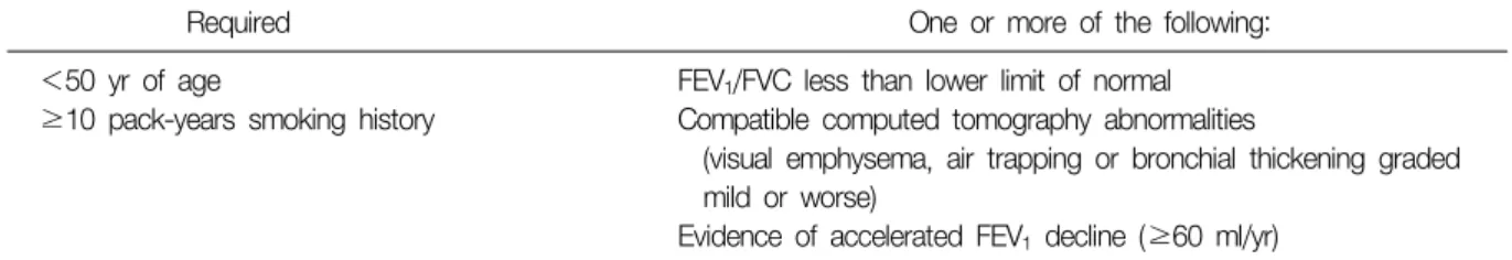
<0.7)은 없지만 만성 기관지염 증상이 있거나, 영상, 폐기능 등의 이상이 동반된 경우, 혹은 폐기능 감소 속도가 빨라서 COPD로 진행할 위험성이 있는 COPD 전단계 환자군이다. COPD의 예방과 치료에 대한 근본적이고 획기적인 패러다임의 변화는 이런 환자들을 대상으로 접근해야 가능할 것으로 기대되고 있다. 그러나 이 분야의 정의에 대해서 연구자들 간의 의견이 서로 다른 상황이며 조기 COPD라는 용어 내에 여러 가지 개념이 혼재하고 있다. Martinez 등은 이런 문제를 인식하고 COPD 위험군인 조기 COPD에 대한 통일된 개념을 제안하였다 (Table 1)5.
3. COPD 조기 진단
COPD는 진단되지 않고 간과되기 쉬운 질환이다. 대부분 고령의 흡연 환자이므로 COPD 증상을 흡연의 결과 로 당연하게 받아들이기 때문이다. 또한 나이가 들고 질환이 진행함에 따라 활동 능력이 감소하므로 호흡곤란 증상을 인지하지 못하는 경우도 많다. 결과적으로 대부분의 환자가 진단되지 않고 있다. 가장 효과적인 방법은 선별검사일 것이지만 모든 인구에 대한 선별검사는 비용 효과 면에서 유용성을 입증하기 힘들고 연구도 거의 없는 상태이므로 권고하지 않는다. 하지만 위험군을 적극적으로 조기 발견하여 치료하는 것은 권고하고 있다.
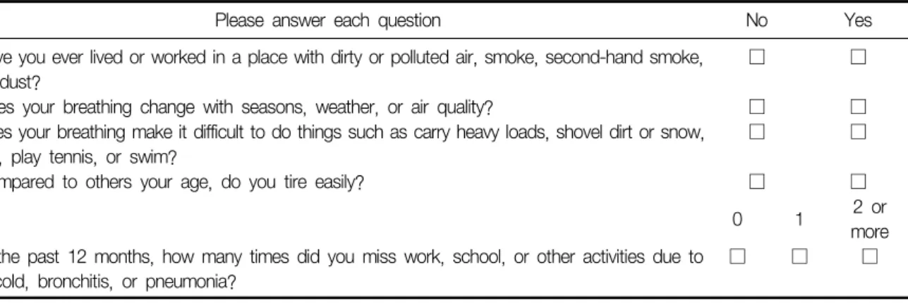
위험군을 조기 발견하기 위해서는 일차 진료의의 역할이 매우 크지만 이들은 대개 COPD 질환에 대한 관심이 적고 전문성이 부족하여 진단에 어려움을 겪게 된다. Di Marco 등은 COPD 조기 발견 확대를 위한 방법에 대하여 호흡기 전문의를 대상으로 설문 조사하여 보고하였는데 호흡기 전문의들은 COPD 조기 발견이 중요하다 고 인식하고 있었고 조기 발견을 위한 일차 진료의의 역할로는 선별 설문조사 혹은 간단한 폐기능 검사도구를 이용하여 COPD 의심 환자를 진단하고 이를 전문의에게 의뢰하는 방법이 가장 적절할 것으로 제시하였다6. 한편, 우선적으로 치료를 요하며 치료의 효과가 가장 클 것으로 예상되는 악화 위험군을 일차 의료기관에서 간단하게 선별할 수 있는 설문 지표를 개발한 연구도 있다. Martinez 등이 제시한 설문표는 기존의 여러 연구에 서 제시한 호흡기 증상 외에 지난 일 년간의 악화 병력을 설문에 포함하고 악화에 가중 점수를 부과하였다(Table 2). 연구에서 정의한 악화 위험군은 FEV1 60% 이하 혹은 전년도 1회 이상의 악화가 있는 COPD 환자이고 대조군은 악화가 없으며 FEV1이 60% 초과하는 그룹이다. 최고 호기 기류(peak expiratory flow)와 설문을 병합 한 경우 악화 위험군 진단의 민감도는 89%, 특이도는 78.1%에 이른다고 보고하였다7.
일차 진료의에 의한 조기 발견을 격려하는 방법 외에 다른 질환으로 진료받는 환자들에게 폐기능 검사를
Required One or more of the following:
<50 yr of age
≥10 pack-years smoking history
FEV1/FVC less than lower limit of normal Compatible computed tomography abnormalities
(visual emphysema, air trapping or bronchial thickening graded mild or worse)
Evidence of accelerated FEV1 decline (≥60 ml/yr)
Exclusion criteria include other known chronic lung diseases, including interstitial lung diseases, but not asthma (This table is cited from reference 5).
Table 1. Components of operational definition of early COPD
적극적으로 시행하여 조기 진단율을 높이고자 하는 방법도 있다. 대표적인 것이 수술 전 평가이며, Choi 등이 발표한 국내 연구에 의하면 전체 술 전 평가 환자 중 15.6%가 COPD 환자였으나 이들 중 이미 진단받은 이들은 5.5%에 불과하였다8.
4. 조기 COPD 평가
현재의 COPD 진단 기준에 속하지 않지만 COPD 전단계인 환자를 조기 진단하기 위한 다양한 검사법이 연구되고 있다. 크게 세가지 분야인데 증상, 폐기능 그리고 영상이다. 가장 오래 전부터 연구된 것은 만성 기관지염으로 증상은 있으나 기류제한이 없는(FEV1/FVC≥0.7) 그룹이다. 이들이 모두 COPD로 진행하지는 않는다는 것과 진단된 COPD 환자 중 기관지염이 없었던 환자도 상당 부분이라는 것이 이미 알려져 있으며 따라서 이들이 COPD 위험군인지에 대해서는 논란이 있어 왔다. 최근 통계적 선택 오차를 최소화하기 위해서 영국 일정 지역에 거주하는 동일 연령의 인구를 대상으로 한 장기간의 코호트 연구결과가 발표되었다. 이 연구 에서 만성 기관지염 증상은 흡연 여부에 크게 영향을 받는다는 것이 다시 한번 입증되었다. 하지만 만성 기관지 염이 흡연자 중에서도 특히 중년에 나타나는 경우, 비흡연자에게 나타나는 경우, 여러 번 보고되어 장기간 지속되었을 것으로 예상되는 환자에서 COPD 진행 위험성이 높은 것으로 나타나 기관지염 증상이 있으나 폐기 능이 정상인 그룹에 대한 관심이 증가하고 있다9.
폐기능 검사에 대한 연구도 활발하다. 기류제한은 없으나 FEV1이 80% 미만인 환자들이 있는데 이들을 PRISm (preserved ratio impaired spirometry)으로 명명하였으며 이들이 조기 COPD인지에 대한 연구가 활발히 진행되 고 있다. 한편 현재의 폐활량 검사법을 개선시켜서 기류제한은 없어도(FEV1/FVC≥0.7) 이미 진행되고 있는 소기도 질환을 진단하고자 하는 시도가 있다. Bhatt 등은 폐활량 검사의 용적 시간 곡선에서 시간에 따른 용적 증가 속도를 측정한 “PARAMETER D”라는 지표를 개발하였는데 Parameter D는 기류제한이 없지만 영상에서 이미 기능적 소기도 질환이 진행된 환자의 결과와 높은 일치도를 나타내 COPD 조기 발견 가능성을 보여주었다10. 같은 그룹에서 용적 기류곡선을 이용한 peak index라는 지표도 개발되었는데 parameter D와 마찬가지로 기류 제한이 없는 환자에서 소기도 질환을 조기에 발견할 수 있다고 보고하였다11.
Harvey 등은 폐활량 검사 결과가 정상인 1,500여명의 흡연자를 대상으로 폐확산능이 감소된 군과 정상인 군으로 나누어서 4년간 관찰한 결과를 발표하였다. 양 군 간에 호흡기 증상이나 흉부 CT의 폐기종 소견 등은 유의한 차이가 없었으나 폐 확산능이 감소되어 있던 군에서 4년 후 COPD로 진단된 환자가 많았다(22% vs 3%)12.
음파 진동을 이용하여 소기도 저항을 측정할 수 있는 forced oscillation technique/impulse oscillometry sys-
Please answer each question No Yes
1. Have you ever lived or worked in a place with dirty or polluted air, smoke, second-hand smoke, or dust?
□ □
2. Does your breathing change with seasons, weather, or air quality? □ □ 3. Does your breathing make it difficult to do things such as carry heavy loads, shovel dirt or snow,
jog, play tennis, or swim?
□ □
4. Compared to others your age, do you tire easily? □ □
0 1 2 or
more 5. In the past 12 months, how many times did you miss work, school, or other activities due to
a cold, bronchitis, or pneumonia?
□ □ □
Table 2. CAPTURE (chronic obstructive pulmonary disease assessment in primary care to identify undiagnosed respiratory disease and exacerbation risk) questionnaire (This table is cited from reference 7)
tem은 폐활량 검사보다 조기에 소기도 이상을 발견할 수 있고 검사 방법이 까다롭지 않아서 노인에게도 쉽게 적용 가능한 유용한 검사법이다. 아직 표준화되어 있지 않고 해석이 까다롭다는 점이 단점이지만 이를 극복하면 폐활량 검사법을 대신하여 조기 COPD 진단에 유용할 것으로 기대된다.
흉부 CT 촬영 기법 및 분석 기법이 발달하면서 기류제한 소견이 없는 흡연자 중에서 폐기종의 유무 및 중증도 를 계산할 수 있고 소기도 벽의 두께 측정을 통한 소기도 질환 진단이 가능해졌다. 또한 흡기와 호기 시에 얻은 폐영상을 분석하여 폐기종과 기류 저류 소견(air trapping)을 구분할 수 있고 이를 통해 기능적 소기도 질환의 정도를 파악할 수 있다. 이렇게 계산된 기능적 소기도 질환은 폐기능 감소 속도와 연관이 있다고 보고되 기도 하였다13. 최근 COPDGene 코호트 연구에서는 분석기법 없이 정성적 방법 즉 visual estimation으로 평가한 폐기종 소견이 사망률과 연관있다고 보고한 바 있어 흉부 CT는 유용한 검사법으로 주목받고 있다14.
5. 요약 및 결론
조기 COPD 진단과 평가는 COPD 질환의 예후를 바꾸기 위한 중요한 과제이다. 진단되지 않고 있는 경증 환자들을 조기 발견하여 금연을 비롯한 약물 치료를 제공하는 것은 환자의 증상을 완화시킬 뿐만 아니라 예후를 향상시킬 수 있다. 또한 기류제한이 없는 흡연자 혹은 비흡연자들을 대상으로 COPD 진행가능성이 높은 지표를 찾고자 하는 노력이 지속되고 있다. 이들에 대한 진단과 평가는 만성 기관지염 증상, 폐기능 검사의 새로운 지표들 및 음파 진동법, 흉부 CT를 이용한 폐기종 및 소기도 질환 진단 등을 통해 이루어지고 있으며 향후 임상 적용 가능성이 높을 것으로 기대된다.
References
1. Zhou Y, Zhong NS, Li X, Chen S, Zheng J, Zhao D, et al. Tiotropium in early-stage chronic obstructive pulmo- nary disease. N Engl J Med 2017;377:923-35.
2. Fletcher C, Peto R. The natural history of chronic airflow obstruction. Br Med J 1977;1:1645-8.
3. Vestbo J, Edwards LD, Scanlon PD, Yates JC, Agusti A, Bakke P, et al. Changes in forced expiratory volume in 1 second over time in COPD. N Engl J Med 2011;365:1184-92.
4. Tantucci C, Modina D. Lung function decline in COPD. Int J Chron Obstruct Pulmon Dis 2012;7:95-9.
5. Martinez FJ, Han MK, Allinson JP, Barr RG, Boucher RC, Calverley PMA, et al. At the root: defining and halting progression of early chronic obstructive pulmonary disease. Am J Respir Crit Care Med 2018;197:1540-51.
6. Di Marco F, Balbo P, de Blasio F, Cardaci V, Crimi N, Girbino G, et al. Early management of COPD: where are we now and where do we go from here? A Delphi consensus project. Int J Chron Obstruct Pulmon Dis 2019;14:353-60.
7. Martinez FJ, Mannino D, Leidy NK, Malley KG, Bacci ED, Barr RG, et al. A new approach for identifying patients with undiagnosed chronic obstructive pulmonary disease. Am J Respir Crit Care Med 2017;195:748-56.
8. Choi SM, Lee J, Park YS, Lee CH, Lee SM, Yim JJ, et al. Prevalence and global initiative for chronic obstructive lung disease group distribution of chronic obstructive pulmonary disease detected by preoperative pulmonary function test. PLoS One 2015;10:e0115787.
9. Allinson JP, Hardy R, Donaldson GC, Shaheen SO, Kuh D, Wedzicha JA. The presence of chronic mucus hypersecretion across adult life in relation to chronic obstructive pulmonary disease development. Am J Respir Crit Care Med 2016;193:662-72.
10. Bhatt SP, Bhakta NR, Wilson CG, Cooper CB, Barjaktarevic I, Bodduluri S, et al. New spirometry indices for detecting mild airflow obstruction. Sci Rep 2018;8:17484.
11. Bhatt SP, Bodduluri S, Raghav V, Bhakta NR, Wilson CG, Kim YI, et al. The peak index: spirometry metric for airflow obstruction severity and heterogeneity. Ann Am Thorac Soc 2019;16:982-9.
12. Harvey BG, Strulovici-Barel Y, Kaner RJ, Sanders A, Vincent TL, Mezey JG, et al. Risk of COPD with obstruction in active smokers with normal spirometry and reduced diffusion capacity. Eur Respir J 2015;46:1589-97.
13. Bhatt SP, Soler X, Wang X, Murray S, Anzueto AR, Beaty TH, et al. Association between functional small airway disease and FEV1 decline in chronic obstructive pulmonary disease. Am J Respir Crit Care Med 2016;194:178-84.
14. Lynch DA, Moore CM, Wilson C, Nevrekar D, Jennermann T, Humphries SM, et al. CT-based visual classification of emphysema: association with mortality in the COPDGene study. Radiology 2018;288:859-66.
III Does Earlier Intervention Have Better Outcomes in COPD?
강지은, 이재승
울산대학교 의과대학 서울아산병원 호흡기내과
Chronic obstructive pulmonary disease (COPD) is one of the major health problems worldwide and a treatment strategy to prevent or slow down disease progression is highly needed. This review discusses the current evidence for early intervention in COPD. Smoking cessation is the most effective intervention for altering the natural course of COPD. It has been shown to reduce the lung function decline rate as well as mortality. On the contrary, no studies have confirmed the same effect with pharmacotherapy. Few post-hoc and subgroup analyses showed potential benefits of bronchodilator treatment, but only limited evidence exists. Further studies are required to advocate early pharmacotherapy in COPD.
Key Words: COPD, Early intervention, Smoking cessation, Bronchodilator Corresponding author: Jae Seung Lee, M.D., Ph.D.
Department of Pulmonary and Critical Care Medicine, Asan Medical Center, University of Ulsan College of Medicine, 88, Olympic-ro 43-gil, Songpa-gu, Seoul 05505, Korea
Tel: +82-2-3010-3994, Fax: +82-2-3010-6968, E-mail: [email protected]
1. 서론
만성폐쇄성폐질환(chronic obstructive pulmonary disease, COPD)은 전 세계적으로 사망의 주된 원인 중 하나이며1 인구 고령화로 인해 유병률 또한 계속 증가할 것으로 생각된다2. 대개의 만성 질환은 조기에 진단하고 적절한 조치를 취해 질병의 악화와 합병증을 예방하는 것이 치료의 핵심이다. 그렇지만 COPD는 여전히 진단과 치료를 받고 있는 환자의 비율이 낮은 질환으로3-5 기침, 객담과 같은 호흡기 증상이 발생해도 흡연에 의한 변화로 간과하고 적절한 진단과 치료를 받지 않는 환자가 많다6,7. COPD는 진단 수년 전부터 서서히 진행하여 질병 초기부터 생리학적 변화를 동반한다8-11. 질병의 발생 전 혹은 초기에 적극적인 치료를 통해 질병으로의 이환과 진행을 예방할 수 있다면 COPD로 인한 사회 경제적 부담을 줄일 수 있을 것으로 예상할 수 있다.
하지만 현재 COPD의 치료는 질병의 자연 경과를 고려하는 접근법보다는 증상과 악화를 바탕으로 결정되고 있다1. 본 종설에서는 초기에 적극적인 치료가 실제로 환자의 예후를 개선시키는지 COPD의 조기 중재에 대한 현재까지의 근거들에 대해 다루고자 한다.
2. 조기 중재의 필요성
비록 개인차가 있기는 하지만 COPD 환자는 정상인에 비해 폐기능이 빨리 감소한다는 특징을 가지고 있다12. Fletcher와 Peto가 제시한 COPD 자연 경과 모델에 따르면 폐기능은 시간이 지남에 따라 감소 속도가 더 빨라지 는 것으로 나타나지만13, 최근 연구들에 의하면 오히려 질병 초기에 폐기능이 더 빨리 감소하는 것으로 밝혀졌
다14-16. Tantucci와 Modina의 보고에 의하면15 연간 1초간 노력성 폐활량(forced expiratory volume in 1 second, FEV1) 감소 속도는 중등증(moderate) COPD에서 47∼79 mL/year로, 중증(56∼59 mL/year)이나 고도중증 (<35 mL/year) 환자의 감소 속도에 비해 더 컸다. 이러한 폐기능 감소 속도를 고려하면 중증 COPD로 진행하기 전 조기에 적극적인 중재를 통해 폐기능 감소를 예방하는 것이 중요할 것이다.
한편 경증 기류 제한을 보이는 초기 COPD 환자에서도 호흡기 증상으로 삶의 질 저하와17,18 운동 능력 저하가 동반될 수 있다는 것이 알려져 있다19,20. 기류 제한이 경증-중등증으로 심하지 않은 환자에서도 호흡기 증상이 동반되는 경우에는 사망률이 증가함이 보고되었다21,22. 뿐만 아니라 유의한 기류 제한이 없음에도 불구하고 (FEV1/forced vital capacity>0.7) COPD와 비슷한 호흡기 증상과 악화가 발생할 수 있다는 것 역시 알려져 있다. Woodruff 등은 흡연력이 있으나 폐기능 검사에서 COPD의 진단기준에는 해당하지 않는 현 흡연자와 과거 흡연자 중 약 50%가 만성 호흡기 증상을 동반하는 것을 보여주었다23. 호흡기 증상이 있는 경우에는 악화의 위험이 더 높고 운동 능력 역시 감소하는 것으로 나타났다. 고해상도 CT에서는 기관지벽 두께가 증가되어 있어 COPD로 진단되지 않았음에도 불구하고 이미 병리학적 변화가 발생하고 있다는 것을 간접적으로 확인할 수 있었다. 이러한 결과는 명백한 COPD 진단으로 진행하기 전, 그리고 심각한 병태생리학적 변화가 진행하기 전에 경과를 바꾸기 위한 조기 중재가 필요함을 시사한다.
3. COPD 치료와 조기 중재 관련 현재까지의 근거
1) 금연
흡연은 COPD 발생의 가장 주된 원인일 뿐 아니라 지속적인 폐기능 감소를 유발하여 COPD의 중증도를 악화시키는 위험인자이다1. 금연은 COPD 질병 경과를 바꿀 수 있는 유일한 방법이다1,24. Lung Health Study25에 서는 금연을 하게 되면 흡연을 지속하는 환자에 비해 폐기능 감소 속도가 절반으로 감소하는 것을 보여주었고, 특히 금연의 시기가 빠를수록 폐기능 감소 속도를 빨리 역전시킬 수 있는 것이 알려져 있다26. 따라서 금연은 COPD 예방을 위해서 뿐만 아니라 이미 COPD가 진단된 환자에서도 강조되어야 하는 핵심 치료이다.
2) 약물 치료
금연과 달리 약물 치료를 통해 COPD의 진행을 막거나 진행 속도를 늦추는 것에 대한 확실한 근거는 아직까지 미약하다. COPD 진행을 대변하는 지표로서 FEV1 감소 속도에 대한 흡입제의 효과는 COPD 연구의 오랜 관심사 였다. 초기 연구들은 흡입 스테로이드제가 COPD 예후 개선에 도움이 되는지 알아보고자 하였지만 유의한 결과를 얻지는 못하였다27-29. EUROSCOP study27와 Copenhagen City Heart study28는 inhaled budesonide가 폐기능 감소 속도를 줄일 수 있는지 분석하였지만 위약과 비교하여 유의한 차이를 밝히지 못하였다. 이후에도 여러 임상 시험에서 폐기능 감소에 대한 약제의 효과를 알아보았는데, TORCH study는 COPD 환자에서 salme- terol/fluticasone의 효과를 분석한 대규모 장기 임상 시험이었다30. 이 연구에서는 통계적으로 유의하지는 않았 지만 salmeterol/fluticasone 치료가 위약 대비 사망이 감소하는 경향을 보여주었다. 더불어 사후분석으로 salme- terol, fluticasone, salmeterol/fluticasone 치료에 따른 FEV1 감소 속도에 관련한 내용이 발표되었고, 세 가지 약제 모두 위약 대비 FEV1 감소 속도를 줄여주었으며 그 중에서도 복합제의 FEV1 감소 속도가 가장 낮은 것으로 나타났다31. 또 다른 대규모 연구인 UPLIFT는 tiotropium이 COPD 환자에게 미치는 영향을 알아보고자 한 임상 시험이었다32. 연구 결과 tiotropium이 위약과 비교하여 FEV1 감소를 유의하게 줄여주지는 못하는 것으로 나타 났지만, 하위그룹 분석으로 중등증 COPD 환자만을 대상으로 분석한 결과에서는 tiotropium을 투여한 환자에서 연간 FEV1 감소 속도가 대조군에 비해 유의하게 낮았다(43 vs. 49 mL/year)33. SUMMIT study에서도 이차 평가 항목으로 FEV1 감소 속도를 분석하였다. 중등증 COPD 환자를 대상으로 했던 이 연구에서는 fluticasone/vi- lanterol과 fluticasone 흡입제가 위약과 비교하여 연간 FEV1 감소 속도를 유의하게 줄여주는 것으로 보고하였 다34. 이러한 결과는 흡입제 치료가 폐기능 감소 속도를 늦추어 COPD 진행을 지연시킬 수 있다는 근거로 간주할
수 있기는 하지만, 이 결과들은 COPD 환자에서 조기에 약물 치료를 시작하는 것이 질병의 진행을 늦추는지 알아보기 위해 설계된 연구의 직접적인 결과가 아니라는 점을 고려해서 해석해야 한다. FEV1 감소 속도에 대한 영향은 사후분석, 하위그룹분석, 이차 평가 항목 등으로만 확인되었고, 또한 폐기능 감소가 개선된 정도 역시 통계적으로 유의하기는 하였지만 위약과 비교하여 임상적으로 큰 의미가 있을 정도의 수치는 아니었다는 점이 한계로 남아있다35. 따라서 아직까지는 폐기능 감소를 역전시키거나 유의하게 지연시키는 약물 치료에 대한 확실한 근거는 부족하다고 생각된다.
초기 COPD 환자에서 적극적인 약물 치료를 통해 장기적인 질병의 경과를 바꿀 수 있는지, 즉 약물 치료를 조기에 시행하는 것이 늦게 시작하는 것보다 환자 예후를 개선시키는지에 대해서도 근거가 분명하지 않다.
많은 임상 시험이 중등증 이상의 COPD 환자를 대상으로 진행되었기 때문에, 특히 기류 제한이 심하지 않은 경증 환자에서 약물 치료의 효과에 대한 연구는 그 수가 많지 않다. 최근 초기 COPD 환자에서 tiotropium 투여를 위약과 비교한 결과가 발표되었다36. 이 연구는 경증-중등증 COPD 환자를 대상으로 하였는데, tio- tropium을 투여 받은 환자는 위약 대비 기관지확장제 투여 후 FEV1의 연간 감소 속도가 유의하게 감소하였고 (29 vs. 51 mL/year) 급성 악화의 발생도 유의하게 낮았다. 이러한 결과는 약물 치료를 통한 조기 중재가 질병의 경과를 호전시킬 수 있다는 가능성을 제시한다. 하지만 이 효과가 tiotropium 외 다른 약물에도 적용되는지에 대해서는 연구가 부족하다. 최근 널리 사용되고 있는 이중 기관지확장제는 단일 기관지확장제에 비해 폐기능과 증상, 삶의 질 개선에 있어 대체로 우월한 결과를 보여주었는데37-39, 이러한 약제를 초기 COPD 환자에게 적극적 으로 사용하는 것이 장기 예후 개선에 도움이 되는지에 대해서도 추가적인 연구가 필요하겠다.
현재 Global Initiative for COPD에서는 폐기능과 무관하게 환자의 증상과 악화를 바탕으로 한 치료 가이드라 인을 제시하고 있어 약물 치료를 통해 COPD 발병을 예방하고 진행을 늦추는 데에는 한계가 있다. 또한 COPD 진단 기준에 맞지 않지만 호흡기 증상이 있는 환자들의 치료 가이드라인 역시 분명하지 않다는 점도 앞으로 해결해야 할 숙제이다.
4. 결론
COPD 환자에서 조기 중재를 통해 질병의 진행 속도를 늦추고 자연 경과를 바꿀 수 있다면 가장 이상적인 치료일 것이다. 금연은 COPD 발생과 진행을 예방할 수 있는 가장 효과적인 치료로, 금연을 빨리 할수록 폐기능 감소를 막을 수 있다. 반면 여러 대규모 임상 시험에서 흡입제의 효과를 분석하였으나 약물 치료는 아직까지 폐기능의 악화 속도를 뚜렷하게 감소시킨다는 근거가 부족하고, 조기에 치료를 시작하는 것이 예후를 개선시킨 다는 근거도 분명하지 않다. 그렇지만 일부 연구에서는 흡입제 투여가 FEV1 감소 속도를 완화시켰다는 결과를 보여주었고, 또한 경증 COPD에서 tiotropium을 투여하였을 때 폐기능 감소 속도가 줄어들었다는 보고도 있어 약물 치료를 통한 조기 중재가 질병의 경과를 호전시킬 수 있다는 가능성을 제시하였다. 조기 중재를 통한 예후 개선의 근거 마련을 위해서는 추가적인 연구가 더 필요하겠다.
References
1. Global Initiative for Chronic Obstructive Lung Disease. Global strategy for the diagnosis, management, and prevention of chronic obstructive pulmonary disease [Internet]. 2019 [cited 2019 Dec 7]. Available from:
https://goldcopd.org/wp-content/uploads/2018/11/GOLD-2019-v1.7-FINAL-14Nov2018-WMS.pdf.
2. Chapman KR, Mannino DM, Soriano JB, Vermeire PA, Buist AS, Thun MJ, et al. Epidemiology and costs of chronic obstructive pulmonary disease. Eur Respir J 2006;27:188-207.
3. Buist AS, McBurnie MA, Vollmer WM, Gillespie S, Burney P, Mannino DM, et al. International variation in the prevalence of COPD (the BOLD Study): a population-based prevalence study. Lancet 2007;370:741-50.
4. Damarla M, Celli BR, Mullerova HX, Pinto-Plata VM. Discrepancy in the use of confirmatory tests in patients
hospitalized with the diagnosis of chronic obstructive pulmonary disease or congestive heart failure. Respir Care 2006;51:1120-4.
5. Make B, Dutro MP, Paulose-Ram R, Marton JP, Mapel DW. Undertreatment of COPD: a retrospective analysis of US managed care and Medicare patients. Int J Chron Obstruct Pulmon Dis 2012;7:1-9.
6. Barnett M. Chronic obstructive pulmonary disease: a phenomenological study of patients' experiences. J Clin Nurs 2005;14:805-12.
7. Rennard S, Decramer M, Calverley PM, Pride NB, Soriano JB, Vermeire PA, et al. Impact of COPD in North America and Europe in 2000: subjects' perspective of Confronting COPD International Survey. Eur Respir J 2002;20:799-805.
8. Hogg JC, Chu F, Utokaparch S, Woods R, Elliott WM, Buzatu L, et al. The nature of small-airway obstruction in chronic obstructive pulmonary disease. N Engl J Med 2004;350:2645-53.
9. Deesomchok A, Webb KA, Forkert L, Lam YM, Ofir D, Jensen D, et al. Lung hyperinflation and its reversibility in patients with airway obstruction of varying severity. COPD 2010;7:428-37.
10. Peinado VI, Pizarro S, Barberà JA. Pulmonary vascular involvement in COPD. Chest 2008;134:808-14.
11. Elbehairy AF, Ciavaglia CE, Webb KA, Guenette JA, Jensen D, Mourad SM, et al. Pulmonary gas exchange abnormalities in mild chronic obstructive pulmonary disease. Implications for dyspnea and exercise intolerance.
Am J Respir Crit Care Med 2015;191:1384-94.
12. Lange P, Celli B, Agustí A, Boje Jensen G, Divo M, Faner R, et al. Lung-function trajectories leading to chronic obstructive pulmonary disease. N Engl J Med 2015;373:111-22.
13. Fletcher C, Peto R. The natural history of chronic airflow obstruction. Br Med J 1977;1:1645-8.
14. Bridevaux PO, Gerbase MW, Probst-Hensch NM, Schindler C, Gaspoz JM, Rochat T. Long-term decline in lung function, utilisation of care and quality of life in modified GOLD stage 1 COPD. Thorax 2008;63:768-74.
15. Tantucci C, Modina D. Lung function decline in COPD. Int J Chron Obstruct Pulmon Dis 2012;7:95-9.
16. Vestbo J, Edwards LD, Scanlon PD, Yates JC, Agusti A, Bakke P, et al. Changes in forced expiratory volume in 1 second over time in COPD. N Engl J Med 2011;365:1184-92.
17. Coultas DB, Mapel D, Gagnon R, Lydick E. The health impact of undiagnosed airflow obstruction in a national sample of United States adults. Am J Respir Crit Care Med 2001;164:372-7.
18. Mathers CD, Loncar D. Projections of global mortality and burden of disease from 2002 to 2030. PLoS Med 2006;3:e442.
19. Ofir D, Laveneziana P, Webb KA, Lam YM, O'Donnell DE. Mechanisms of dyspnea during cycle exercise in symptomatic patients with GOLD stage I chronic obstructive pulmonary disease. Am J Respir Crit Care Med 2008;177:622-9.
20. O'Donnell DE, Maltais F, Porszasz J, Webb KA, Albers FC, Deng Q, et al. The continuum of physiological impairment during treadmill walking in patients with mild-to-moderate COPD: patient characterization phase of a randomized clinical trial. PLoS One 2014;9:e96574.
21. Mannino DM, Buist AS, Petty TL, Enright PL, Redd SC. Lung function and mortality in the United States: data from the First National Health and Nutrition Examination Survey follow up study. Thorax 2003;58:388-93.
22. Ekberg-Aronsson M, Pehrsson K, Nilsson JA, Nilsson PM, Löfdahl CG. Mortality in GOLD stages of COPD and its dependence on symptoms of chronic bronchitis. Respir Res 2005;6:98.
23. Woodruff PG, Barr RG, Bleecker E, Christenson SA, Couper D, Curtis JL, et al. Clinical significance of symptoms in smokers with preserved pulmonary function. N Engl J Med 2016;374:1811-21.
24. Anthonisen NR, Skeans MA, Wise RA, Manfreda J, Kanner RE, Connett JE; Lung Health Study Research Group.
The effects of a smoking cessation intervention on 14.5-year mortality: a randomized clinical trial. Ann Intern Med 2005;142:233-9.
25. Anthonisen NR, Connett JE, Kiley JP, Altose MD, Bailey WC, Buist AS, et al. Effects of smoking intervention and the use of an inhaled anticholinergic bronchodilator on the rate of decline of FEV1. The Lung Health Study. JAMA 1994;272:1497-505.
26. Kohansal R, Martinez-Camblor P, Agustí A, Buist AS, Mannino DM, Soriano JB. The natural history of chronic airflow obstruction revisited: an analysis of the Framingham offspring cohort. Am J Respir Crit Care Med 2009;180:3-10.
27. Pauwels RA, Löfdahl CG, Laitinen LA, Schouten JP, Postma DS, Pride NB, et al. Long-term treatment with inhaled budesonide in persons with mild chronic obstructive pulmonary disease who continue smoking.
European Respiratory Society Study on Chronic Obstructive Pulmonary Disease. N Engl J Med 1999;340:1948-53.
28. Vestbo J, SØrensen T, Lange P, Brix A, Torre P, Viskum K. Long-term effect of inhaled budesonide in mild and moderate chronic obstructive pulmonary disease: a randomised controlled trial. Lancet 1999;353:1819-23.
29. Lung Health Study Research Group, Wise R, Connett J, Weinmann G, Scanlon P, Skeans M. Effect of inhaled triamcinolone on the decline in pulmonary function in chronic obstructive pulmonary disease. N Engl J Med 2000;343:1902-9.
30. Calverley PM, Anderson JA, Celli B, Ferguson GT, Jenkins C, Jones PW, et al. Salmeterol and fluticasone propio- nate and survival in chronic obstructive pulmonary disease. N Engl J Med 2007;356:775-89.
31. Celli BR, Thomas NE, Anderson JA, Ferguson GT, Jenkins CR, Jones PW, et al. Effect of pharmacotherapy on rate of decline of lung function in chronic obstructive pulmonary disease: results from the TORCH study.
Am J Respir Crit Care Med 2008;178:332-8.
32. Tashkin DP, Celli B, Senn S, Burkhart D, Kesten S, Menjoge S, et al. A 4-year trial of tiotropium in chronic obstructive pulmonary disease. N Engl J Med 2008;359:1543-54.
33. Decramer M, Celli B, Kesten S, Lystig T, Mehra S, Tashkin DP; UPLIFT investigators. Effect of tiotropium on outcomes in patients with moderate chronic obstructive pulmonary disease (UPLIFT): a prespecified subgroup analysis of a randomised controlled trial. Lancet 2009;374:1171-8.
34. Calverley PMA, Anderson JA, Brook RD, Crim C, Gallot N, Kilbride S, et al. Fluticasone furoate, vilanterol, and lung function decline in patients with moderate chronic obstructive pulmonary disease and heightened cardiovascular risk. Am J Respir Crit Care Med 2018;197:47-55.
35. Rennard SI, Drummond MB. Early chronic obstructive pulmonary disease: definition, assessment, and preven- tion. Lancet 2015;385:1778-88.
36. Zhou Y, Zhong NS, Li X, Chen S, Zheng J, Zhao D, et al. Tiotropium in early-stage chronic obstructive pulmo- nary disease. N Engl J Med 2017;377:923-35.
37. Wedzicha JA, Decramer M, Ficker JH, Niewoehner DE, Sandström T, Taylor AF, et al. Analysis of chronic obstructive pulmonary disease exacerbations with the dual bronchodilator QVA149 compared with glyco- pyrronium and tiotropium (SPARK): a randomised, double-blind, parallel-group study. Lancet Respir Med 2013;
1:199-209.
38. Buhl R, Maltais F, Abrahams R, Bjermer L, Derom E, Ferguson G, et al. Tiotropium and olodaterol fixed-dose combination versus mono-components in COPD (GOLD 2-4). Eur Respir J 2015;45:969-79.
39. Decramer M, Anzueto A, Kerwin E, Kaelin T, Richard N, Crater G, et al. Efficacy and safety of umeclidinium plus vilanterol versus tiotropium, vilanterol, or umeclidinium monotherapies over 24 weeks in patients with chronic obstructive pulmonary disease: results from two multicentre, blinded, randomised controlled trials.
Lancet Respir Med 2014;2:472-86.