© 2010 The Korean Academy of Medical Sciences.
This is an Open Access article distributed under the terms of the Creative Commons Attribution Non-Commercial License (http://creativecommons.org/licenses/by-nc/3.0) which permits unrestricted non-commercial use, distribution, and reproduction in any medium, provided the original work is properly cited.
pISSN 1011-8934 eISSN 1598-6357
Use of a Tunneling Technique to Achieve a Lower Defibrillation Threshold during Implantable Cardioverter Defibrillator
Implantation via the Right Subclavian Vein
A 56-yr-old man with aborted sudden cardiac death underwent implantable cardioverter defibrillator (ICD) implantation. While the ICD was being implanted, a left subclavian venogram failed to visualize the left subclavian vein, which was attributed to likely prolonged indwelling of the left subclavian sheath for venous access. Accordingly, the right subclavian vein was punctured and the ICD lead was diverted from the right side area to the active Can in the left pectoral area by tunneling over the sternum for high defibrillation threshold. The approach used in this case may be considered in patients who had difficult left subclavicular venous access and it may be prudent to save the left subclavian vein for ICD implantation in patients with fatal tachyarrhythmia.
Key Words: Defibrillators, Implantable; Electric Countershock Jin-Bae Kim1, Boyoung Joung2,
Moon-Hyoung Lee2, and Sung-Soon Kim2 Cardiology Division, Departmet of Internal Medicine1, Kyung Hee University College of Medicine, Seoul;
Cardiology Division, Yonsei Cardiovascular Center and Cardiovascular Research Institute2, Yonsei University College of Medicine, Seoul, Korea Received: 14 September 2009
Accepted: 21 December 2009 Address for Correspondence:
Moon-Hyoung Lee, M.D.
Cardiology Division, Yonsei Cardiovascular Center and Cardiovascular Research Institute, Yonsei University College of Medicine, 262 Seongsan-no, Seodaemun-gu, CPO Box 8044, Seoul 120-752, Korea
Tel: +82-2-2228-8460, Fax: +82.2-393-2041 E-mail: [email protected]
DOI: 10.3346/jkms.2010.25.10.1526 • J Korean Med Sci 2010; 25: 1526-1528
CASE REPORT
Cardiovascular Disorders
INTRODUCTION
Left transvenous pectoral implantation with an active Can has become the standard for implantable cardioverter defibrillator (ICD) device placement to secure a lower defibrillation thresh- old (DFT). However, some patients require implantation in the right pectoral region for a variety of reasons such as left pectoral infection, left subclavian access difficulty, left pectoral hardware, or left mastectomy (1). Right-sided pectoral implantation is fea- sible but may be associated with an unacceptably high DFT. In this report, we describe a case of ICD implantation in the left pectoral side, by tunneling after inserting a lead to the right sub- clavian vein, because of left subclavian vein obstruction.
CASE REPORT
A 56-yr-old man collapsed without chest pain after playing bad- minton on January 29, 2007. His friend witnessed the collapse and promptly performed cardiopulmonary resuscitation. The Emergency Rescue Service was called, and on arrival, after de- termining the presence of ventricular fibrillation, applied an automatic external defibrillator.
Defibrillation shock was delivered twice and sinus rhythm recovered. After admission, his mental state was markedly dis-
oriented with retrograde amnesia. His speech was appropriate but unable to perform 7 serial subtractions. Physical examina- tion was unremarkable. A profile of routine chemistry did not reveal any specific abnormalities except for slightly increased cardiac enzymes (CK: 1289 IU/L, CK-MB: 13.6 ng/mL) which might be due to repeated cardioversions. The electrocardiogram revealed normal sinus rhythm with left ventricular hypertrophy by voltage criteria and small Q waves in inferior leads. There was no ventricular preexcitation or QT prolongation. He had been on medication (Losartan, Thiazide, Glimepiride, Metformin) for hypertension and diabetes over the last 5 yr. According to de- tailed history, the patient have had exertion angina in the last 2 yr but did not seek for medical advice. The prolonged telemetry monitoring revealed an episode of non-sustained monomor- phic ventricular tachycardia (8 beats, right bundle branch block morphology, cycle length 340 ms). Echocardiography and cor- onary angiogram were performed to evaluate the structural heart disease. An echocardiography revealed inferior wall akinesia and an ejection fraction of 42%. A coronary angiography dem- onstrated a chronic total occlusion of the proximal right coro- nary artery and patent left coronary artery without stenosis. Per- cutaneous coronary intervention with stent was performed in the right coronary artery without complications. During an elec- trophysiologic study, programmed electrical stimulation with
Kim J-B, et al. • ICD Implantation with Tunneling
http://jkms.org 1527
DOI: 10.3346/jkms.2010.25.10.1526
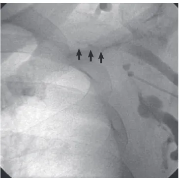
single or double ventricular extrastimuli reproducibly induced polymorphic ventricular tachycardia with hemodynamic com- promise. In view of the inducible ventricular tachycardia ob- served during electrophysiologic study and aborted sudden car- diac death without an acute coronary event, we decided to im- plant a single chamber ICD for the secondary prevention of sud- den cardiac death. However, while the ICD was being implant- ed, a left subclavian venogram failed to visualize the left subcla- vian vein, and the retrograde femoral catheter could not be ad- vanced to the vein (Fig. 1), which was attributed to likely pro- longed indwelling of the left subclavian sheath for venous ac- cess. Accordingly, the right subclavian vein was punctured, and
a defibrillating ventricular dual coil lead for an ICD (Vitality VR 1870, Guidant, St. Paul, MN, USA) was inserted. Lead measure- ments revealed ventricular sensing at 9.0 mV, a pacing threshold of 0.8 V/0.5 ms, and an impedance of 420 Ohms. Subsequently, a defibrillation test using a biphasic waveform was conducted in the dual coil system configuration (conventional configura- tion; RV-→SVC++CAN+).
Initially, an active Can was positioned at the right pectoral site. A DFT test of the active Can that revealed high energy (31J) to terminate the induced ventricular fibrillation. Polarity rever- sal, different shock configuration (RV-→CAN+) and lead repo- sitioning were not helpful at reducing DFT (31J).
Therefore, after fixation of the lead sleeve at left pectoralis fascia, the ICD lead was diverted from the right side area to the ICD in the left pectoral area via tunnel made by tunneling tool (Medtronic, Minneapolis, MN) over the sternum. Adaptor of lead extension was not necessary because lead length was long enough. A repeat defibrillation test revealed a lower DFT of 11J, and the active Can was successfully implanted in a pocket on the left pectoral side without complication (Fig. 2). During 2 yr follow up after ICD implantation, patients had a no episode of tachyarrhythmia and recent lead measurements revealed ven- tricular sensing at 10.2 mV, a pacing threshold of 0.8 V/0.5 ms, and an impedance of 439 Ohms.
DISCUSSION
As the ICD pulse generator is used as one of the electrode, the active Can is routinely being placed in the left side pectoral po- sition and the intracardiac ring electrode in RV, which theoreti- cally includes more ventricular myocardium. Accordingly, the left subclavian route has been used for ICD lead placement.
However, the subclavian vein route may sometimes be difficult to gain access to heart. In cases of critical inflammation at the Fig. 1. Venogram showed no visible left subclavian vein due to total occlusion of left
subclavian vein after prolonged catheter indwelling (arrows).
Fig. 2. (A) Chest PA after ICD implanta
tion. Arrowheads indicate ICD lead. (B) Left lateral view after ICD implantation.
A B
Kim J-B, et al. • ICD Implantation with Tunneling
1528 http://jkms.org DOI: 10.3346/jkms.2010.25.10.1526
left pectoral side, left subclavian vein obstruction or preexisting device at the left pectoral side, right sided route is an alternative.
Gold et al. (2) reported DFT were higher on the right side and a higher mortality in patients with right sided ICDs and Ovadia et al. (3) showed that the ipsilateral supraclavicular approach is feasible and safe in cases of subclavian vein obstruction, but this approach can only be adopted after confirming the length of the obstructive segment by venography. Therefore, an alter- native strategy is required when the subclavian vein is totally occluded.
Previously we reported a case (4) in which a preexisting left side pulse generator was repositioned by subcutaneous tunnel- ing over the sternum. By using same subcutaneous tunneling technique, a permanent defibrillation lead was diverted to left pectoral side to achieve the lower DFT in this patient.
The approach used in this case might be considered in pa- tients who had difficult left subclavicular venous access and it may be prudent to save the left subclavian vein for ICD implan- tation in patients with fatal tachyarrhythmia.
REFERENCES
1. Cannon BC, Friedman RA, Fenrich AL, Fraser CD, McKenzie ED, Kertesz NJ. Innovative techniques for placement of implantable cardioverter-de- fibrillator leads in patients with limited venous access to the heart. Pac- ing Clin Electrophysiol 2006; 29: 181-7.
2. Gold MR, Shih HT, Herre J, Breiter D, Zhang Y, Schwartz M; Low Energy Safety Study Investigators. Comparison of defibrillation efficacy and survival associated with right versus left pectoral placement for implant- able defibrillators. Am J Cardiol 2007; 100: 243-6.
3. Ovadia M, Cooper RS, Parnell VA, Dicapua D, Vatsia SK, Vlay SC. Trans- venous pacemaker insertion ipsilateral to chronic subclavian vein ob- struction: an operative technique for children and adults. Pacing Clin Electrophysiol 2000; 23 (11 Pt 1): 1585-93.
4. Choung B, Lee D, Ahn S, Lee M, Kim M, Kim S, Kim S. Repositioning of pacemaker generator due to therapeutic radiation: a tunneling method.
Korean Circ J 1998; 28: 1620-3.