Original Article
Sung-Eun Kim ,
1*Eun Sun Jang ,
2*Moran Ki ,
3Geum-Youn Gwak ,
4Kyung-Ah Kim ,
5Gi-Ae Kim ,
6Do Young Kim ,
7Dong Joon Kim ,
8Man Woo Kim ,
9Yun Soo Kim,
10Young Seok Kim ,
11In Hee Kim ,
12Chang Wook Kim,
13Ho Dong Kim ,
14Hyung Joon Kim,
15Neung Hwa Park ,
16Soon Koo Baik ,
17Jeong Ill Suh ,
18Byung-Cheol Song ,
19Il Han Song ,
20Jong Eun Yeon ,
21Byung Seok Lee,
22Youn Jae Lee,
23Young Kul Jung,
24Woo Jin Chung ,
25Sung Bum Cho ,
26Eun-Young Cho,
27Hyun Chin Cho ,
28Gab Jin Cheon,
29Hee Bok Chae,
30DaeHee Choi ,
31Sung-Kyu Choi,
32Hwa Young Choi ,
2Won Young Tak ,
33Jeong Heo,
34and Sook-Hyang Jeong
21 Department of Internal Medicine, Hallym University Sacred Heart Hospital, Hallym University College of Medicine, Anyang, Korea
2 Department of Internal Medicine, Seoul National University Bundang Hospital, Seoul National University College of Medicine, Seongnam, Korea
3 Department of Cancer Control and Population Health, Graduate School of Cancer Science and Policy, National Cancer Center, Goyang, Korea
4Department of Medicine, Samsung Medical Center, Sungkyunkwan University School of Medicine, Seoul, Korea
5Department of Internal Medicine, Inje University Ilsan Paik Hospital, Goyang, Korea
6 Health Screening and Promotion Center, Asan Medical Center, University of Ulsan College of Medicine, Seoul, Korea
7Department of Internal Medicine, Yonsei University College of Medicine, Seoul, Korea
8Department of Internal Medicine, Hallym University College of Medicine, Chuncheon, Korea
9Department of Internal Medicine, Chosun University School of Medicine, Gwangju, Korea
10Department of Internal Medicine, Gachon University, College of Medicine, Incheon, Korea
11 Department of Internal Medicine, Soonchunhyang University Bucheon Hospital, Soonchunhyang University College of Medicine, Bucheon, Korea
12 Department of Internal Medicine, Chonbuk National University Hospital, Chonbuk National University College of Medicine, Jeonju, Korea
13 Department of Internal Medicine, Uijeongbu St. Mary's Hospital, The Catholic University of Korea, College of Medicine, Uijeongbu, Korea
14Department of Internal Medicine, St. Carollo General Hospital, Suncheon, Korea
15 Department of Internal Medicine, Chung-Ang University Hospital, Chung-Ang University College of Medicine, Seoul, Korea
16Department of Internal Medicine, University of Ulsan College of Medicine, Ulsan, Korea
17Department of Internal Medicine, Yonsei University Wonju College of Medicine, Wonju, Korea
18 Department of Internal Medicine, Dongguk University Gyeongju Hospital, Dongguk University College of Medicine, Gyeongju, Korea
19Department of Internal Medicine, Jeju National University College of Medicine, Jeju, Korea
20Department of Internal Medicine, Dankook University Hospital, Cheonan, Korea
21 Department of Internal Medicine, Korea University Guro Hospital, Korea University College of Medicine, Seoul, Korea
22Department of Internal Medicine, Chungnam National University College of Medicine, Daejeon, Korea
23 Department of Internal Medicine, Inje University Busan Paik Hospital, Inje University College of Medicine, Busan, Korea
24 Department of Internal Medicine, Korea University Ansan Hospital, Korea University College of Medicine, Ansan, Korea
25Department of Internal Medicine, Keimyung University School of Medicine, Daegu, Korea
26Department of Internal Medicine, Chonnam National University Hwasun Hospital, Hwasun, Korea
27Department of Internal Medicine, Wonkwang University College of Medicine, Iksan, Korea
Chronic Hepatitis B Infection Is
Significantly Associated with Chronic Kidney Disease: a Population-based, Matched Case-control Study
Received: Mar 9, 2018 Accepted: Jun 14, 2018 Address for Correspondence:
Sook-Hyang Jeong, MD
Department of Internal Medicine, Seoul National University Bundang Hospital, Seoul National University, College of Medicine, 82 Gumi-ro, 173-beon-gil, Bundang-gu, Seongnam 13620, Republic of Korea.
E-mail: [email protected]
*Sung Eun Kim and Eun Sun Jang contributed equally to this study.
© 2018 The Korean Academy of Medical Sciences.
This is an Open Access article distributed under the terms of the Creative Commons Attribution Non-Commercial License (https://
creativecommons.org/licenses/by-nc/4.0/) which permits unrestricted non-commercial use, distribution, and reproduction in any medium, provided the original work is properly cited.
ORCID iDs Sung-Eun Kim
https://orcid.org/0000-0001-6236-780X Eun Sun Jang
https://orcid.org/0000-0003-4274-2582 Moran Ki
https://orcid.org/0000-0002-8892-7104 Geum-Youn Gwak
https://orcid.org/0000-0002-6453-3450 Kyung-Ah Kim
https://orcid.org/0000-0002-6128-6407
Nephrology
Gi-Ae Kim
https://orcid.org/0000-0002-5002-0822 Do Young Kim
https://orcid.org/0000-0002-8327-3439 Dong Joon Kim
https://orcid.org/0000-0002-5792-1500 Man Woo Kim
https://orcid.org/0000-0003-0776-919X Young Seok Kim
https://orcid.org/0000-0002-7113-3623 In Hee Kim
https://orcid.org/0000-0003-3863-7907 Ho Dong Kim
https://orcid.org/0000-0002-1899-1629 Neung Hwa Park
https://orcid.org/0000-0002-5648-9189 Soon Koo Baik
https://orcid.org/0000-0001-6245-2537 Jeong Ill Suh
https://orcid.org/0000-0002-3040-8766 Byung-Cheol Song
https://orcid.org/0000-0002-8319-3375 Il Han Song
https://orcid.org/0000-0003-3975-6342 Jong Eun Yeon
https://orcid.org/0000-0002-0510-7371 Woo Jin Chung
https://orcid.org/0000-0002-3736-1067 Sung Bum Cho
https://orcid.org/0000-0001-9816-3446 Hyun Chin Cho
https://orcid.org/0000-0001-7750-474X DaeHee Choi
https://orcid.org/0000-0002-8956-7518 Hwa Young Choi
https://orcid.org/0000-0002-8195-9250 Won Young Tak
https://orcid.org/0000-0002-1914-5141 Sook-Hyang Jeong
https://orcid.org/0000-0002-4916-7990 Funding
This study was supported by a grant of the Korea Health Technology R&D Project through the Korea Health Industry Development Institute (KHIDI), funded by the Ministry of Health & Welfare, Republic of Korea (Grant No.
HC15C1193).
Disclosure
The authors have no potential conflicts of interest to disclose.
Author Contributions
Conceptualization: Jeong SH. Data curation:
Kim SE, Jang ES, Ki M, Gwak GY, Kim KA, Kim GA, Kim DY, Kim DJ, Kim MW, Kim YS, Kim YS, Kim IH, Kim CW, Kim HD, Kim HJ, Park NH,
ABSTRACT
Background: Hepatitis B virus (HBV) infection leads to hepatic and extrahepatic
manifestations including chronic kidney disease (CKD). However, the association between HBV and CKD is not clear. This study investigated the association between chronic HBV infection and CKD in a nationwide multicenter study.
Methods: A total of 265,086 subjects who underwent health-check examinations in 33 hospitals
from January 2015 to December 2015 were enrolled. HBV surface antigen (HBsAg) positive cases (n = 10,048), and age- and gender-matched HBsAg negative controls (n = 40,192) were identified. CKD was defined as a glomerular filtration rate (GFR) < 60 mL/min/1.73 m
2or proteinuria as at least grade 2+ of urine protein.
Results: HBsAg positive cases showed a significantly higher prevalence of GFR
< 60 mL/min/1.73 m
2(3.3%), and proteinuria (18.9%) than that of the controls (2.6%, P < 0.001, and 14.1%, P < 0.001, respectively). In the multivariate analysis, HBsAg positivity was an independent factor associated with GFR < 60 mL/min/1.73 m
2along with age, blood levels of albumin, bilirubin, anemia, and hemoglobin A1c (HbA1c). Likewise, HBsAg positivity was an independent factor for proteinuria along with age, male, blood levels of bilirubin, protein, albumin, and HbA1c. A subgroup analysis showed that HBsAg positive men but not women had a significantly increased risk for GFR < 60 mL/min/1.73 m
2.
Conclusion: Chronic HBV infection was significantly associated with a GFR < 60 mL/min/1.73 m2
and proteinuria (≥ 2+). Therefore, clinical concern about CKD in chronic HBV infected
patients, especially in male, is warranted.
Keywords: Hepatitis B Virus; Chronic Renal Insufficiency; Glomerular Filtration Rate;
Proteinuria
INTRODUCTION
Globally, 240 million people representing 5%–6% of the world population are chronically infected with hepatitis B virus (HBV).
1Although the prevalence of HBV infection is decreasing in Korea, it is still a serious public health problem of liver-related morbidity and mortality.
2Chronic HBV infection predominantly manifests as hepatic diseases such as chronic hepatitis, liver cirrhosis, and hepatocellular carcinoma. However, extrahepatic manifestations including renal diseases, polyarteritis nodosa or dermatologic diseases such as popular acrodermatitis occur in 10%–20% of HBV infected patients even in the absence of hepatic disease.
3,4Recognition of these extrahepatic manifestations is important for the comprehensive care of HBV infected patients, especially in the era of highly effective antiviral therapy.
28 Department of Internal Medicine, Gyeongsang National University Hospital, Gyeongsang National University College of Medicine, Jinju, Korea
29 Department of Internal Medicine, GangNeung Asan Hospital, University of Ulsan College of Medicine, Gangneung, Korea
30Department of Internal Medicine, Chungbuk National University College of Medicine, Cheongju, Korea
31Department of Internal Medicine, Kangwon National University School of Medicine, Chuncheon, Korea
32Department of Internal Medicine, Chonnam National University Medical School, Gwangju, Korea
33Department of Internal Medicine, Kyungpook National University College of Medicine, Daegu, Korea
34Department of Internal Medicine, Pusan National University School of Medicine, Busan, Korea
Baik SK, Suh JI, Song BC, Song IH, Yeon JE, Lee BS, Lee YJ, Jung YK, Chung WJ, Cho SB, Cho EY, Cho HC, Cheon GJ, Chae HB, Choi DH, Choi SK, Choi HY, Tak WY, Heo J. Formal analysis: Kim SE, Jang ES. Investigation: Kim SE, Jang ES, Ki M, Gwak GY, Kim KA, Kim GA, Kim DY, Kim DJ, Kim MW, Kim YS, Kim YS, Kim IH, Kim CW, Kim HD, Kim HJ, Park NH, Baik SK, Suh JI, Song BC, Song IH, Yeon JE, Lee BS, Lee YJ, Jung YK, Chung WJ, Cho SB, Cho EY, Cho HC. Writing - original draft: Kim SE, Jang ES, Jeong SH. Writing - review & editing: Kim SE, Jang ES, Jeong SH.
HBV-related renal diseases could lead to chronic kidney disease (CKD)
5,6and subsequent end-stage renal disease (ESRD).
7,8It can be classified as immune complex glomerulonephritis (membranous nephropathy or membranoproliferative glomerulonephritis) and immune complex-related vasculitis (polyarteritis nodosa
9or cryoglobulinemic vasculitis
8). Although the pathogenic mechanism behind these renal manifestations is not clear,
10deposition of HBV-related immune complexes,
11HBV replication in the renal tubular epithelium which may promote apoptosis of renal tubular cells,
12HBV-related insulin resistance,
13and oxidative stress
14may contribute to renal dysfunction.
Even though HBV-related CKD and ESRD cases are encountered in the clinic, there are only a few studies on the association between HBV infection and the development of CKD at a population level. Moreover, those studies showed striking disparate results on the role of HBV infection in CKD.
15-18Thus, the aim of this study was to investigate the association between chronic HBV infection and the prevalence of CKD defined as a decreased estimated glomerular filtration rate (GFR) or proteinuria in a population-based, nationwide study in Korea.
METHODS
Study design and data acquisition
This was a multicenter, retrospective, cross-sectional, and matched case-control study. A total of 265,086 subjects who underwent a health-check examination including hepatitis B virus surface antigen (HBsAg) status were enrolled nationwide in 33 hospitals from January 1, 2015 to December 31, 2015. After exclusion of 1,325 people who showed anti- hepatitis C virus (HCV) antibody positivity, 10,048 subjects who showed HBsAg positivity were identified (cases). For the purpose of comparison, 40,192 HBsAg negative subjects (controls) were randomly selected in the same dataset who were 1:4 matched for age by decades and gender using the R program (R Foundation, Vienna, Austria; www.r-project.
org/) version 3.3.3.
Demographic data, including age, gender, resident address, height, and weight were obtained from an electronically extracted database for the health-check examinee at each hospital. The laboratory results of the blood chemistry obtained after overnight fasting included the fasting plasma glucose, serum protein, albumin, bilirubin, cholesterol, alanine aminotransferase (ALT), hemoglobin A1c (HbA1c), urea nitrogen, creatinine, thyroid stimulating hormone and complete blood cell count as well as an assay for the HBsAg and anti-HCV antibody. Urine analysis was performed with spontaneously voided clean-caught mid-stream fresh urine.
Urine protein was measured using an immediate semi-quantitative urine protein dipstick test and graded as negative, trace, 1+, 2+, 3+, or 4+.
Serum HBsAg was measured mainly using chemiluminescent microparticle immunoassay with Abbott Architect assay and electrochemiluminescence immunoassay. Dipstick urine analysis was performed using the Urisys 2400 cassette strip, which was read by Urisys 2400 automated analyzer (Roche, Mannheim, Germany) in 50% of the hospitals, and various ways in the remaining hospitals. Serum creatinine level was measured using the compensated kinetic alkaline picrate (Jaffe reaction) in 70% of the hospitals, so that the estimated GFR was calculated with the Modification of Diet in Renal Disease equation
19:
estimated GFR = 175 × serum creatinine
−1.154× age
−0.203× 0.742 (if female)
Although each hospital has different testing methods, the quality control of all the measuring method was certified by clinical laboratory accreditation program, which was operated by the Korean Society of Laboratory Medicine.
Definition of the CKD, proteinuria, and anemia
The stages of CKD were classified into 6 categories (G1 to G6) by the clinical practice guideline Kidney Disease: Improving Global Outcomes 2012
20: G1, the participants had a GFR of at least 90 mL/min/1.73 m
2; G2, a GFR of 60–89 mL/min/1.73 m
2; G3a, a GFR of 45–59 mL/min/1.73 m
2; G3b, a GFR of 30–44 mL/min/1.73 m
2; G4a, a GFR of 15–29 mL/min/1.73 m
2, and G5a, a GFR less than 15 mL/min/1.73 m
2. In this study, a decreased GFR was defined as a GFR < 60 mL/min/1.73 m
2(GFR categories G3a–G5), and the proteinuria was defined as the presence of urine protein of at least grade 2+ in the urine protein dipstick test. Therefore, CKD was defined as a GFR < 60 mL/
min/1.73 m
2or proteinuria at least grade 2+. Anemia defined as serum hemoglobin level < 13 g/dL in men, and < 12 g/dL in women according to World Health Organization criteria.
Statistical analyses
All data were expressed as the means ± standard deviation or as a percentage. The continuous variables were analyzed with t-tests, and categorical variables were investigated with
Pearson's χ
2tests. Conditional logistic regression model analyses were used to determine the independent factors associated with the decreased GFR and proteinuria. P < 0.05 was considered to indicate a statistical significance. The statistical analyses were performed using SPSS version 24.0 (SPSS, Inc., Chicago, IL, USA).
Ethics statement
The present study protocol was reviewed and approved by the Institutional Review Board of each hospital (Seoul National University Bundang Hospital approval No. B-1602/336-101).
The data were analyzed anonymously. Informed consents were waived because this was a retrospective data analysis study.
RESULTS
Comparison of the clinical characteristics between the HBsAg positive cases and the age- and gender-matched controls
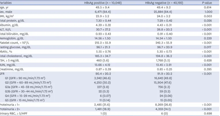
The clinical characteristics of a total 50,240 subjects consisting of 10,048 HBsAg positive cases and 40,192 control subjects are summarized in Table 1. In the case group, the mean age was 49.5 years with male proportion of 64.4%. The mean ALT level was higher in the case group than in the control group (30.7 IU/L vs. 26.8 IU/L; P < 0.001), while the serum albumin level was lower in the case group compared with the control group (4.39 g/dL vs. 4.43 g/dL; P < 0.001).
The mean serum cholesterol level (185.3 mg/dL) and platelet count (212.2 × 10
9/L) were significantly lower in the case group compared with those of the control group (194.8 mg/dL [P < 0.001] and 242.3 × 10
9/L [P < 0.001], respectively). While the mean serum blood urea nitrogen level in the case group (13.66 mg/dL) was significantly higher than in the case group (13.45 mg/dL, P < 0.001), the mean serum creatinine level was not significantly different between the case and control group. However, the prevalence of GFR < 60 mL/min/1.73 m
2in the case group (3.3%) was significantly higher than in the control group (2.6%, P < 0.001).
Moreover, the prevalence of proteinuria (≥ +2) in the case group (18.9%) was significantly
higher than in the control group (14.1%, P < 0.001). Therefore, HBsAg positive cases showed
a significantly higher frequency of CKD than the HBsAg negative controls.
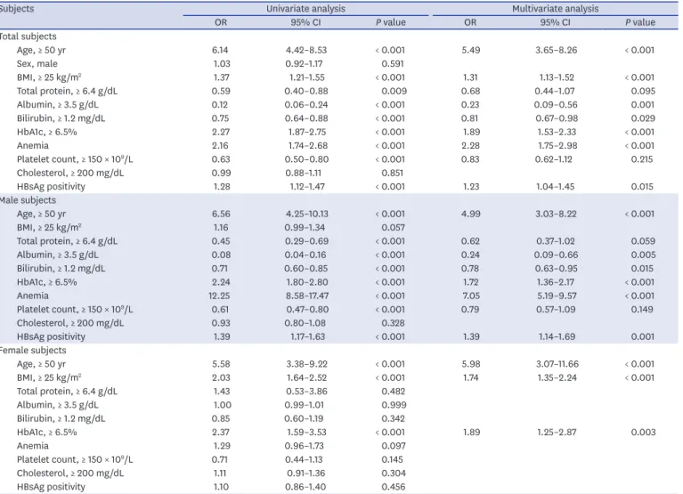
Association between HBsAg positivity and GFR < 60 mL/min/m
2In the univariate analysis, HBsAg positivity, age (≥ 50 years), body mass index (BMI) (≥ 25 kg/m
2), serum protein (≥ 6.4 g/dL), serum albumin (≥ 3.5 g/dL), serum bilirubin (≥ 1.2 mg/dL), HbA1c (≥ 6.5%), and anemia were the candidate variables for the multivariate analysis (P < 0.05). In the multivariate analysis, age (odd ratio [OR], 5.49; 95% confidence interval [CI], 3.65–8.26;
P < 0.001), HbA1c (OR, 1.89; 95% CI, 1.53–2.33; P < 0.001), BMI (OR, 1.31; 95% CI, 1.13–1.52;
P < 0.001), HBsAg positivity (OR, 1.23; 95% CI, 1.0–-1.45; P = 0.015), serum albumin (OR, 0.23;
95% CI, 0.09–0.56; P = 0.001), serum bilirubin (OR, 0.81; 95% CI, 0.67–0.98; P = 0.029) and anemia (OR, 2.28; 95% CI, 1.75–2.98; P < 0.001) were the independent factors for GFR < 60 mL/min/m
2(Table 2 and Fig. 1).
To clarify the sex-specific association between HBsAg positivity and CKD, we conducted a subgroup analysis by sex. HBsAg positivity was an independent risk factor for GFR < 60 mL/
min/m
2in male (OR, 1.39; 95% CI, 1.14–1.69; P = 0.001) but not in female. In contrast, BMI was an independent factor for GFR < 60 mL/min/m
2in female but not in male (Table 2).
Association between HBsAg positivity and proteinuria (≥ 2+)
In the univariate analysis, HBsAg positivity, age (≥ 50 years), male, serum protein (≥ 6.4 g/dL), serum albumin (≥ 3.5 g/dL), serum bilirubin (≥ 1.2 mg/dL), HbA1c (≥ 6.5%), and cholesterol (≥ 200 mg/dL) were the candidate variables for the multivariate analysis (P < 0.05). In the multivariate analysis, HBsAg positivity (OR, 1.41; 95% CI, 1.32–1.51; P < 0.001) was the independent risk factor of proteinuria along with age (OR, 1.78; 95% CI, 1.62–1.95; P < 0.001), HbA1c (OR, 1.44; 95% CI, 1.29–1.60; P < 0.001), male (OR, 0.82; 95% CI, 0.77–0.87; P < 0.001), bilirubin (OR, 0.77; 95% CI, 0.71–0.84; P < 0.001), albumin (OR, 0.45; 95% CI, 0.23–0.85;
Table 1. Baseline characteristics of the subjects at study entry by serostatus of HBsAg
Variables HBsAg positive (n = 10,048) HBsAg negative (n = 40,192) P value
Age, yr 49.5 ± 9.4 49.4 ± 9.2 0.614
Sex, male 6,471 (64.4) 25,884 (64.4) 1.000
BMI, kg/m2 23.9 ± 3.2 24.0 ± 3.2 0.003
Total protein, g/dL 7.30 ± 0.44 7.29 ± 0.42 0.026
Albumin, g/dL 4.39 ± 0.32 4.43 ± 0.31 < 0.001
ALT, IU/L 30.7 ± 37.2 26.8 ± 20.2 < 0.001
Total bilirubin, mg/dL 0.93 ± 0.43 0.91 ± 0.40 < 0.001
Hemoglobin, g/dL 14.56 ± 1.50 14.54 ± 1.50 0.239
Platelet count, × 109/L 212.2 ± 55.9 242.3 ± 55.9 < 0.001
Fasting glucose, mg/dL 96.1 ± 21.3 96.7 ± 20.9 0.017
HbA1c, % 5.55 ± 0.76 5.30 ± 0.73 < 0.001
Total cholesterol, mg/dL 185.3 ± 34.7 194.8 ± 36.2 < 0.001
TSH, ≥ 5 mg/dL 460 (5.6) 1,768 (5.5) 0.628
BUN, mg/dL 13.66 ± 4.12 13.45 ± 3.91 < 0.001
Creatinine, mg/dL 0.87 ± 0.39 0.85 ± 0.25 0.395
GFR 90.4 ± 20.2 91.9 ± 20.2 < 0.001
G1 (GFR ≥ 90 mL/min/1.73 m2) 3,942 (46.6) 16,642 (49.8)
G2 (GFR = 60–89 mL/min/1.73 m2) 4,250 (50.2) 15,904 (47.6)
G3a (GFR = 45–59 mL/min/1.73 m2) 237 (2.8) 756 (2.3)
G3b (GFR = 30–44 mL/min/1.73 m2) 22 (0.3) 59 (0.2)
G4 (GFR = 15–29 mL/min/1.73 m2) 6 (0.07) 24 (0.06)
G5 (GFR < 15 mL/min/1.73 m2) 11 (0.14) 15 (0.05)
Proteinuria ≥ 1+ 2,485 (31.6) 8,269 (26.8) < 0.001
Proteinuria ≥ 2+ 1,481 (18.9) 4,359 (14.1) < 0.001
Urinary RBC, ≥ 5/HPF 1 (0) 6 (0) 0.698
Data are shown as mean ± standard deviation or number (%).
HbsAg = hepatitis B virus surface antigen, BMI = body mass index, ALT = alanine aminotransferase, HbA1c = hemoglobin A1c, TSH = thyroid stimulating hormone, BUN = blood urea nitrogen, GFR = glomerular filtration rate, RBC = red blood cell.
P < 0.001), and total protein (OR, 0.69; 95% CI, 0.56–0.85; P < 0.001) (Table 3). Fig. 2 shows a forest plot of the independent risk factors associated with the proteinuria (≥ 2+). In the subgroup analysis by sex, HBsAg positivity was an independent factor associated with proteinuria in both male and female (Table 3). No significant difference was found in the relationship between HBsAg positivity and the CKD (eGFR < 60 mL/min/m
2or proteinuria 1+) in both male and female subjects (Supplementary Table 1).
DISCUSSION
In this nationwide multicenter study, we demonstrated that HBsAg positive cases showed a significantly higher frequency (3.3%) of a GFR < 60mL/min/m
2than that of the HBsAg negative, age-sex matched controls (2.6%), and they also showed a higher frequency of proteinuria ≥ 2+ (18.9%) than that of the controls (14.1%). Moreover, HBsAg positivity was an independent factor not only for a GFR < 60 mL/min/m
2but also for proteinuria in the multivariable logistic regression analysis. Interestingly, the subgroup analysis showed that the association between HBsAg positivity and a GFR < 60 mL/min/m
2was only confirmed in male but not in female.
Table 2. Univariate and multivariate logistic analyses of decreased GFR (< 60 mL/min/1.73 m2) with subgroup analysis by sex
Subjects Univariate analysis Multivariate analysis
OR 95% CI P value OR 95% CI P value
Total subjects
Age, ≥ 50 yr 6.14 4.42–8.53 < 0.001 5.49 3.65–8.26 < 0.001
Sex, male 1.03 0.92–1.17 0.591
BMI, ≥ 25 kg/m2 1.37 1.21–1.55 < 0.001 1.31 1.13–1.52 < 0.001
Total protein, ≥ 6.4 g/dL 0.59 0.40–0.88 0.009 0.68 0.44–1.07 0.095
Albumin, ≥ 3.5 g/dL 0.12 0.06–0.24 < 0.001 0.23 0.09–0.56 0.001
Bilirubin, ≥ 1.2 mg/dL 0.75 0.64–0.88 < 0.001 0.81 0.67–0.98 0.029
HbA1c, ≥ 6.5% 2.27 1.87–2.75 < 0.001 1.89 1.53–2.33 < 0.001
Anemia 2.16 1.74–2.68 < 0.001 2.28 1.75–2.98 < 0.001
Platelet count, ≥ 150 × 109/L 0.63 0.50–0.80 < 0.001 0.83 0.62–1.12 0.215
Cholesterol, ≥ 200 mg/dL 0.99 0.88–1.11 0.851
HBsAg positivity 1.28 1.12–1.47 < 0.001 1.23 1.04–1.45 0.015
Male subjects
Age, ≥ 50 yr 6.56 4.25–10.13 < 0.001 4.99 3.03–8.22 < 0.001
BMI, ≥ 25 kg/m2 1.16 0.99–1.34 0.057
Total protein, ≥ 6.4 g/dL 0.45 0.29–0.69 < 0.001 0.62 0.37–1.02 0.059
Albumin, ≥ 3.5 g/dL 0.08 0.04–0.16 < 0.001 0.24 0.09–0.66 0.005
Bilirubin, ≥ 1.2 mg/dL 0.71 0.60–0.85 < 0.001 0.78 0.63–0.95 0.015
HbA1c, ≥ 6.5% 2.24 1.80–2.80 < 0.001 1.72 1.36–2.17 < 0.001
Anemia 12.25 8.58–17.47 < 0.001 7.05 5.19–9.57 < 0.001
Platelet count, ≥ 150 × 109/L 0.61 0.47–0.80 < 0.001 0.79 0.57–1.09 0.149
Cholesterol, ≥ 200 mg/dL 0.93 0.80–1.08 0.328
HBsAg positivity 1.39 1.17–1.63 < 0.001 1.39 1.14–1.69 0.001
Female subjects
Age, ≥ 50 yr 5.58 3.38–9.22 < 0.001 5.98 3.07–11.66 < 0.001
BMI, ≥ 25 kg/m2 2.03 1.64–2.52 < 0.001 1.74 1.35–2.24 < 0.001
Total protein, ≥ 6.4 g/dL 1.43 0.53–3.86 0.482
Albumin, ≥ 3.5 g/dL 1.00 0.99–1.01 0.999
Bilirubin, ≥ 1.2 mg/dL 0.85 0.60–1.19 0.342
HbA1c, ≥ 6.5% 2.37 1.59–3.53 < 0.001 1.89 1.25–2.87 0.003
Anemia 1.29 0.96–1.73 0.097
Platelet count, ≥ 150 × 109/L 0.71 0.44–1.13 0.145
Cholesterol, ≥ 200 mg/dL 1.11 0.91–1.36 0.304
HBsAg positivity 1.10 0.86–1.40 0.456
GFR = glomerular filtration rate, OR = odds ratio, CI = confidence interval, BMI = body mass index, HbA1c = hemoglobin A1c, HbsAg = hepatitis B virus surface antigen.
A 2-year multicenter cross-sectional, single arm French study (Hepatitis and Renal Parameters Evaluation) showed that renal abnormality was highly prevalent in treatment- naïve chronic hepatitis B patients (n = 280): a GFR < 60 mL/min/1.73 m
2in 3.5% (4/73) and proteinuria ≥ 1+ in 38.1% (59/155). It was similar with the results of our study: a decreased GFR in 3.3% and proteinuria ≥ 1+ in 30.9% among the HBsAg positive cases.
21The HBsAg, HBV core antigen or HBV e antigen was detected within immune complex deposits in kidney tissues from patients with HBV associated glomerulonephritis (HBGN). Moreover, the sera of HBV-infected patients induced the apoptosis of human kidney proximal tubular epithelial cells by up-regulation of Fas.
12The circulating Cu/Zn superoxide dismutase levels, suggesting an enhanced oxidative stress were higher in hepatitis B patients undergoing renal replacement therapy than in patients without hepatitis B.
14Serum HBV-DNA levels were associated with proteinuria in HBGN,
22and antiviral therapy against HBV, such as lamivudine with or without immunosuppressive therapy, resulted in a complete resolution of membranous nephropathy in 75%–80% of patients.
23-25Therefore, these findings support the role of HBV infection in the development of renal injury.
However, several clinical studies on the association between the HBV infection and CKD showed highly controversial results. Lee et al.
17reported that HBV infection was not associated with CKD in a Taiwanese community based cross-sectional study that included health check examinees (n = 54,996) from April to November 2004, while HCV infection was associated with CKD. However, the same authors reported that HBV infection, but not HCV infection, was associated with CKD in elderly patients > 60 years of age, which was not consistent with their previous study.
18A Japanese single center health check examinee study (n = 12,535) during 2004 and 2006 showed that HBV infection (n = 130, 1.0%) was
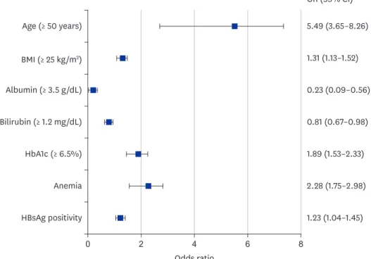
Age (≥ 50 years)
OR (95% CI) 5.49 (3.65–8.26) 1.31 (1.13–1.52) 0.23 (0.09–0.56) 0.81 (0.67–0.98) 1.89 (1.53–2.33) 2.28 (1.75–2.98) 1.23 (1.04–1.45) BMI (≥ 25 kg/m2)
Albumin (≥ 3.5 g/dL) Bilirubin (≥ 1.2 mg/dL) HbA1c (≥ 6.5%) Anemia HBsAg positivity
Odds ratio
0 2 4 6 8
Fig. 1. Forest plot of the impact of the risk factors for a decreased GFR (GFR < 60 mL/min/m2). Age (P < 0.001), HbA1c (P < 0.001), BMI (P < 0.001), HBsAg positivity (P = 0.015), serum albumin (P < 0.001), serum bilirubin (P = 0.029), and anemia (P < 0.001) were the independent factors for a decreased GFR.
GFR = glomerular filtration rate, HbA1c = hemoglobin A1c, BMI = body mass index, HbsAg = hepatitis B virus surface antigen, OR = odds ratio, CI = confidence interval.
not associated with a lower GFR and albuminuria, while HCV infection (n = 72, 0.6%) was associated after adjusting for age, sex, systolic blood pressure and fasting glucose. However, further adjustment for either homeostasis model assessment insulin resistance or serum ALT abolished the association between HCV infection and albuminuria.
15A Chinese single center health check examinee study (n = 15,600) from 2009–2012 showed that there was no association between either HBV or HCV infection and CKD; however, the presence of hypertension, diabetes, albumin level, triglyceride level and total cholesterol level were independent factors, while age and sex were not significant factors.
26In contrast to the above cross-sectional studies, a longitudinal 13-year nationwide Taiwan cohort study using the National Health Insurance Research Database (n = 88,980) from 1996–2010 showed that untreated chronic HBV infection was associated with an increased risk of CKD (adjusted hazard ratio [HR], 2.58; 95% CI, 1.95–3.42), and the association was significant in male of any age and female under the age of 50 but not in female aged 50 years or older.
6The same authors also reported that untreated chronic HBV infection is associated with an increased risk of ESRD (12-year cumulative incidence, 1.9%) compared with a non-HBV cohort (0.49%) (adjusted HR, 3.85; 95% CI, 2.83–4.50; P < 0.001).
7Another single-center cohort study from Hong Kong that included 2,838 patients with type 2 diabetes enrolled during 1995–1999 showed
Table 3. Univariate and multivariate logistic analyses of proteinuria (≥ 2+) with subgroup analysis by sexSubjects Univariate analysis Multivariate analysis
OR 95% CI P value OR 95% CI P value
Total subjects
Age, ≥ 50 yr 1.81 1.66–1.98 < 0.001 1.78 1.62–1.95 < 0.001
Sex, male 0.85 0.80–0.90 < 0.001 0.82 0.77–0.87 < 0.001
BMI, ≥ 25 kg/m2 0.96 0.91–1.02 0.219
Total protein, ≥ 6.4 g/dL 0.56 0.46–0.67 < 0.001 0.69 0.56–0.85 < 0.001
Albumin, ≥ 3.5 g/dL 0.27 0.16–0.47 < 0.001 0.45 0.23–0.85 0.014
Bilirubin, ≥ 1.2 mg/dL 0.73 0.68–0.79 < 0.001 0.77 0.71–0.84 < 0.001
HbA1c, ≥ 6.5% 1.46 1.35–1.57 < 0.001 1.44 1.29–1.60 < 0.001
Anemia 1.23 0.98–1.29 0.092
Platelet, ≥ 150 × 109/L 1.12 0.97–1.28 0.126
Cholesterol, ≥ 200 mg/dL 1.06 1.00–1.12 0.037 1.06 0.99–1.13 0.054
HBsAg positivity 1.41 1.32–1.51 < 0.001 1.41 1.32–1.51 < 0.001
Male subjects
Age, ≥ 50 yr 1.95 1.73–2.19 < 0.001 1.70 1.49–1.94 < 0.001
BMI, ≥ 25 kg/m2 1.12 1.04–1.20 0.003 1.15 1.06–1.24 0.001
Total protein, ≥ 6.4 g/dL 0.62 0.49–0.80 < 0.001 0.78 0.59–1.03 0.075
Albumin, ≥ 3.5 g/dL 0.20 0.10–0.38 < 0.001 0.33 0.15–0.76 0.009
Bilirubin, ≥ 1.2 mg/dL 0.75 0.69–0.81 < 0.001 0.73 0.67–0.81 < 0.001
HbA1c, ≥ 6.5% 1.66 1.47–1.87 < 0.001 1.48 1.30–1.67 < 0.001
Anemia 1.66 1.14–2.41 0.008 1.16 0.75–1.77 0.505
Platelet, ≥ 150 × 109/L 1.09 0.92–1.29 0.305
Cholesterol, ≥ 200 mg/dL 0.99 0.93–1.07 0.867
HBsAg positivity 1.44 1.33–1.56 < 0.001 1.47 1.34–1.61 < 0.001
Female subjects
Age, ≥ 50 yr 1.67 1.46–1.92 < 0.001 1.73 1.50–1.99 < 0.001
BMI, ≥ 25 kg/m2 0.76 0.68–0.86 < 0.001 0.71 0.63–0.81 < 0.001
Total protein, ≥ 6.4 g/dL 0.46 0.34–0.63 < 0.001 0.46 0.33–0.63 < 0.001
Albumin, ≥ 3.5 g/dL 0.59 0.19–1.85 0.368
HbA1c, ≥ 6.5% 1.17 0.93–1.47 0.173
Bilirubin, ≥ 1.2 mg/dL 0.75 0.64–0.88 0.001 0.72 0.61–0.86 < 0.001
Anemia 0.96 0.83–1.12 0.627
Platelet count, ≥ 150 × 109/L 1.12 0.87–1.46 0.374
Cholesterol, ≥ 200 mg/dL 1.20 1.09–1.31 < 0.001 1.11 1.00–1.22 0.042
HBsAg positivity 1.37 1.23–1.52 < 0.001 1.45 1.29–1.62 < 0.001
OR = odds ratio, CI = confidence interval, BMI = body mass index, HbA1c = hemoglobin A1c, HbsAg = hepatitis B virus surface antigen.
that chronic HBV infection was associated with an increased risk of ESRD (adjusted HR, 1.53;
95% CI, 1.11–18.58; P = 0.036) during a median follow-up of 3.5 years.
27All of the above 3 published longitudinal study results were similar with our cross-sectional study results showing that chronic HBV infection was significantly associated with a decreased GFR (adjusted OR, 1.22;
95% CI, 1.03–1.43) and with proteinuria (adjusted OR, 1.35; 95% CI, 1.27–1.43).
Previous studies have shown that age, diabetes mellitus, hypertension, male, anemia, and obesity are independent risk factors for CKD,
28,29which was partially concordant with our results; old age, high BMI, high HbA1c level, anemia, and hypoalbuminemia, as well as HBsAg positivity, were independent factors associated with GFR < 60 mL/min/m
2. In the subgroup analysis by sex, HBsAg positivity remained an independent factor for GFR < 60 mL/min/m
2in male but not in female. This finding is consistent with a previous cross-sectional study.
30It may be related to the higher susceptibility of chronic HBV infection and the higher risk of HBV-related liver injury in male than in female.
31Moreover, the renal protective role of estrogen was also demonstrated previously
32: the course of kidney disease is more modest in female than in male,
33,34and estrogen replacement therapy can help to improve renal functions in menopausal women.
35Screening proteinuria by dipstick is often the first approach to evaluate the kidneys and provides a sensitive test for renal disease from early to advanced stages.
36In this study, HBsAg positivity was an independent factor for proteinuria not only among all the subjects but also in both male and female in the subgroup analysis.
Although the present study could not provide information on the HBV-DNA level and HBV genotype, almost all (95%–99%) of the Korean hepatitis B patients exhibited the HBV
Age (≥ 50 years)
OR (95% CI) 1.78 (1.62–1.95) 0.82 (0.77–0.87) 0.69 (0.56–0.85) 0.45 (0.23–0.85) 0.77 (0.71–0.84) 1.44 (1.29–1.60) 1.41 (1.32–1.51) Male
Albumin (≥ 3.5 g/dL) Protein (≥ 6.4 g/dL)
Bilirubin (≥ 1.2 mg/dL) HbA1c (≥ 6.5%) HBsAg positivity
Odds ratio
0 0.5 1.0 1.5 2.0
Fig. 2. Forest plot of the impact of the risk factors for proteinuria (≥ 2+). HBsAg positivity (P < 0.001) was the independent risk factor of proteinuria along with age (P < 0.001), HbA1c (P < 0.001), male gender (P < 0.001), bilirubin (P < 0.001), albumin (P = 0.014), and total protein (P < 0.001).
HbsAg = hepatitis B virus surface antigen, HbA1c = hemoglobin A1c, OR = odds ratio, CI = confidence interval.
genotype C
37which is more virulent and has a lower antiviral response to interferon than that of genotype B.
38,39Lei et al.
40reported that individuals with HBV genotype C may be more susceptible to HBGN and severe clinical manifestation. Additionally, individuals with HBV genotype C may be susceptible to renal damage due to a high viral load.
There are several limitations in this study. Because of the nature of the data, information on the serum HBV-DNA level and antiviral therapy against HBV was not available. Even though some portions of the cases may have had received antiviral drugs with complete control of HBV replication, HBsAg positivity remained as an independent factor for CKD or proteinuria in this study. Second, this study had a cross-sectional design with a single measurement for the GFR and proteinuria which do not provide the causal relationship between HBV infection and CKD. Third, because our case group was defined as HBsAg positive individuals, very rare case of acute hepatitis B may be not excluded in this group. However, majority of acute hepatitis B cases in adults showed a symptomatic presentation, so that they may go doctor's clinic rather than health-check examination center. Fourth, the selection bias would be existed in our study because it is likely that subjects with relatively good health and economic conditions underwent health-check examination. Furthermore, the GFR was not determined by a direct measurement, and the estimated GFR may not accurately reflect renal function in some patients with a severe liver disease because these patients had decreased muscle and creatinine production. Lastly, information on hypertension, diabetes, medication, and liver images such as abdomen ultrasonography and/or abdomen computed tomography was not available, and we could not assess potential confounders associated with the decreased GFR or proteinuria. Despite these limitations, to our knowledge, this is the first study to report a population level association between chronic HBV infection and CKD in Korea with a large sample size and comprehensive laboratory results.
In conclusion, this large, nationwide, multicenter study indicates that HBV infection is significantly associated with CKD. Especially, male sex may affect the association between HBV infection and CKD. Therefore, physicians should pay attention to the renal function as well as the liver function of chronic hepatitis B patients in the era of effective antiviral therapy.
SUPPLEMENTARY MATERIAL
Supplementary Table 1
Univariate and multivariate logistic analyses of decreased GFR (< 60 mL/min/1.73m
2) or proteinuria (≥ 1+) with subgroup analysis by sex
Click here to view
REFERENCES
1. Zampino R, Boemio A, Sagnelli C, Alessio L, Adinolfi LE, Sagnelli E, et al. Hepatitis B virus burden in developing countries. World J Gastroenterol 2015;21(42):11941-53.
PUBMED | CROSSREF
2. Cho EJ, Kim SE, Suk KT, An J, Jeong SW, Chung WJ, et al. Current status and strategies for hepatitis B control in Korea. Clin Mol Hepatol 2017;23(3):205-11.
PUBMED | CROSSREF
3. Pyrsopoulos NT, Reddy KR. Extrahepatic manifestations of chronic viral hepatitis. Curr Gastroenterol Rep 2001;3(1):71-8.
PUBMED | CROSSREF
4. Fabrizi F, Donato FM, Messa P. Association between hepatitis B virus and chronic kidney disease: a systematic review and meta-analysis. Ann Hepatol 2017;16(1):21-47.
PUBMED | CROSSREF
5. Nakahara K, Takahashi H, Okuse C, Shigefuku R, Yamada N, Murao M, et al. Membranous nephropathy associated with chronic hepatitis B occurring in a short period after acute hepatitis B virus infection.
Intern Med 2010;49(5):383-8.
PUBMED | CROSSREF
6. Chen YC, Su YC, Li CY, Hung SK. 13-year nationwide cohort study of chronic kidney disease risk among treatment-naïve patients with chronic hepatitis B in Taiwan. BMC Nephrol 2015;16(1):110.
PUBMED | CROSSREF
7. Chen YC, Su YC, Li CY, Wu CP, Lee MS. A nationwide cohort study suggests chronic hepatitis B virus infection increases the risk of end-stage renal disease among patients in Taiwan. Kidney Int 2015;87(5):1030-8.
PUBMED | CROSSREF
8. Kupin WL. Viral-associated GN: Hepatitis B and other viral infections. Clin J Am Soc Nephrol 2017;12(9):1529-33.
PUBMED | CROSSREF
9. Bae YD, Choi HJ, Lee JC, Park JJ, Lee YJ, Lee EB, et al. Clinical features of polyarteritis nodosa in Korea. J Korean Med Sci 2006;21(4):591-5.
PUBMED | CROSSREF
10. Han SH. Extrahepatic manifestations of chronic hepatitis B. Clin Liver Dis 2004;8(2):403-18.
PUBMED | CROSSREF
11. Bhimma R, Coovadia HM. Hepatitis B virus-associated nephropathy. Am J Nephrol 2004;24(2):198-211.
PUBMED | CROSSREF
12. Deng CL, Song XW, Liang HJ, Feng C, Sheng YJ, Wang MY. Chronic hepatitis B serum promotes apoptotic damage in human renal tubular cells. World J Gastroenterol 2006;12(11):1752-6.
PUBMED | CROSSREF
13. Chen CL, Yang HI, Yang WS, Liu CJ, Chen PJ, You SL, et al. Metabolic factors and risk of hepatocellular carcinoma by chronic hepatitis B/C infection: a follow-up study in Taiwan. Gastroenterology
2008;135(1):111-21.
PUBMED | CROSSREF
14. Pawlak K, Pawlak D, Mysliwiec M. Hepatitis intensified oxidative stress, MIP-1beta and RANTES plasma levels in uraemic patients. Cytokine 2004;28(6):197-204.
PUBMED | CROSSREF
15. Ishizaka N, Ishizaka Y, Seki G, Nagai R, Yamakado M, Koike K. Association between hepatitis B/C viral infection, chronic kidney disease and insulin resistance in individuals undergoing general health screening. Hepatol Res 2008;38(8):775-83.
PUBMED | CROSSREF
16. Cai J, Fan X, Mou L, Gao B, Liu X, Li J, et al. Association of reduced renal function with hepatitis B virus infection and elevated alanine aminotransferase. Clin J Am Soc Nephrol 2012;7(10):1561-6.
PUBMED | CROSSREF
17. Lee JJ, Lin MY, Yang YH, Lu SN, Chen HC, Hwang SJ. Association of hepatitis C and B virus infection with CKD in an endemic area in Taiwan: a cross-sectional study. Am J Kidney Dis 2010;56(1):23-31.
PUBMED | CROSSREF
18. Lin MY, Chiu YW, Lee CH, Yu HY, Chen HC, Wu MT, et al. Factors associated with CKD in the elderly and nonelderly population. Clin J Am Soc Nephrol 2013;8(1):33-40.
PUBMED | CROSSREF
19. Levey AS, Coresh J, Greene T, Marsh J, Stevens LA, Kusek JW, et al. Expressing the Modification of Diet in Renal Disease Study equation for estimating glomerular filtration rate with standardized serum creatinine values. Clin Chem 2007;53(4):766-72.
PUBMED | CROSSREF
20. Kidney Disease: Improving Global Outcomes (KDIGO) CKD Work Group. KDIGO 2012 clinical practice guideline for the evaluation and management of chronic kidney disease. Kidney Int Suppl 2013;3:1-150.
21. Amet S, Bronowicki JP, Thabut D, Zoulim F, Bourliere M, Mathurin P, et al. Prevalence of renal abnormalities in chronic HBV infection: the HARPE study. Liver Int 2015;35(1):148-55.
PUBMED | CROSSREF
22. Wei RB, Li P, Wu J, Zhang XG, Yin Z, Shi SZ, et al. Clinicopathological analysis on hepatitis B virus- associated glomerulonephritis in 205 patients. Zhonghua Shi Yan He Lin Chuang Bing Du Xue Za Zhi 2010;24(6):464-7.
PUBMED
23. Wang WN, Wu MY, Ma FZ, Sun T, Xu ZG. Meta-analysis of the efficacy and safety of nucleotide/nucleoside analog monotherapy for hepatitis B virus-associated glomerulonephritis. Clin Nephrol 2016;85(1):21-9.
PUBMED | CROSSREF
24. Yi Z, Jie YW, Nan Z. The efficacy of anti-viral therapy on hepatitis B virus-associated glomerulonephritis: a systematic review and meta-analysis. Ann Hepatol 2011;10(2):165-73.
PUBMED
25. Yang Y, Ma YP, Chen DP, Zhuo L, Li WG. A meta-analysis of antiviral therapy for hepatitis B virus- associated membranous nephropathy. PLoS One 2016;11(9):e0160437.
PUBMED | CROSSREF
26. Zeng Q, Gong Y, Dong S, Xiang H, Wu Q. Association between exposure to hepatitis B virus and chronic kidney disease in China. J Int Med Res 2014;42(5):1178-84.
PUBMED | CROSSREF
27. Cheng AY, Kong AP, Wong VW, So WY, Chan HL, Ho CS, et al. Chronic hepatitis B viral infection independently predicts renal outcome in type 2 diabetic patients. Diabetologia 2006;49(8):1777-84.
PUBMED | CROSSREF
28. Mehdi U, Toto RD. Anemia, diabetes, and chronic kidney disease. Diabetes Care 2009;32(7):1320-6.
PUBMED | CROSSREF
29. Ramkumar N, Cheung AK, Pappas LM, Roberts WL, Beddhu S. Association of obesity with inflammation in chronic kidney disease: a cross-sectional study. J Ren Nutr 2004;14(4):201-7.
PUBMED | CROSSREF
30. Senghore T, Su FH, Lin YS, Chu FY, Yeh CC. Association between hepatitis B infection chronic kidney disease in university students receiving physical check-ups: a cross-sectional study. J Exp Clin Med 2013;5(5):181-6.
CROSSREF
31. Huang YT, Jen CL, Yang HI, Lee MH, Su J, Lu SN, et al. Lifetime risk and sex difference of hepatocellular carcinoma among patients with chronic hepatitis B and C. J Clin Oncol 2011;29(27):3643-50.
PUBMED | CROSSREF
32. Gava AL, Freitas FP, Meyrelles SS, Silva IV, Graceli JB. Gender-dependent effects of aging on the kidney.
Braz J Med Biol Res 2011;44(9):905-13.
PUBMED | CROSSREF
33. Dubey RK, Jackson EK. Estrogen-induced cardiorenal protection: potential cellular, biochemical, and molecular mechanisms. Am J Physiol Renal Physiol 2001;280(3):F365-88.
PUBMED | CROSSREF
34. Kaygusuz I, Gumus II, Yuvaci HU, Kasapoğlu B, Carlioglu A. Does hormone replacement therapy have beneficial effects on renal functions in menopausal women? Arch Gynecol Obstet 2012;285(6):1643-6.
PUBMED | CROSSREF
35. Gross ML, Adamczak M, Rabe T, Harbi NA, Krtil J, Koch A, et al. Beneficial effects of estrogens on indices of renal damage in uninephrectomized SHRsp rats. J Am Soc Nephrol 2004;15(2):348-58.
PUBMED | CROSSREF
36. Agarwal R, Panesar A, Lewis RR. Dipstick proteinuria: can it guide hypertension management? Am J Kidney Dis 2002;39(6):1190-5.
PUBMED | CROSSREF
37. Kim H, Jee YM, Song BC, Shin JW, Yang SH, Mun HS, et al. Molecular epidemiology of hepatitis B virus (HBV) genotypes and serotypes in patients with chronic HBV infection in Korea. Intervirology 2007;50(1):52-7.
PUBMED | CROSSREF
38. Kao JH, Chen PJ, Lai MY, Chen DS. Genotypes and clinical phenotypes of hepatitis B virus in patients with chronic hepatitis B virus infection. J Clin Microbiol 2002;40(4):1207-9.
PUBMED | CROSSREF
39. Lavanchy D. Worldwide epidemiology of HBV infection, disease burden, and vaccine prevention. J Clin Virol 2005;34 Suppl 1:S1-3.
PUBMED | CROSSREF
40. Lei X, Gao X, Yang J, Sun Y, Sai Y, You W, et al. The genotype C could play a key role in hepatitis B virus associated nephritis among the northwest Chinese children. Eur J Intern Med 2013;24(8):835-8.
PUBMED | CROSSREF