© 2017 The Korean Academy of Medical Sciences.
This is an Open Access article distributed under the terms of the Creative Commons Attribution Non-Commercial License (http://creativecommons.org/licenses/by-nc/4.0) which permits unrestricted non-commercial use, distribution, and reproduction in any medium, provided the original work is properly cited.
pISSN 1011-8934 eISSN 1598-6357
MERS-CoV Infection in a Pregnant Woman in Korea
Middle East respiratory syndrome (MERS) is a lethal respiratory disease — caused by MERS- coronavirus (MERS-CoV) which was first identified in 2012. Especially, pregnant women can be expected as highly vulnerable candidates for this viral infection. In May 2015, this virus was spread in Korea and a pregnant woman was confirmed with positive result of MERS-CoV polymerase chain reaction (PCR). Her condition was improved only with conservative treatment. After a full recovery of MERS, the patient manifested abrupt vaginal bleeding with rupture of membrane. Under an impression of placenta abruption, an emergent cesarean section was performed. Our team performed many laboratory tests related to MERS-CoV and all results were negative. We report the first case of MERS-CoV infection during pregnancy occurred outside of the Middle East. Also, this case showed relatively benign maternal course which resulted in full recovery with subsequent healthy full-term delivery without MERS-CoV transmission.
Keywords: Middle East Respiratory Syndrome; Coronavirus; Pregnancy; Term Newborn Soo Young Jeong,1* Se In Sung,2*
Ji-Hee Sung,3 So Yoon Ahn,2 Eun-Suk Kang,4 Yun Sil Chang,2 Won Soon Park,2 and Jong-Hwa Kim1
1Department of Obstetrics and Gynecology, Samsung Medical Center, Sungkyunkwan University School of Medicine, Seoul, Korea; 2Department of Pediatrics, Samsung Medical Center, Sungkyunkwan University School of Medicine, Seoul, Korea;
3Department of Obstetrics and Gynecology, Kangbuk Samsung Hospital, Sungkyunkwan University School of Medicine, Seoul, Korea; 4Department of Laboratory Medicine and Genetics, Samsung Medical Center, Sungkyunkwan University School of Medicine, Seoul, Korea
* Soo Young Jeong and Se In Sung contributed equally to this work.
Received: 17 March 2017 Accepted: 1 July 2017 Address for Correspondence:
Jong-Hwa Kim, MD
Department of Obstetrics and Gynecology, Samsung Medical Center, Sungkyunkwan University School of Medicine, 81 Irwon-ro, Gangnam-gu, Seoul 06351, Korea E-mail: [email protected]
https://doi.org/10.3346/jkms.2017.32.10.1717 • J Korean Med Sci 2017; 32: 1717-1720
INTRODUCTION
Middle East respiratory syndrome (MERS) is a lethal respiratory disease caused by MERS-coronavirus (MERS-CoV) and occurs mostly in the Middle East, initially by cam- el-to-human transmission, and then by human-to-human transmission. However, the disease was spread to other continents, probably by an index case, with subsequent pandemic outbreaks through human-to-human transmission through droplets and contact. During these respiratory viral outbreaks, pregnant women can be expected as highly vulnerable candidates for infection (1).
A MERS outbreak occurred in Korea in 2015 with 186 infections, including 38 deaths (2,3). We experienced a case of a Korean pregnant woman who was confirmed for a MERS-CoV infection via a polymerase chain reaction (PCR) test. This is the first case of a MERS-positive pregnancy reported outside the Middle East and is also the first case of having been exposed and confirmed on 3rd trimester of pregnancy with full-recov- ery and successful full-term delivery.
CASE DESCRIPTION
On May 27, 2015, the patient’s mother was exposed to the 14th MERS patient, had a fe- ver starting from June 3 and was diagnosed with MERS on June 7. While febrile, she had been in close contact with her daughter, a 39-year-old pregnant woman (gravida 2 para 1). On June 8 (35 weeks and 4 days of gestational age [GA]), this pregnant woman visited the emergency room complaining of mild myalgia. Based on this contact history with a MERS patient and her symptoms, a MERS-CoV PCR test was performed and the re- sult was found to be positive on June 9. Starting from June 9, the patient developed dyspnea and sputum production. Although chest auscultation was normal, the oxygen saturation (SpO2) was 95% in room air and chest radiography showed diffuse opacity in the left lower lung area compared to a previously obtained radiographic image. The laboratory findings included a leukocyte count of 5,570/mm3 (normal range 4,000–
10,000/mm3), with a differential of 71.4% segmented neutrophils, 20.5% lymphocytes, CASE REPORT
Infectious Diseases, Microbiology & Parasitology
1 / 1 CROSSMARK_logo_3_Test
2017-03-16 https://crossmark-cdn.crossref.org/widget/v2.0/logos/CROSSMARK_Color_square.svg
Jeong SY, et al. • MERS in Pregnancy
1718 http://jkms.org https://doi.org/10.3346/jkms.2017.32.10.1717 Table 2. Characteristics and outcomes of MERS-CoV infection during pregnancy
Case (Ref. No.) Country reported GA on confirmation for infection Maternal course/outcome Fetal or neonatal outcome/GA at delivery
1 (15) Jordan 5 mon No ICU/alive Stillborn/5 mon
2 (14) United Arab Emirates 32 wk ICU/dead Alive/33 wk
3 (12) Saudi Arabia 32 wk ICU/alive Alive/32 wk
4 (13) Saudi Arabia 34 wk No ICU/alive Stillborn/34 wk
5 (13) Saudi Arabia 38 wk ICU/dead Alive/38 wk
6 (13) Saudi Arabia 24 wk ICU/dead Dead/25 wk
7 (13) Saudi Arabia 22 wk No ICU/alive Alive/term
8 (13) Saudi Arabia 23 wk ICU/alive Alive/term
9* Korea 35 wk No ICU/alive Alive/term (37 wk)
MERS-CoV = Middle East respiratory syndrome-coronavirus, GA = gestational age, ICU = required intubation and intensive unit care.
*current study.
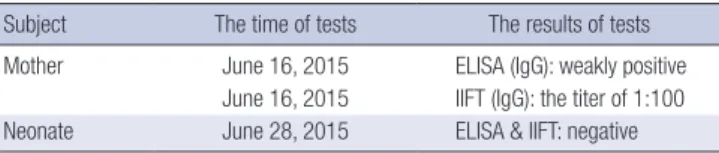
Table 1. Maternal and neonatal antibody tests for MERS
Subject The time of tests The results of tests Mother June 16, 2015 ELISA (IgG): weakly positive
June 16, 2015 IIFT (IgG): the titer of 1:100 Neonate June 28, 2015 ELISA & IIFT: negative MERS = Middle East respiratory syndrome, ELISA = enzyme-linked immunosorbent assay, IgG = immunoglobulin G, IIFT = indirect immunofluorescence test.
Fig. 1. A gross finding of placenta. Placenta abruption was observed as dark blood clot on the maternal side of placenta.
and 7.9% monocytes; and C-reactive protein level of 1.95 mg/
dL (normal range 0–0.3 mg/dL). She was given supplemental oxygen for hypoxia and conservative treatment, with hydration and pain control. The antiviral agents used in other severe MERS- CoV patients were not used in this patient, because her symp- toms and laboratory findings were not severe. Also, there was no evidence of any potential harm to the fetus and pregnant woman related to those drugs.
After several days, her dyspnea and myalgia improved. The SpO2 was 98% in room air and chest radiography showed inter- val improvement. On June 19 and 21, MERS-CoV PCR was per- formed and the results were negative. She had no symptoms related to MERS.
On June 23, the patient manifested abrupt vaginal bleeding with rupture of membranes. A fist-sized blood clot was found through speculum examination and she had abdominal pain.
Fetal cardiotocography showed no deceleration, but a variabili- ty of fetal heart rate changed from moderate to minimal. With an impression of placental abruption, her obstetrical team de- cided on emergent cesarean delivery. A 3,140 g male newborn was delivered at 37 weeks and 5 days of gestation. Apgar scores at 1 and 5 minutes were 9 and 9, respectively. As expected, about
10% placental abruption was found (Fig. 1). After delivery, the baby was immediately moved to the airborne infection isola- tion room (AIIR) and received an initial care with all health care personnel (HCP) completely protected according to the Cen- ters for Disease Control and Prevention (CDC) guidelines (4).
MERS-CoV PCR tests and antibody tests were performed with umbilical cord blood and placenta, and all results were negative.
A systematic testing procedure for coronavirus infection, in- cluding chest radiograph and serial reverse transcription (RT)- PCR assays with peripheral blood and nasopharyngeal swab, did not demonstrate the presence of MERS-CoV in the new- born. MERS-CoV antibody tests were performed with mother and newborn sera on June 16 and June 28, respectively (5). In the mother’s serum, immunoglobulin G (IgG) was detected, al- beit weakly, (0.302) via enzyme-linked immunosorbent assay (ELISA; Euroimmun AG, Luebeck, Germany), and via indirect immunofluorescence test (IIFT; Euroimmun AG) with a titer of 1:100. IgM and IgA were not detected through ELISA and the plaque reduction neutralization test (PRNT) result was below the cutoff value. However, MERS antibodies for IgG, IgM, and IgA were not detected in the newborn’s blood samples (Table 1).
The patient and her newborn baby were discharged in stable condition on June 30 with no clinical abnormalities on follow- up at the outpatient clinic.
DISCUSSION
MERS-CoV was first isolated from a patient who died from a se- vere respiratory illness in Jeddah, Saudi Arabia in June 2012 (6).
Since then, more than 1,698 confirmed cases were reported to
Jeong SY, et al. • MERS in Pregnancy
http://jkms.org 1719
https://doi.org/10.3346/jkms.2017.32.10.1717
the World Health Organization (WHO). Clinical features of MERS are variable, and infected patients can be asymptomatic or have an acute febrile illness, upper respiratory tract disease, or even multiple organ failure resulting in death (7-11). However, there are limited data about the clinical features of MERS-CoV infec- tion during pregnancy and the perinatal outcome of patients diagnosed with MERS-CoV infection. To our best knowledge, there have been 9 reported cases in which pregnant patients had positive laboratory results for MERS-CoV including this case (12-15) (Table 2). Unlike other cases, this case is not only the first MERS-CoV infection during pregnancy occurred out- side of the Middle East, but also the first case of MERS confirmed on 3rd trimester of pregnancy showing good outcome of both mother and baby.
Currently, an exposure time to this virus during pregnancy and a severity of maternal disease could be expected to affect the perinatal outcome. However, there is limited knowledge about the clinical implications of MERS-CoV infection on the maternal, fetal, and placental aspects of pregnancy. From the maternal aspect, there is no epidemiologic data regarding whe- ther pregnant women are more susceptible to MERS. Also, it is unknown whether MERS-CoV infected pregnant women have a more severe disease course compared with the non-pregnant population. In our case, she showed a mild disease course. She had low level of IgG antibody by ELISA and IIFT but not detect- able neutralization activity by PRNT. It has been suggested that neutralizing antibodies are produced at low levels and are po- tentially short-lived after mild or asymptomatic MERS-CoV in- fection (16,17). From the fetal aspect, it is unclear whether MERS was a causative factor in the stillbirth or preterm birth. Fetal specimen and/or placenta were not available for evaluation in the previous cases. As pregnancy alters maternal pulmonary function and consumes more oxygen, severe respiratory illness during pregnancy results in maternal hypoxemia. Maternal hy- poxemia can be associated with poor fetal oxygenation, which eventually could lead to preterm birth or stillbirth. Also, altered immune responses during pregnancy could affect the fetal out- come (13). From the placental aspect, there have been no re- ports of MERS causing pathology of the placenta including in- farction, insufficiency, or villus placentitis. Our case showed placenta abruption clinically, which can be caused by maternal infection. There is no evidence of a relationship between MERS- CoV and placenta disorder. However, the possibility that this vi- rus may be a cause of placenta abruption should be of concern.
Lastly, the remaining question was whether the virus could cross the placenta causing significant infection in the fetus, and whether MERS could cause vertical transmission. Camel-to- human transmission, and human-to-human transmission via contact, droplet, and possibly airborne routes are the known modes of transmission (18,19). However, there are no data about perinatal transmission of MERS-CoV. Moreover, if the mother
mounts an appropriate immune response to produce enough neutralizing antibodies without serious conditions, passive an- tibodies transferred from mother to fetus may have a protective effect on the fetus. There is only one case reporting the mother’s serologic data previously (15), in which stillbirth occurred at approximately 5 months of gestation, although the mother had MERS-CoV antibody by ELISA (titer 1:1,600), immunofluores- cent antibody (IFA), and microneutralization titer assay (titer 1:80). In our case, although the mother had IgG antibody (titer 1:100 by IIFT), antibody was not detected in neonatal serum.
This finding may provoke different interpretations in regard to the role of maternal antibodies in the fetus or to transmission of maternal antibodies, necessitating more data in the future. To know whether prenatal transmission of MERS-CoV can occur, collection of samples including amniotic fluid, placenta, and umbilical cord is needed from an infected pregnant patient.
Further studies with a larger sample size will help in under- standing of the pathophysiology and perinatal outcome of MERS during pregnancy and the optimal mode of delivery.
ACKNOWLEDGMENT
We thank Dr. Christian Drosten and Dr. Marcel A. Muller in In- stitute of Virology, University of Bonn Medical Center for per- forming immunoglobulin A (IgA) enzyme-linked immunosor- bent assay (ELISA) and plaque reduction neutralization test (PRNT). We also thank Prof. Kyong Ran Peck in Division of In- fectious Disease, Department of Medicine, Samsung Medical Center, Sungkyunkwan University School of Medicine for pro- viding the reagent and practical help for us to obtain the anti- body test results.
DISCLOSURE
The authors have no potential conflicts of interest to disclose.
AUTHOR CONTRIBUTION
Data curation: Jeong SY, Sung SI. Supervision: Kim JH. Writing - original draft: Jeong SY, Sung SI. Writing - review & editing: Sung JH, Ahn SY, Kang ES, Chang YS, Park WS, Kim JH.
ORCID
Soo Young Jeong https://orcid.org/0000-0001-8789-901X Se In Sung https://orcid.org/0000-0002-8717-6142 Ji-Hee Sung https://orcid.org/0000-0002-9658-2898 So Yoon Ahn https://orcid.org/0000-0002-1821-3173 Eun-Suk Kang https://orcid.org/0000-0001-6386-6520 Yun Sil Chang https://orcid.org/0000-0001-9201-2938 Won Soon Park https://orcid.org/0000-0002-8245-4692
Jeong SY, et al. • MERS in Pregnancy
1720 http://jkms.org https://doi.org/10.3346/jkms.2017.32.10.1717 Jong-Hwa Kim https://orcid.org/0000-0003-4356-0561
REFERENCES
1. Hardy JM, Azarowicz EN, Mannini A, Medearis DN Jr, Cooke RE. The ef- fect of Asian influenza on the outcome of pregnancy, Baltimore, 1957–
1958. Am J Public Health Nations Health 1961; 51: 1182-8.
2. Cho SY, Kang JM, Ha YE, Park GE, Lee JY, Ko JH, Lee JY, Kim JM, Kang CI, Jo IJ, et al. MERS-CoV outbreak following a single patient exposure in an emergency room in South Korea: an epidemiological outbreak study.
Lancet 2016; 388: 994-1001.
3. Hsieh YH. 2015 Middle East Respiratory Syndrome Coronavirus (MERS- CoV) nosocomial outbreak in South Korea: insights from modeling. PeerJ 2015; 3: e1505.
4. Centers for Disease Control and Prevention (US). Interim infection pre- vention and control recommendations for hospitalized patients with Mid- dle East Respiratory Syndrome Coronavirus (MERS-CoV) [Internet]. Avail- able at http://www.cdc.gov/coronavirus/mers/infection-prevention-con- trol.html [accessed on 1 May 2017].
5. Ko JH, Lee JY, Baek JY, Seok H, Park GE, Lee JY, Cho SY, Ha YE, Kang CI, Kang JM, et al. Serologic evaluation of MERS screening strategy for health- care personnel during a hospital-associated outbreak. Infect Control Hosp Epidemiol 2017; 38: 234-8.
6. Zaki AM, van Boheemen S, Bestebroer TM, Osterhaus AD, Fouchier RA.
Isolation of a novel coronavirus from a man with pneumonia in Saudi Arabia. N Engl J Med 2012; 367: 1814-20.
7. Al-Hameed F, Wahla AS, Siddiqui S, Ghabashi A, Al-Shomrani M, Al-Tha- qafi A, Tashkandi Y. Characteristics and outcomes of Middle East Respi- ratory Syndrome Coronavirus patients admitted to an intensive care unit in Jeddah, Saudi Arabia. J Intensive Care Med 2016; 31: 344-8.
8. Choi WS, Kang CI, Kim Y, Choi JP, Joh JS, Shin HS, Kim G, Peck KR, Chung DR, Kim HO, et al. Clinical presentation and outcomes of Middle East Respiratory Syndrome in the Republic of Korea. Infect Chemother 2016;
48: 118-26.
9. Ki M. 2015 MERS outbreak in Korea: hospital-to-hospital transmission.
Epidemiol Health 2015; 37: e2015033.
10. Lee J. Better understanding on MERS Corona Virus outbreak in Korea. J Korean Med Sci 2015; 30: 835-6.
11. Madani TA. Case definition and management of patients with MERS Coro- navirus in Saudi Arabia. Lancet Infect Dis 2014; 14: 911-3.
12. Alserehi H, Wali G, Alshukairi A, Alraddadi B. Impact of Middle East Re- spiratory Syndrome Coronavirus (MERS-CoV) on pregnancy and peri- natal outcome. BMC Infect Dis 2016; 16: 105.
13. Assiri A, Abedi GR, Al Masri M, Bin Saeed A, Gerber SI, Watson JT. Middle East Respiratory Syndrome Coronavirus infection during pregnancy: a report of 5 cases from Saudi Arabia. Clin Infect Dis 2016; 63: 951-3.
14. Malik A, El Masry KM, Ravi M, Sayed F. Middle East Respiratory Syndrome Coronavirus during pregnancy, Abu Dhabi, United Arab Emirates, 2013.
Emerg Infect Dis 2016; 22: 515-7.
15. Payne DC, Iblan I, Alqasrawi S, Al Nsour M, Rha B, Tohme RA, Abedi GR, Farag NH, Haddadin A, Al Sanhouri T, et al. Stillbirth during infection with Middle East Respiratory Syndrome Coronavirus. J Infect Dis 2014; 209:
1870-2.
16. Drosten C, Meyer B, Müller MA, Corman VM, Al-Masri M, Hossain R, Ma- dani H, Sieberg A, Bosch BJ, Lattwein E, et al. Transmission of MERS-coro- navirus in household contacts. N Engl J Med 2014; 371: 828-35.
17. Payne DC, Iblan I, Rha B, Alqasrawi S, Haddadin A, Al Nsour M, Alsanouri T, Ali SS, Harcourt J, Miao C, et al. Persistence of antibodies against Mid- dle East Respiratory Syndrome Coronavirus. Emerg Infect Dis 2016; 22:
1824-6.
18. Durai P, Batool M, Shah M, Choi S. Middle East Respiratory Syndrome Coronavirus: transmission, virology and therapeutic targeting to aid in outbreak control. Exp Mol Med 2015; 47: e181.
19. Milne-Price S, Miazgowicz KL, Munster VJ. The emergence of the Middle East Respiratory Syndrome Coronavirus. Pathog Dis 2014; 71: 121-36.