53 게재결정: 2008년 3월 19일
*교신저자: 서영호, 506-705, 광주보훈병원 소화기내과, Phone: 062-602- 6107, Fax: 062-602-6988, E-mail: endoscopist@kornet.net
전남의대학술지 제44권 제1호 Chonnam Medical Journal Vol. 44, No. 1, pp. 53∼56
점막하 종양 양상으로 발현된 속발성 위결핵 1예
광주 보훈병원 소화기내과, 1영상의학과, 2병리과, 3상무병원 내과
정안덕ㆍ서영호*ㆍ박상현ㆍ박정수ㆍ조상철ㆍ이남훈ㆍ이봉규ㆍ주소영ㆍ고준석1ㆍ고향미2ㆍ박철주3
A Case of Secondary Gastric Tuberculosis Mimicking Submucosal Tumor
An Doc Jung, Young Ho Seo*, Sang Hyun Park, Jung Su Park, Sang Cheol Cho, Nam Hun Lee, Bong Kyu Lee, So Young Ju, Jun Seok Ko
1, Hyang Mi Ko
2and Cheol Ju Park
3Departments of Internal Medicine, 1Radiology and 2Pathology, Gwangju Veterans Hospital,
3Department of Internal Medicine, Sangmu Hospital, Gwangju, Korea
Gastrointestinal tuberculosis has steadily decreased. But, it can occasionally be found in South Korea.
The prevalence of gastric tuberculosis is lower than the other gastrointestinal tuberculosis. It is usually secondary to pulmonary or intestinal tuberculosis. Most commonly, it is presented as an ulcerative lesion on the lesser curvature of the antrum, it’s clinical feature being similar to that of peptic ulcer disease. Very rarely, however, it mimics submucosal tumor. And only few such cases have been reported. We report here with a case of gastric tuberculosis, which was initially presented as a submucosal tumor.
Keywords: Tuberculosis; Neoplasms; Ultrasonography
서 론
위결핵은 폐나 기타 장기의 결핵에 주로 이차적으로 발생 하는1 매우 드문 질환으로 대부분 전정부 소만의 궤양성 병 변으로 관찰되고 점막하 종양 양상으로 발현되는 경우는 극 히 드물다. 또한 위결핵의 진단이 어려워 수술 후 조직학적 검사를 통해 가능하였으나 본 예에서는 내시경, 전산화단층 촬영 및 항결핵제 복용 후 진단할 수 있었다. 이에 저자들은 점막하 종양 양상으로 발현된 위결핵 1예를 경험하였기에 보고하는 바이다.
증 례
27세 여자가 1개월 전부터 지속되는 상복부 동통을 주소 로 타원에 내원하였다. 과거력이나 가족력에서 특이소견은 없었으며, 약 3개월 전부터 객담을 동반하지 않은 기침 및 피로감을 호소하였다. 신체 검사에서 상복부 압통 외에 특이 소견은 없었으며, 활력 징후 및 검사실 소견은 백혈구 11,800/mm3 (분절형 호중구 65%, 림프구 32%, 호산구 1%, 단핵구 1%), 적혈구 침강 속도 23 mm/hr로 증가되었 으나, 혈청 생화학 검사 및 암표지자 검사는 음성이었다. 또 한 기침을 호소하여 시행한 단순 흉부 방사선 검사에서도 특이소견은 보이지 않았다.
본원 내원 10일 전 소화성 궤양을 의심하여 시행한 상부 소화관 내시경검사에서 정상이었으나, 보존적 치료에 증상 호전을 보이지 않았다. 그리고 내원 전날 시행한 복부 초음3
54 전남의대학술지 제44권 제1호 2008
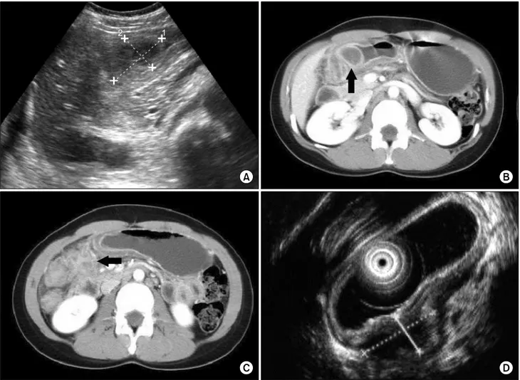
Fig. 1. (A) There is hypoechoic mass (about 3.0×4.0 cm) on the gastric antrum in transabdominal ultrasonogarphy. (B&C) There is a submucosal tumor (arrow on B) with central low attenuation and several lymphadenopathies (arrow on C) in perigastric area on contrast enhanced on abdominal CT. (D) Endoscopic ultrasonography shows hypoechoic submucosal tumor with intact overlying mucosa and central ulceration. CT: Computed Tomography.
파에서 위벽에 30∼40 mm 가량의 저에코의 종괴가 발견되 어(Fig. 1A) 복부 전산화단층촬영을 시행하였다. 검사에서 위 전정부에 위벽의 비후를 동반한 점막하 종양(Fig. 1B) 및 위 주위 림프절의 종대가 관찰되었다(Fig. 1C). 추적 상 부위장관 검사를 시행하여 전정부 후벽에 위치한 점막하 종 양 양상의 병변이 관찰되어(Fig. 2A) 확진을 위해 본원으로 전원 되었다. 본원에서 시행한 초음파 내시경에서는(EUS:
endoscopic ultrasonography)(Fig. 1D) 위 전정부 후벽에 정상 점막으로 둘러 싸였으나 중심부 궤양을 동반한 약 2.5 cm의 점막하 종양과 유사한 병변이 관찰되었고, 개구부에서 백색 농성 액체가 누출되었다. 병변은 주변 점막의 전반적인 비후를 동반하였고, 위벽의 모든 층을 침윤하고 범위가 잘 구분되지 않는 낭성의 저에코성 종괴였으며, 십이지장의 하 행부까지 퍼져있어 복강 내 농양이 의심 되었다. 위 세척액
흡인액에서 Gram Stain 및 AFS (Acid-Fast Stain), 배양 검사, TB PCR (Tuberculosis polymerase chain reaction) 등을 시행하였으며, 브러시를 이용한 세포진 검사 및 중심성 궤양 내부에서 조직 검사를 시행하였다. 위 흡인액 검사에서 AFB stain은 음성이었으나, TB PCR이 양성이었다. 또한 조직검사에서 육아종성 염증이 발견되어 결핵을 의심하였 고, 조직에서 동시에 시행한 TB PCR에서도 양성으로 보여 결핵성 위장관염을 확진할 수 있었다.
환자는 3개월 전부터 피로, 기침이 지속되었으나 단순 흉 부촬영에서 폐병변이 확인되지 않아 원발 부위를 확인하기 위해 흉부 전산화단층촬영을 시행하였다. 흉부 전산화단층 촬영에서는 폐상엽에 공동성 병변을 동반한 활동성 결핵이 (Fig. 2C) 확인되어 폐결핵의 전파로 속발성 위결핵이 발생 했을 것으로 추정하였다.
정안덕 외 10인: 점막하 종양 양상으로 발현된 속발성 위결핵 1예 55
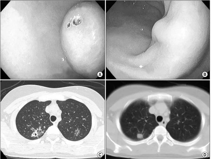
Fig. 2. Endoscopic findings of stomach (A&B). (A) Initial finding shows a submucosal tumor with intact overlying mucosa and central ulceration at the posterior wall of antrum. (B) It shows only a hypertrophic lesion on a follow-up endoscopy. Chest CT findings (C&D). (C) Initial finding shows a tree-in-bud pattern and cavitary lesion containing consolidation at the posterior segment of both upper lobe. (D) The cavitary lesion was changed to a small nodule (D) on a follow-up chest CT.
폐·위결핵 진단 하에 항결핵제 isoniazid, rifampin, ethambutol과 pyrazinamide를 이용한 6개월 단기요법을 시행하였다. 추적 내시경 검사에서 점막하 종양 양상으로 보 였던 병변은 점막의 비후 만 관찰되었고(Fig. 2B) 복부 전 산화단층촬영에서도 위주변의 림프절 종대가 소실되었다.
추적 흉부 전산화단층촬영에서도 공동성 병변은 소결절 형 태로 관찰되었다(Fig. 2D). 환자는 추적 검사에서 항결핵 요법 완료 후 결핵의 치유를 확인하였으며 현재 합병증 없 이 관찰 중이다.
고 찰
위결핵은 위장관 결핵 중에서도 상대적으로 빈도가 낮으
며2 점막하 종양 양상으로 발현하는 경우는 매우 드물다. 그 이유로 위는 위산, 위점막 저항성, 위 내용물의 빠른 통과 시간, 그리고 임파계가 비교적 적어서 드물다고 생각되며3 호발 부위인 전정부가 상대적으로 임파조직이 많은 점을 들 수 있겠다.
위결핵의 발생경로는 원발성으로 경구를 통해 들어간 결 핵균이 직접 위점막을 침범하거나 속발성으로 혈관 및 임파 선을 통해서, 또는 결핵에 이환된 인접 장기로부터 직접 전 이로도 발생할 수 있으며 이중 혈관을 통해 감염되는 경우 가 가장 흔한 것으로 생각된다.2
위결핵 진단은 병력과 신체 검사, 방사선 및 내시경 소견 외에 병변의 조직병리학적 검사에서 건락성 육아종과 결핵 균을 증명하는 것이 도움이 된다.4
현미경적 소견은 중심 괴사 및 섬유화를 갖는 육아종으로
56 전남의대학술지 제44권 제1호 2008
주위의 상피양 세포와 임파구 및 형질 세포의 침윤이 동반 된다. 하지만 진단은 매우 어려워 대부분 병소에서 결핵균 검출은 용이하지 않고 Toole과 Porpatoridis5는 위결핵 절 제 표본의 1/3에서 결핵균을 관찰했다고 하였다. 그리고 위 액이나 대변에서 결핵균이 동정되면 진단적일 수 있으나 폐 결핵이 동반된 경우에는 폐결핵에 의한 이차적 소견일 가능 성이 있어 진단율이 떨어진다.6 하지만 TB PCR은 조직이 나 배양 검사 음성 시 위장관 결핵의 진단에 유용하다고 알 려져 있다.7
위내시경 소견은 크게 궤양형, 증식형, 결절형 병변으로 분류될 수 있고 궤양형이 가장 많으며 주로 위각와 유문부 사이의 소만측에 호발하고8 대만, 유문부, 후벽, 분문의 순으 로 발생한다. 궤양성 병변은 그 주변이 불규칙하고 기저면은 건락성 궤사 또는 결절이 드물게 동반되는 것이 특징이다.1 증식형은 두 번째로 흔한 형태로 주로 전정부에 호발하고 위벽은 고리형으로 비후되며 섬유화를 일으켜 유문 협착을 일으키기도 한다. 결절형은 주로 속립성 결핵의 말기에 관찰 되며 단독 결핵종 형태는 점막하에 침범이 국한되면서 증식 성으로 발생한다.1,9
본 예에서는 전정부 후벽측에 궤양을 동반한 점막하 종양 양상으로 발현하였고 궤양성 개구부에서 조직 검사, 조직 TB PCR, AFS 및 내부 흡인액의 TB PCR을 시행하였으며 조직 검사에서 건락성 육아종이나 AFS에서 결핵의 특징적 인 소견은 발견되지 않았다. 그러나 병소에서 시행한 TB PCR에서 양성 소견을 보이고 항결핵제 투여 후 병변이 소 실된 점으로 미루어 활동성 폐결핵의 전파로 인한 속발성 위결핵 및 주위 임파선 종대가 발생했을 것으로 생각되었 다.7
현재 우리나라에서 위결핵 치료의 적절한 요법이나 기간 에 대한 기준은 없으며 폐결핵에 준하는 4제 6개월 요법이
추천된다. 위결핵은 진단이 매우 어려워 위암, 점막하 종양 또는 위궤양으로 오인되어10 수술 후 대부분이 진단되었다.3 하지만 본 예에서는 임상 증상, 단기간의 추적 내시경 검사 및 전산화단층촬영 등으로 속발성 위결핵을 진단하였고 배 농 후 증상이 호전되었으며 항결핵제 복용 후 완치를 확인 하였다.
References
1. Lee DJ, Shon SH, Chin YJ, Lim CY, Song IH, Kim JW, et al. A case of primary gastric tuberculosis diagnosed as a submucosal tumor.
Korean J Gastrointest Endosc 1998;18:567-72.
2. al Karawi MA, Mohamed AE, Yasawy MI, Graham DY, Shariq S, Ahmed AM, et al. Protean manifestation of gastrointestinal tuberculo- sis: report on 130 patients. J Clin Gastroenterol 1995;20:225-32.
3. Esterman GB, Balfour DC. The stomach and duodenum. Philadelphia:
WB Saunders, 1935.
4. Broders AC. Tuberculosis of the stomach with report a case of multiple tuberculosis ulcer. Surg Gynec & Obst 1917;24:490.
5. Toole H, Porpatoridis J. Contribution to the study of gastric tuber- culosis. Rev Gastroenterol 1950;17:125-36.
6. Choi HY, Lee JW, Lee JS, Kim YK, Lee JH, Kim I, et al. A case of tuberculosis affecting stomach and duodenum simutaneously, mimicking malignant tumor. Korean J Gastrointest Endosc 2004;29:
142-46.
7. Kim KM, Lee A, Choi KY, Lee KY, Kwak JJ. Intestinal tuberculosis:
clinicopathologic analysis and diagnosis by endoscopic biopsy. Am J Gastroeneterol 1998;93:606-9.
8. Subei I, Attar B, Schmitt G, Levendoglu H. Primary gastric tuber- culosis: a case report and literature review. Am J Gastroenterol 1987;82:769-72.
9. McGee GS, Williams LF, Potts J, Barnwell S, Sawyers JL. Gastroin- testinal tuberculosis: resurgence of an old pathogen. Am J Surg 1989;55:16-20.
10. Ostrum HW, Serber W. Tuberculosis of stomach and duodenum. Am J Roentgenol 1948;60:315-22.