Received on June 16, 2014. Revised on July 24, 2014. Accepted on July 30, 2014.
CC This is an open access article distributed under the terms of the Creative Commons Attribution Non-Commercial License (http://creativecommons.org/licenses/by-nc/3.0) which permits unrestricted non-commercial use, distribu- tion, and reproduction in any medium, provided the original work is properly cited.
*Corresponding Author. Hye-Young Kim, Department of Medical Science, Seoul National University College of Medicine and Hospital, 103 Daehak-ro, Jongno-gu, Seoul, Korea. Tel: 82-2-740-8970; Fax: 82-2-743-5530; E-mail: hykim11@
snu.ac.kr
Abbreviations: ILCs, innate lymphoid cells; AHR, airway hyperreactivity; BAL, broncho alveolar lavage; NCR, natural cyto- toxicity receptor; Lti, Lymphoid Tissue inducer; RORα, retinoic acid receptor-related orphan receptor α; RORγt, retinoic acid receptor-related orphan gamma t; ID2, inhibitor of DNA binding 2; OVA, ovalbumin; HDM, house dust mite; RSV, respiratory syncytial virus; TSLP, Thymic stromal lymphopoietin
The Roles of Innate Lymphoid Cells in the Development of Asthma
Yeonduk Woo1, Dongjin Jeong1, Doo Hyun Chung1 and Hye Young Kim2*
1Laboratory of Immune Regulation, Department of Biomedical Sciences, Seoul National University College of Medicine, Seoul 110-744,
2Department of Medical Science, Seoul National University College of Medicine and Hospital, Seoul 110-744, Korea
Asthma is a common pulmonary disease with several differ- ent forms. The most studied form of asthma is the allergic form, which is mainly related to the function of Th2 cells and their production of cytokines (IL-4, IL-5, and IL-13) in associa- tion with allergen sensitization and adaptive immunity.
Recently, there have been many advances in understanding non-allergic asthma, which seems to be related to environ- mental factors such as air pollution, infection, or even obesity. Cells of the innate immune system, including macro- phages, neutrophils, and natural killer T cells as well as the newly described innate lymphoid cells, are effective pro- ducers of a variety of cytokines and seem to play important roles in the development of non-allergic asthma. In this re- view, we focus on recent findings regarding innate lymphoid cells and their roles in asthma.
[Immune Network 2014;14(4):171-181]
Keywords: Airway hyperreactivity, Asthma, Innate lymphoid cells, Allergic asthma, Non-allergic asthma
INTRODUCTION
Traditionally, most scientists believed that asthma was an im- munological disease mainly mediated by Th2 cells and the adaptive immune system (1). Indeed, Th2 cells do play a crit-
ical role in allergic asthma, mostly through their production of an array of cytokines: IL-4, a switch factor for IgE; IL-5, a growth and differentiation factor for eosinophils; and IL-13, a direct causative agent of airway hyperreactivity (AHR) that causes contraction of airway epithelial cells and airway smooth muscle cells. However, over the past few years, it has become clear that asthma is actually a heterogeneous and complex disease, and other immune cells as well as Th2 cells can play major roles in inducing asthma (2). For example, therapeutic trials targeting Th2 cytokines benefitted only a portion of asthma patients, demonstrating the involvement of other immune cells in asthma (3).
The most commonly studied form of asthma is allergic asth- ma, which is triggered by continuous exposure to allergens.
As mentioned above, Th2 cells and eosinophils are at the cen- ter of this pathogenesis. The other type of asthma, non-aller- gic asthma, is mainly associated with exposure to environ- mental factors such as air pollution, ozone, cigarette smoke, diesel particles, viral infection, stress, and nutritional diseases such as obesity (2). Neutrophils and other innate immune cells in the airway seem to play key roles in inducing non-al- lergic asthma (4-7). Genetic factors also seem to influence the development of intrinsic forms of asthma. This heterogeneity suggests that in addition to the allergic and non-allergic forms of asthma, there may be even other forms of asthma medi-
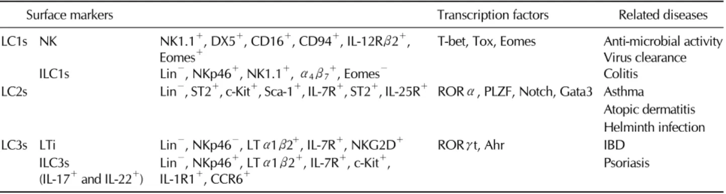
Table I. Surface markers, transcription factors and related diseases of innate lymphoid cells
Surface markers Transcription factors Related diseases
ILC1s NK ILC1s
NK1.1+, DX5+, CD16+, CD94+, IL-12Rβ2+, Eomes+
Lin−, NKp46+, NK1.1+, α4β7+, Eomes−
T-bet, Tox, Eomes Anti-microbial activity Virus clearance Colitis ILC2s Lin−, ST2+, c-Kit+, Sca-1+, IL-7R+, ST2+, IL-25R+ RORα, PLZF, Notch, Gata3 Asthma
Atopic dermatitis Helminth infection
ILC3s LTi Lin−, NKp46−, LTα1β2+, IL-7R+, NKG2D+ RORγt, Ahr IBD
ILC3s
(IL-17+ and IL-22+) Lin−, NKp46+, LTα1β2+, IL-7R+, c-Kit+,
IL-1R1+, CCR6+ Psoriasis
ated by immune cells that regulate and shape pulmonary in- flammation (2). For example, recently identified innate lym- phoid cells (ILCs) were found to regulate asthma through pathways that do not involve Th2 cells. In this review, we focus on the role of ILCs in inducing several forms of asthma as well as recent findings regarding these newly described im- mune cells.
INNATE LYMPHOID CELLS
ILCs are non-T, non-B effector cells that show high levels of effector cell functions (mainly cytokine production) upon activation. ILCs were first identified in the fat associated lym- phoid cluster (FALC) and the intestinal tract (8-10), and short- ly thereafter they were also identified in the lung (11-13).
These cells were shown to be involved in tissue homeostasis, repair, and remodeling as well as innate immunity (14). As non-T, non-B effector cells, ILCs lack rearranged anti- gen-specific receptors (T cell and B cell receptors) and are therefore antigen nonspecific, but they react rapidly to a wide range of innate signals. Given that ILCs produce a great varie- ty and amount of cytokines, it is surprising that they were unidentified earlier, but this might have been due to the focus of immunologists on adaptive immune cells or to technical limits on the ability to identify different cell populations.
ILCs have been classified into three subsets−type 1(ILC1s), type 2(ILC2s), and type 3(ILC3s)−based on their cytokine production (Table I) (15). The prototypic ILC1s is the NK cell, which produces IFN-γ and expresses T-bet (T-box tran- scription factor) (16,17). ILC2s (previously called natural help- er cells or nuocytes) were discovered more recently; these cells produce IL-5, IL-9, and IL-13, a set of cytokines similar to those produced by Th2 cells (8,9). ILC2s require the tran-
scription factors RORα (retinoic acid receptor-related orphan receptor α) (18,19) and GATA3 (20). Among type 3 ILCs (ILC3s), there are at least three different subtypes: (a) lym- phoid tissue inducer (LTi) cells, which produce both IL-17 and IL-22 (21); (b) IL-17-producing cells, which are active in the gut of patients with colitis (22) and in obesity-related forms of asthma (23); and (c) IL-22-producing cells, which are present in the skin (24,25) and gut (26).
Although there are different subsets of ILCs, all share a common lymphoid progenitor, which has been identified as Lin−IL-7Rα+Kit+/lowSca-1+/low, and they also share common developmental pathways (14,19,27). All ILCs require IL-7 sig- naling and Id2, a transcriptional repressor which regulates Notch signaling, for proper development (28-31). These find- ings indicate that ILCs might develop from a common progen- itor and then differentiate into different subsets based on the situation. However, several studies have also suggested that additional pathways and plasticity might exist in ILC lineage establishment (31).
ROLES OF ILC1s IN THE DEVELOPMENT OF ASTHMA
ILC1s
ILC1s express the transcription factor T-bet and produce IFN-γ, a type 1 cytokine. Through their production of IFN-γ, ILC1s show antimicrobial activity and are involved in the clearance of intracellular infections. Compared to ILC2s and Ilc3s, ILC1s have not been well defined. They currently in- clude IL-7Rα− NK cells, because NK cells also express T-bet and produce IFN-γ; however, it is not clear whether a non-NK, IL-7Rα+ ILC1 lineage exists. A recent study identi- fied a distinct ILC1 lineage in the intestinal lamina propria that
expresses NKp46, NK1.1, and T-bet but lacks Eomesodermin, RORγt, and fate map. Interestingly, this newly described sub- set of ILC1s is the main producer of IFN-γ and TNF-α in response to IL-12 and intestinal infection by Toxoplasma gon- dii (32). Therefore, these results suggest that there might be an additional ILC1 subset distinct from NK cells.
ILC1s in asthma
Although the ILC1 lineages have not been well characterized, NK cells belong to the ILC1 family and have been studied thoroughly in the past. Therefore, we will focus on NK cells in the following discussion as analogous to the role of ILC1s in the development of asthma. In the context of asthma, NK cells have not been studied extensively and their roles are still not clear. This might be due to limitations of the methods used for specific depletion of NK cells. However, recent stud- ies have shown that NK cells play an important role in the development of several forms of asthma. NK cells are respon- sible for lung inflammation during viral infections (33,34) and may regulate the pathogenesis of virus-induced asthma. For example, when humans and mice are infected with rhinovi- ruses, which cause a range of respiratory diseases, sig- nificantly higher amounts of IL-15 and IL-15Rα are detected in fluid from the nasal mucosa and in bronchial tissues (35).
Since IL-15 is essential for the activation of NK cells as well as the development of ILC1s, it is possible that ILC1s are in- volved in the development of virus-associated asthma.
The role of NK cells in allergic asthma has been better studied. Korsgren et al. showed that depletion of NK cells using anti-NK1.1 antibody reduced asthma symptoms. In mice depleted of NK cells, eosinophilic infiltration and aller- gen-specific IgE production in the lung were greatly reduced (36). However, depletion of NK cells using anti-NK1.1 anti- body can also deplete natural killer T cells, which are essen- tial for the development of AHR. Therefore, it was not clear whether NK cells themselves could regulate the development of asthma. However, more recent studies of ovalbumin (OVA)-induced AHR have shown that NK cells are more nu- merous in OVA-challenged airways. Moreover, perforin-de- ficient mice failed to develop OVA-induced AHR, suggesting that NK cells are essential for priming OVA-induced AHR (37). Another study was done with a house dust mite (HDM)-induced asthma model. Allergic airway inflammation was induced by intranasal administration of HDM extract in wild-type mice and in NKG2D-deficient mice, which lack the receptor that activates NK cells. NKG2D-deficient mice were
resistant to the development of HDM-induced AHR and showed reduced levels of eosinophils, serum IgE, and Th2 cells. Furthermore, transfer of NK cells into NKG2D-deficient mice restored HDM-induced airway inflammation, suggesting that NK cells might be critical for the development of aller- gen-induced AHR (38). Taken together, these studies suggest potential roles of ILC1s in the pathogenesis of asthma.
ROLES OF ILC2s IN THE DEVELOPMENT OF ASTHMA
ILC2s
ILC2s were first reported among ILC lineages as non-T, non-B cells that proliferated upon administration of IL-25 (39,40) and induced Th2-like immune responses such as increased serum IgE level and eosinophil infiltration in the digestive tract.
Non-T, non-B lymphocytes producing IL-5 and IL-13 were al- so detected in the lungs of patients with asthma, but their specific functions were unclear at the time (41). Then, in 2010, three different groups characterized ILCs that produced large quantities of IL-5 and IL-13 in response to IL-25 and IL-33 (8,9,42). The initial report indicated that these cells were found in lymphoid compartments of adipose tissue and in gut-associated lymphoid tissue. Although these cells had different surface markers and functions, they also shared some common surface markers and cytokine secretions (43).
Now, a consensus has been reached to refer to all of these cells as ILC2s (8,9,40,44).
The cytokines IL-7, IL-25, and IL-33 seem to be important for the development of ILC2s. Transcription factors such as Id2, RORα, and GATA3 as well as Notch signaling also con- tribute to the development of ILC2s (18,19,27). Although there were subtle differences between the cells described in the previous reports, it seems that mouse ILC2s generally ex- press CD25, CD90, and variable amounts of CD117 and CD127 and play critical roles in type 2 immune responses.
In addition, ILC2s express variable amounts of CD278, ST2 (IL-1RL1), and IL-17BR.
ILC2s in virus-induced asthma
Th2 cells are believed to be at the center of allergic asthma pathology. Thus, previous studies of asthma paid little atten- tion to other cell types. However, there is evidence that other cell types are important for inducing asthma. In human pa- tients with asthma, viral infections can precipitate virtually all asthma symptoms regardless of the presence of allergy, sug-
gesting that innate cells rather than adaptive cells (such as Th2 cells) are critical for this type of asthma. For example, Il1rl1−/− mice, which lack the IL-33 receptor, developed near-normal AHR responses after sensitization and challenge with OVA. However, when Il1rl1−/− mice were infected with influenza A virus (H1N1), AHR was greatly reduced (11).
Even in the absence of adaptive immunity (e.g., in Rag2−/−
mice), AHR was induced by influenza A virus. This airway hyperreactivity depended on ILC2s expressing c-Kit, Sca-1, Thy1.2 (CD90), and ST2 (11). Moreover, depletion of ILC2s in Rag2−/− mice using anti-Thy1.2 antibody abolished the in- fluenza-induced AHR response. These responses were de- pendent on IL-13 production, since adoptive transfer of IL-13- secreting ILC2s restored influenza-induced AHR in Il13−/−
recipients. In another study, Hong et al. infected 6-day-old neonatal mice with rhinovirus and found that the virus pro- moted IL-25 production, which in turn induced activation of ILC2s. In addition, proliferation of IL-13-secreting ILC2s was observed in rhinovirus-infected neonatal mice but not in adult mice. Treatment with anti-IL-25 neutralizing antibody attenu- ated ILC2 proliferation, mucous hypersecretion, and airway responsiveness (45). It has also been suggested that infection with respiratory syncytial virus might result in the subsequent development of asthma, independent of allergen challenge (46).
The studies described above show that ILC2s play a patho- logical role during virus-induced asthma. However, another study has suggested that ILC2s may also contribute to lung homeostasis. In mice infected with influenza virus (PR8), ILCs accumulated in the lung, and depletion of ILCs resulted in loss of airway epithelial integrity, diminished lung function, and impaired airway remodeling. ILCs seem to contribute to lung homeostasis by producing amphiregulin (47). Taken to- gether, these results indicate that ILC2s may have a dual func- tion, either inducing inflammation or restoring airway cell in- tegrity depending on the strength of the pathogen and the length of infection.
ILC2s in allergic airway disease
As mentioned previously, ILC2s secrete high levels of IL-5 and IL-13, which suggests they might play an important role in allergic asthma as well as virus-induced asthma, since type 2 cytokines are believed to play a critical role in the develop- ment of allergen-induced AHR. In contrast to the virus mod- el, Il1rl1−/− mice had normal AHR responses after sensitiza- tion and challenge with OVA, suggesting that ILC2s may not
be required for allergen-induced AHR, although ILC2s clearly accumulated in the lungs during allergen challenge (11).
Using an OVA sensitization and airway challenge method, Kearly et al. showed that persistent AHR was correlated with the continued presence of Th2 cells (48). They found that the ST2-IL-33 pathway was important, since antibody against ST2 reduced IL-4 and IL-13 production as well as allergic in- flammation and AHR. They concluded that Th2 cells are the primary cell type that cause AHR. However, ILC2s also rely on the ST2-IL-33 pathway, therefore, they are capable of in- ducing type 2 allergic airway inflammation.
While studies with allergen-induced asthma models have focused on allergen-specific Th2 cells, ILC2s also contribute to type 2 allergic airway disease. Treatment with recombinant IL-25 or IL-33 induced the proliferation of IL-5- and IL-13- producing ILC2s in the lungs and mediastinal lymph nodes (49-53). Klein et al. showed that both ILC2s and Th2 cells are the major sources of IL-5 and IL-13 in HDM-induced and OVA-induced asthma (53). Halim et al. showed that intranasal administration of protease-containing allergens (papain) in- duced eosinophilia and mucus hyperproduction in Rag1−/−
mice (which have ILCs) but not Rag2−/− Il2rg−/− mice (which lack all types of ILCs). Adoptive transfer of ILC2s en- abled Rag2−/− Il2rg−/− mice to respond to papain. Papain damages stromal cells, which can then release IL-33 and thy- mic stromal lymphopoietin (TSLP), which in turn can activate ILC2s (54). Wilhelm et al. administered papain into the lungs and found IL-9-producing ILC2s, a new subset of ILC2s (55).
These IL-9-producing cells required IL-2 and IL-33 (but not IL-25) to produce IL-5 and IL-13, and blockade of IL-9 re- sulted in reduced expression of IL-5 and IL-13, suggesting that ILC2s may initially produce IL-9 and then mature into IL-5- and IL-13-producing cells. In a papain-induced asthma mod- el, ILC2s were the main source of type 2 cytokine production.
Another ubiquitous fungal allergen, Alternaria alternata, al- so induced IL-33 production in the lung, which in turn in- duced the proliferation of Lin−CD25+CD44hi ILC2s. The pro- duction of IL-5 and IL-13 by ILC2s in response to A. alternata was nearly abolished in Il1rl1−/− mice, suggesting that ILC2s mediate asthma in an IL-33-dependent manner (50). While all of these studies together suggest that ILC2s play essential roles in allergen-induced airway disease, at this point it is un- clear whether ILC2s alone are sufficient for inducing allergic asthma. Further study of the precise requirement for ILC2s in allergic lung disease is needed.
ROLES OF ILC3s IN THE DEVELOPMENT OF ASTHMA ILC3s
RORγt is a ligand-dependent nuclear hormone receptor ex- pressed in several cell types such as Th17 cells and the type 3 ILCs (ILC3s) (29,56,57). RORγt-dependent ILCs can be div- ided into at least three different subpopulations based on their functional characteristics. LTi cells, the first sub- population to be identified among the type 3 ILCs, represent the prototypic cell type of the RORγt+ family of ILCs (58).
These cells are RORγt+ and IL-7Rα+ and are mainly involved in secondary lymphoid organ formation (59). Approximately half of fetal LTi cells express CD4, while half of them do not, suggesting that there might be a precursor-progeny relation- ship between CD4− and CD4+ LTi cells (60). The second group of ILC3s is the IL-17-producing ILC3s. These cells were found in the intestine of fetal mouse (61) and in human fetal lymph nodes (62). IL-17-producing ILC3s are also present in the intestine of adult mice (10) and humans (63) under in- flammatory conditions. IL-17-producing ILC3s do not express NKp46, suggesting that these cells are distinct from IL-22-producing NKp46+ cells. The third group of ILC3s is the IL-22-producing ILC3s. IL-22-producing ILC3s express the natural cytotoxicity receptor (NCR), which is NKp44 in hu- mans (64) and NKp46 in mice (65,66). Owing to their NK-as- sociated receptor expression, the ILC3 and ILC1 (NK cells) lineages are considered closer to each other than to the ILC2 lineage; however, expression of RORγt and production of type 3 cytokines such as IL-17 and/or IL-22 should be consid- ered standard markers of type 3 ILCs.
ILC3s in obesity-associated asthma
Asthma is one of the representative immune diseases induced by type 2 cytokines produced by Th2 cells and ILC2s. The role of IL-17A or IL-22 in the development of asthma is con- troversial, since IL-17A may either inhibit or exacerbate aller- gic asthma (5,67). However, recent studies have indicated that IL-17A can directly cause AHR (68,69). These studies have shown that cytokine production by Th17 cells or direct administration of recombinant IL-17 can induce airway in- flammation and AHR by inducing contraction of smooth mus- cle cells. Therefore, IL-17 might have a pathogenic role in airway disease, particularly non-allergic asthma.
Obesity is usually associated with several diseases, such as type 2 diabetes mellitus, cardiovascular disease, liver disease, and cancer (70). Interestingly, obesity is also a major risk fac-
tor for the development of asthma, particularly a severe, ste- roid-resistant form of asthma (71,72). Thus, these findings point to unknown mechanisms of non-allergic asthma that differ from those of allergic asthma.
Interestingly, some obese people show asthma symptoms with increasing levels of IL-17A. Recently, a role for IL-17A- producing ILC3s in AHR associated with obesity was pro- posed (23). Kim et al. demonstrated that obese mice fed a high-fat diet spontaneously developed AHR and had signifi- cant numbers of IL-17-producing cells in their lungs. The ma- jority of IL-17A-producing cells were Lin−Thy1.2+Sca-1+CCR6+ ILC3s. In this study, it was first shown that ILC3s were present in the lung and mediated the development of AHR. These IL-17-producing ILC3s were distinct from ILC1s and ILC2s, since they did not express T-bet or produce IFN-γ and did not produce IL-5 or IL-13. Obesity-induced AHR is in- dependent of adaptive immune cells, since Rag1−/− mice fed a high-fat diet became obese, had more IL-17-producing ILC3s in the lungs, and developed AHR. Just as IL-33 and IL-25 are key cytokines that can induce proliferation of ILC2s, IL-1β could promote proliferation of IL-17-producing ILC3s in the lung (but not IL-22-producing ILC3s). Furthermore, adoptive transfer of IL-17-producing ILC3s into Rag2−/−
Il2rg−/− mice (which lack all types of ILCs) restored IL-1β- induced AHR, indicating that IL-17-producing ILC3s by them- selves can induce AHR. The production of IL-1β required ac- tivation of the NLRP3 inflammasome, since Nlrp3−/− mice fed the high-fat diet became obese but did not develop AHR.
Secretion of the active form of IL-1β, which is mediated by the NLRP3 inflammasome, was increased in M1 macrophages and the lungs of obese mice and induced the proliferation of IL-17-producing ILC3s. Moreover, blocking IL-1β signaling with a short treatment with anakinra (an IL-1R antagonist) ab- rogated development of AHR in the obese mice and greatly reduced the number of IL-17-producing ILC3s in the lung.
Although there is only one study so far showing that ILC3s are related to the development of asthma, it is possible that ILC3s may also be involved in the development of other forms of asthma, such as those induced by viruses or aller- gens, since Th17 and Th22 cells are considered pathogenic in asthma. However, the precise roles of each ILC lineage (ILC1s, ILC2s, and ILC3s) remain to be determined.
ILCs IN HUMANS
Although most studies regarding ILCs have been done with
mice, multiple studies in human subjects suggest that ILCs are also important for the human immune system as well, espe- cially under inflammatory conditions. Here we will summa- rize the studies that have been done with ILC1s, ILC2s, and ILC3s in the context of human diseases.
ILC1s in humans were first identified in the gut (73,74).
These cells expressed the transcription factor T-bet and re- sponded to IL-12. ILC1s are now characterized in humans and mice, and immunologists believe that these cells are distinct from NK cells because they do not produce perforin or gran- zyme B and do not express NK cell markers such as CD56, CD16, and CD94 (74). Under the influence of IL-12 (and IL-15), a subset of human ILC1s produce IFN-γ. The fre- quency of this ILC1 subset was greatly increased in inflamed intestine of patients with Crohn’s disease (73,74), which in- dicated a role for IFN-γ-producing ILC1s in the pathogenesis of gut inflammation as well as inflammation of other mucosal sites.
ILC2s are the best-studied subset of ILCs in humans. An ILC2-like cell population was first reported by Allakhverdi et al. in 2009, although ILCs had not been identified at that time. They found non-T, non-B cells producing IL-13 and IL-5 in the sputum of asthmatic subjects but not in normal subjects. They also found that the non-T, non-B CD34+ cells expressed receptors for TSLP and IL-33 and responded to these cytokines very rapidly (which is characteristic of ILC2s).
In addition, the number of these cells increased in response to a specific allergen inhalation challenge (41). Although Allakhverdi et al. did not further characterize this subset, their findings suggest that ILC2s may play an important role in hu- man asthma. The source of the IL-33 and TSLP was not de- fined by this group, although airway epithelial cells and air- way smooth muscle cells may be the key sources of IL-33 and TSLP (75).
Recent studies have also suggested that human ILC2s are im- portant sources of IL-13 and IL-5. These studies identified ILC2s as Lin−, IL-7Rα+ cells expressing CRTH2 and CD161 (76).
ILC2s also seem to be involved in the pathogenesis of several other diseases. For example, ILC2s may important roles in eosi- nophilic airway inflammation resulting in pleural pathology.
Primary spontaneous pneumothorax, the spontaneous pres- ence of air in the pleural space, is one of the most common causes of eosinophilic pleural effusion. Significantly higher concentrations of IL-5 and eotaxin-3 were detected in the pleu- ral fluid of patients with primary spontaneous pneumothorax, and this was associated with the presence of IL-33 and TSLP
(77). ILC2s have also been detected in the skin of patients with atopic dermatitis; these were identified as Lin−IL-7Rα+ST2+ cells, which are dependent on IL-33 or IL-25. Signaling via IL-33 induced type 2 cytokine production and amphiregulin expression by ILC2s and also induced migration of ILC2s (78). Lastly, ILC2s have been found in nasal polyp tissue from patients with chronic rhinosinusitis (79). Since TSLP is in- creased in nasal polyps of these patients (79) and IL-33 pro- duction and ST2 expression are increased in patients with al- lergic rhinitis (80), ILC2s may play an important role in hu- man allergic rhinitis. Taken together, these human reports certainly suggest that the characteristics and functions of hu- man ILC2s are similar to those of murine ILC2s and that these cells play a critical role in the human respiratory tract and skin.
ILC3s have also been investigated in humans, especially in patients with psoriasis. Teumissen et al. found CD117+NCR+ ILC3s in healthy peripheral blood and cultured dermal explants. NCR+ ILC3s produced IL-22 after cytokine stimula- tion. Remarkably, IL-1β plus IL-23 converted dermal NCR− ILC3s to NCR+ ILC3s in ex vivo culture. IL-22 is a key cyto- kine involved in epidermal thickening, suggesting that IL-22-producing NCR+ ILC3s may participate in the pathology of psoriasis (25). Another study tested tissue samples from non-lesional or lesional psoriatic skin and from nickel- and petrolatum-exposed skin of patients with contact allergy to nickel. They found that RORγt+CD56+ ILC3s, which are known to produce IL-22, were elevated in both non-lesional and lesional skin from patients with psoriasis as well as skin from patients with contact allergy to nickel in comparison with healthy skin. These results suggest that IL-22-producing ILC3s contribute to the pathogenesis of psoriasis in humans (81).
Additionally, examination of bronchoalveolar lavage fluid from a small group of patients with severe asthma revealed the presence of IL-17-producing ILC3s, particularly from pa- tients with more severe asthma. IL-17-producing ILC3s can al- so be detected in human bronchoalveolar lavage fluid after in vitro stimulation with IL-2, IL-7, and IL-1β (23). This in- dicates that ILC3s are present in the lungs of patients with asthma and might play an important role in some forms of this disease.
Unlike the study of mice, the study of human has many limitations; however, a growing body of evidence indicates that ILCs are present in the human immune system.
Therefore, more precise studies are needed, since ILCs seem
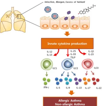
Figure 1. Schematic of innate lymphoid cell function in the development of asthma. Viruses (influenza A virus, rhinovirus, and respiratory syncytial virus), allergens (ovalbumin, human dust mite, papain, etc.), or excess nutrients induce lung epithelial cells to release various cytokines that can amplify the activation of subsets of innate lymphoid cells (ILCs). Release of IL-12 and IL-15 can induce the proliferation and activation of type 1 ILCs producing IFN-γ, which has an antiviral function. In response to IL-25 and IL-33, type 2 ILCs produce large amounts of IL-5, IL-9, and IL-13, which promote eosinophilia (IL-5), bronchial hyperresponsiveness (IL-9 and IL-13), smooth muscle cell contraction (IL-13), and goblet cell hyperplasia (IL-13). By producing various cytokines, ILCs can induce activation of downstream immune cells and intensify the symptoms of asthma.
to play an important role in the development of various im- mune diseases.
CONCLUSION
The discovery of innate lymphoid cells (ILCs) over the past several years has changed our understanding of the principles of immune regulation. ILCs have critical roles in the cyto- kine-mediated regulation of several diseases including auto- immune diseases, infectious diseases, and allergic diseases.
Indeed, it is noteworthy that subsets of ILCs seem to be par- ticularly prevalent at mucosal surfaces (e.g., respiratory tract, skin, and intestine), which are constantly exposed to in- fectious agents in the external environment. The key function of the epithelial barriers of the skin, respiratory tract, and in- testine is to limit exposure to commensal and pathogenic mi-
crobes and maintain tissue homeostasis. However, in addition they are also involved in immune responses through the pro- duction of innate cytokines, which initiate the function of ILCs. ILCs might have evolved to play critical roles in host defense, regulation of inflammation, and tissue repair through the rapid production of large amounts of cytokines.
Studies of ILCs have opened new areas of investigation in allergic diseases and asthma. ILCs are thought to be the main producers of effector cytokines such as IL-5, IL-9, IL-13, IL-17, and IL-22, which are involved in the development of different forms of asthma (Fig. 1). ILC2s in particular play very im- portant roles in the development of asthma by producing large amounts of type 2 cytokines (IL-5, IL-9, and IL-13) in response to IL-25, IL-33, and TSLP. In addition, ILC3s, at least under stress conditions (like obesity), seem to play a critical role in inducing asthma by secreting IL-17 in response to IL-1β. The roles of IL-22-producing ILC3s and of ILC1s in the development of asthma still need to be explored.
Although there have been many new findings regarding ILCs, many unanswered questions remain. However, it is cer- tain that ILCs are important therapeutic targets in several respects. First, ILCs can regulate the adaptive immune response by early production of cytokines. Second, ILCs can interact with various cell types, such as natural killer T cells, mast cells, macrophages, eosinophils, as well as T cells, and regulate their function. Further investigation of this particular cell subset will lead to new ways of targeting many unresolved diseases.
ACKNOWLEDGEMENTS
This work was supported by Research Resettlement Fund for the new faculty of Seoul National University and the Cooperative Research Program of Basic Medical Science and Clinical Science from Seoul National University College of Medicine (800-20140168).
CONFLICTS OF INTEREST
The authors have no financial conflict of interest.
REFERENCES
1. Robinson, D. S., Q. Hamid, S. Ying, A. Tsicopoulos, J.
Barkans, A. M. Bentley, C. Corrigan, S. R. Durham, and A.
B. Kay. 1992. Predominant Th2-like bronchoalveolar T-lym- phocyte population in atopic asthma. N. Engl. J. Med. 326:
298-304.
2. Kim, H. Y., R. H. DeKruyff, and D. T. Umetsu. 2010. The many paths to asthma: phenotype shaped by innate and adaptive immunity. Nat. Immunol. 11: 577-584.
3. Corren, J., R. F. Lemanske, N. A. Hanania, P. E. Korenblat, M. V. Parsey, J. R. Arron, J. M. Harris, H. Scheerens, L. C.
Wu, Z. Su, S. Mosesova, M. D. Eisner, S. P. Bohen, and J.
G. Matthews. 2011. Lebrikizumab Treatment in Adults with Asthma. N. Engl. J. Med. 365: 1038-1098.
4. Johnston, R. A., M. Zhu, Y. M. Rivera-Sanchez, F. L. Lu, T.
A. Theman, L. Flynt, and S. A. Shore. 2007. Allergic airway responses in obese mice. Am. J. Respir. Crit. Care Med. 176:
650-658.
5. Pichavant, M., S. Goya, E. H. Meyer, R. A. Johnston, H. Y.
Kim, P. Matangkasombut, M. Zhu, Y. Iwakura, P. B. Savage, R. H. Dekruyff, S. A. Shore, and D. T. Umetsu. 2008. Ozone exposure in a mouse model induces airway hyperreactivity that requires the presence of natural killer T cells and IL-17.
J. Exp. Med. 205: 385-393.
6. Kim, E. Y., J. T. Battaile, A. C. Patel, Y. You, E. Agapov, M. H. Grayson, L. A. Benoit, D. E. Byers, Y. Alevy, J.
Tucker, S. Swanson, R. Tidwell, J. W. Tyner, J. D. Morton, M Castro, D. Polineni, G. A. Patterson, R. A. Schwendener, J. D. Allard, G. Peltz, and M. J. Holtzman. 2008. Persistent activation of an innate immune response translates respiratory viral infection into chronic lung disease. Nat. Med. 14:
633-640.
7. Wright, R. J. 2005. Stress and atopic disorders. J. Allergy Clin.
Immunol. 116: 1301-1306.
8. Moro, K., T. Yamada, M. Tanabe, T. Takeuchi, T. Ikawa, H.
Kawamoto, J. Furusawa, M. Ohtani, H. Fujii, and S. Koyasu.
2010. Innate production of T(H)2 cytokines by adipose tis- sue-associated c-Kit(+)Sca-1(+) lymphoid cells. Nature 463:
540-544.
9. Neill, D. R., S. H. Wong, A. Bellosi, R. J. Flynn, M. Daly, T. K. Langford, C. Bucks, C. M. Kane, P. G. Fallon, R.
Pannell, H. E. Jolin, and A. N. McKenzie. 2010. Nuocytes represent a new innate effector leukocyte that mediates type-2 immunity. Nature 464: 1367-1370.
10. Buonocore, S., P. P. Ahern, H. H. Uhlig, I. I. Ivanov, D.
R. Littman, K. J. Maloy, and F. Powrie. 2010. Innate lym- phoid cells drive interleukin-23-dependent innate intestinal pathology. Nature 464: 1371-1375.
11. Chang, Y. J., H. Y. Kim, L. A. Albacker, N. Baumgarth, A.
N. McKenzie, D. E. Smith, R. H. Dekruyff, and D. T. Umetsu.
2011. Innate lymphoid cells mediate influenza-induced airway hyper-reactivity independently of adaptive immunity. Nat.
Immunol. 12: 631-638.
12. Kim, H. Y., H. J. Lee, Y. J. Chang, M. Pichavant, S. A. Shore, K. A. Fitzgerald, Y. Iwakura, E. Israel, K. Bolger, J. Faul, R.
H. DeKruyff, and D. T. Umetsu. 2014. Interleukin-17-produc- ing innate lymphoid cells and the NLRP3 inflammasome facili- tate obesity-associated airway hyperreactivity. Nat. Med. 20:
54-61.
13. Monticelli, L. A., G. F. Sonnenberg, M. C. Abt, T. Alenghat, C. G. Ziegler, T. A. Doering, J. M. Angelosanto, B. J.
Laidlaw, C. Y. Yang, T. Sathaliyawala, M. Kubota, D. Turner, J. M. Diamond, A. W. Goldrath, D. L. Farber, R. G. Collman, E. J. Wherry, and D. Artis. 2011. Innate lymphoid cells pro-
mote lung-tissue homeostasis after infection with influenza virus. Nat. Immunol. 12: 1045-1054.
14. Spits, H., and J. P. Di Santo. 2011. The expanding family of innate lymphoid cells: regulators and effectors of immunity and tissue remodeling. Nat. Immunol. 12: 21-27.
15. Bernink, J., J. Mjosberg, and H. Spits. 2013. Th1- and Th2-like subsets of innate lymphoid cells. Immunol. Rev. 252:
133-138.
16. Strowig, T., F. Brilot, and C. Munz. 2008. Noncytotoxic func- tions of NK cells: direct pathogen restriction and assistance to adaptive immunity. J. Immunol. 180: 7785-7791.
17. Vivier, E., D. H. Raulet, A. Moretta, M. A. Caligiuri, L.
Zitvogel, L. L. Lanier, W. M. Yokoyama, and S. Ugolini.
2011. Innate or adaptive immunity? The example of natural killer cells. Science 331: 44-49.
18. Halim, T. Y., A. MacLaren, M. T. Romanish, M. J. Gold, K.
M. McNagny, and F. Takei. 2012. Retinoic-acid-receptor-re- lated orphan nuclear receptor alpha is required for natural helper cell development and allergic inflammation. Immunity 37: 463-474.
19. Wong, S. H., J. A. Walker, H. E. Jolin, L. F. Drynan, E.
Hams, A. Camelo, J. L. Barlow, D. R. Neill, V. Panova, U.
Koch, F. Radtke, C. S. Hardman, Y. Y. Hwang, P. G. Fallon, and A. N. McKenzie. 2012. Transcription factor RORalpha is critical for nuocyte development. Nat. Immunol. 13: 229-236.
20. Hoyler, T., C. S. Klose, A. Souabni, A. Turqueti-Neves, D.
Pfeifer, E. L. Rawlins, D. Voehringer, M. Busslinger, and A.
Diefenbach. 2012. The transcription factor GATA-3 controls cell fate and maintenance of type 2 innate lymphoid cells.
Immunity 37: 634-648.
21. Sonnenberg, G. F., L. A. Monticelli, M. M. Elloso, L. A.
Fouser, and D. Artis. 2011. CD4(+) lymphoid tissue-inducer cells promote innate immunity in the gut. Immunity 34:
122-134.
22. Coccia, M., O. J. Harrison, C. Schiering, M. J. Asquith, B.
Becher, F. Powrie, and K. J. Maloy. 2012. IL-1beta mediates chronic intestinal inflammation by promoting the accumu- lation of IL-17A secreting innate lymphoid cells and CD4(+) Th17 cells. J. Exp. Med. 209: 1595-1609.
23. Kim, H. Y., H. J. Lee, Y.-J. Chang, M. Pichavant, S. A. Shore, K. A. Fitzgerald, Y. Iwakura, E. Israel, K. Bolger, J. Faul, R.
H. DeKruyff, and D. T. Umetsu. 2013. IL-17 producing innate lymphoid cells and the NLRP3 inflammasome facilitate obe- sity-associated airway hyperreactivity. Nature Med. doi:
10.1038/nm.3423
24. Villanova, F., B. Flutter, I. Tosi, K. Grys, H. Sreeneebus, G.
K. Perera, A. Chapman, C. H. Smith, P. Di Meglio, and F.
O. Nestle. 2014. Characterization of innate lymphoid cells in human skin and blood demonstrates increase of NKp44+ ILC3 in psoriasis. J. Invest. Dermatol. 134: 984-991.
25. Teunissen, M. B., J. M. Munneke, J. H. Bernink, P. I. Spuls, P. C. Res, A. Te Velde, S. Cheuk, M. W. Brouwer, S. P.
Menting, L. Eidsmo, H. Spits, M. D. Hazenberg, and J.
Mjosberg. 2014. Composition of Innate Lymphoid Cell Subsets in the Human Skin: Enrichment of NCR ILC3 in Lesional Skin and Blood of Psoriasis Patients. J. Invest.
Dermatol. doi: 10.1038/jid.2014.146
26. Tumanov, A. V., E. P. Koroleva, X. Guo, Y. Wang, A.
Kruglov, S. Nedospasov, and Y. X. Fu. 2011. Lymphotoxin controls the IL-22 protection pathway in gut innate lymphoid cells during mucosal pathogen challenge. Cell Host Microbe 10: 44-53.
27. Zhou, L. 2012. Striking similarity: GATA-3 regulates ILC2 and Th2 cells. Immunity 37: 589-591.
28. Satoh-Takayama, N., S. Lesjean-Pottier, P. Vieira, S. Sawa, G.
Eberl, C. A. Vosshenrich, and J. P. Di Santo. 2010. IL-7 and IL-15 independently program the differentiation of intestinal CD3-NKp46+ cell subsets from Id2-dependent precursors. J.
Exp. Med. 207: 273-280.
29. Cherrier, M., S. Sawa, and G. Eberl. 2012. Notch, Id2, and RORγt sequentially orchestrate the fetal development of lym- phoid tissue inducer cells. J. Exp. Med. 209: 729-740.
30. Possot, C. 2011. Notch signaling is necessary for adult, but not fetal, development of RORγt+ innate lymphoid cells. Nat.
Immunol. 12: 949-958.
31. Hughes, T., E. L. Briercheck, A. G. Freud, R. Trotta, S.
McClory, S. D. Scoville, K. Keller, Y. Deng, J. Cole, N.
Harrison, C. Mao, J. Zhang, D. M. Benson, J. Yu, and M.
A. Caligiuri. 2014. The transcription factor AHR prevents the differentiation of a stage 3 innate lymphoid cell subset to nat- ural killer cells. Cell Rep. doi: 10.1016/j.celrep.2014.05.042 32. Klose, C. S., M. Flach, L. Mohle, L. Rogell, T. Hoyler, K.
Ebert, C. Fabiunke, D. Pfeifer, V. Sexl, D. Fonseca-Pereira, R. G. Domingues, H. Veiga-Fernandes, S. J. Arnold, M.
Busslinger, I. R. Dunay, Y. Tanriver, and A. Diefenbach.
2014. Differentiation of type 1 ILCs from a common progeni- tor to all helper-like innate lymphoid cell lineages. Cell 157:
340-356.
33. Malhotra, A., and A. Shanker. 2011. NK cells: immune cross-talk and therapeutic implications. Immunotherapy 3:
1143-1166.
34. Li, F., H. Zhu, R. Sun, H. Wei, and Z. Tian. 2012. Natural killer cells are involved in acute lung immune injury caused by respiratory syncytial virus infection. J. Virol. 86: 2251- 2258.
35. Jayaraman, A., D. J. Jackson, S. D. Message, R. M. Pearson, J. Aniscenko, G. Caramori, P. Mallia, A. Papi, B. Shamji, M.
Edwards, J. Westwick, T. Hansel, L. A. Stanciu, S. L.
Johnston, and N. W. Bartlett. 2014. IL-15 complexes induce NK- and T-cell responses independent of type I IFN signaling during rhinovirus infection. Mucosal Immunol. doi: 10.1038/
mi.2014.2.
36. Korsgren, M., C. G. Persson, F. Sundler, T. Bjerke, T.
Hansson, B. J. Chambers, S. Hong, L. Van Kaer, H. G.
Ljunggren, and O. Korsgren. 1999. Natural killer cells de- termine development of allergen-induced eosinophilic airway inflammation in mice. J. Exp. Med. 189: 553-562.
37. Mathias, C. B., L. A. Guernsey, D. Zammit, C. Brammer, C.
A. Wu, R. S. Thrall, and H. L. Aguila. 2014. Pro-inflammatory role of natural killer cells in the development of allergic air- way disease. Clin. Exp. Allergy 44: 589-601.
38. Farhadi, N., L. Lambert, C. Triulzi, P. J. Openshaw, N.
Guerra, and F. J. Culley. 2014. Natural killer cell NKG2D and granzyme B are critical for allergic pulmonary inflammation.
J. Allergy Clin. Immunol. 133: 827-835.
39. Fort, M. M., J. Cheung, D. Yen, J. Li, S. M. Zurawski, S.
Lo, S. Menon, T. Clifford, B. Hunte, R. Lesley, T. Muchamuel, S. D. Hurst, G. Zurawski, M. W. Leach, D. M. Gorman, and D. M. Rennick. 2001. IL-25 induces IL-4, IL-5, and IL-13 and Th2-associated pathologies in vivo. Immunity 15: 985-995.
40. Fallon, P. G., S. J. Ballantyne, N. E. Mangan, J. L. Barlow, A. Dasvarma, D. R. Hewett, A. McIlgorm, H. E. Jolin, and A. N. McKenzie. 2006. Identification of an interleukin (IL)-25-dependent cell population that provides IL-4, IL-5, and IL-13 at the onset of helminth expulsion. J. Exp. Med. 203:
1105-1116.
41. Allakhverdi, Z., M. R. Comeau, D. E. Smith, D. Toy, L. M.
Endam, M. Desrosiers, Y. J. Liu, K. J. Howie, J. A. Denburg, G. M. Gauvreau, and G. Delespesse. 2009. CD34+ hemo- poietic progenitor cells are potent effectors of allergic inflammation. J. Allergy Clin. Immunol. 123: 472-478.
42. Saenz, S. A., M. C. Siracusa, J. G. Perrigoue, S. P. Spencer, J. F. Jr. Urban, J. E. Tocker, A. L. Budelsky, M. A.
Kleinschek, R. A. Kastelein, T. Kambayashi, A. Bhandoola, and D. Artis. 2010. IL25 elicits a multipotent progenitor cell population that promotes T(H)2 cytokine responses. Nature 464: 1362-1366.
43. Spits, H., and T. Cupedo. 2012. Innate lymphoid cells:
emerging insights in development, lineage relationships, and function. Annu. Rev. Immunol. 30: 647-675.
44. Price, A. E., H. E. Liang, B. M. Sullivan, R. L. Reinhardt, C.
J. Eisley, D. J. Erle, and R. M. Locksley. 2010. Systemically dispersed innate IL-13-expressing cells in type 2 immunity.
Proc. Natl. Acad. Sci. U. S. A. 107: 11489-11494.
45. Hong, J. Y., J. K. Bentley, Y. Chung, J. Lei, J. M. Steenrod, Q. Chen, U. S. Sajjan, and M. B. Hershenson. 2014. Neonatal rhinovirus induces mucous metaplasia and airways hyper- responsiveness through IL-25 and type 2 innate lymphoid cells. J. Allergy Clin. Immunol. 134(2): 429-439.
46. Blanken, M. O., M. M. Rovers, J. M. Molenaar, P. L.
Winkler-Seinstra, A. Meijer, J. L. Kimpen, L. Bont, and Dutch RSV Neonatal Network. 2013. Respiratory syncytial virus and recurrent wheeze in healthy preterm infants. N. Engl. J. Med.
368: 1791-1799.
47. Monticelli, L. A., G. F. Sonnenberg, M. C. Abt, T. Alenghat, C. G. Ziegler, T. A. Doering, J. M. Angelosanto, B. J.
Laidlaw, C. Y. Yang, T. Sathaliyawala, M. Kubota, D. Turner, J. M. Diamond, A. W. Goldrath, D. L. Farber, R. G. Collman, E. J. Wherry, and D. Artis. 2011. Innate lymphoid cells pro- mote lung-tissue homeostasis after infection with influenza virus. Nat. Immunol. doi: 10.1031/ni.2131.
48. Kearley, J., K. F. Buckland, S. A. Mathie, and C. M. Lloyd.
2009. Resolution of allergic inflammation and airway hyper- reactivity is dependent upon disruption of the T1/ST2-IL-33 pathway. Am. J. Respir. Crit. Care Med. 179: 772-781.
49. Barlow, J. L., A. Bellosi, C. S. Hardman, L. F. Drynan, S.
H. Wong, J. P. Cruickshank, and A. N. McKenzie. 2012.
Innate IL-13-producing nuocytes arise during allergic lung in- flammation and contribute to airways hyperreactivity. J.
Allergy Clin. Immunol. 129: 191-198.
50. Bartemes, K. R., K. Iijima, T. Kobayashi, G. M. Kephart, A.
N. McKenzie, and H. Kita. 2012. IL-33-responsive lineage- CD25+ CD44(hi) lymphoid cells mediate innate type 2 im- munity and allergic inflammation in the lungs. J. Immunol.
188: 1503-1513.
51. Schmitz, J., A. Owyang, E. Oldham, Y. Song, E. Murphy, T.
K. McClanahan, G. Zurawski, M. Moshrefi, J. Qin, X. Li, D.
M. Gorman, J. F. Bazan, and R. A. Kastelein. 2005. IL-33, an interleukin-1-like cytokine that signals via the IL-1 re- ceptor-related protein ST2 and induces T helper type 2-asso- ciated cytokines. Immunity 23: 479-490.
52. Hurst, S. D., T. Muchamuel, D. M. Gorman, J. M. Gilbert, T. Clifford, S. Kwan, S. Menon, B. Seymour, C. Jackson, T.
T. Kung, J. K. Brieland, S. M. Zurawski, R. W. Chapman, G. Zurawski, and R. L. Coffman. 2002. New IL-17 family members promote Th1 or Th2 responses in the lung: in vivo function of the novel cytokine IL-25. J. Immunol. 169:
443-453.
53. Klein Wolterink, R. G., A. Kleinjan, M. van Nimwegen, I.
Bergen, M. de Bruijn, Y. Levani, and R. W. Hendriks. 2012.
Pulmonary innate lymphoid cells are major producers of IL-5 and IL-13 in murine models of allergic asthma. Eur. J.
Immunol. 42: 1106-1116.
54. Halim, T. Y., R. H. Krauss, A. C. Sun, and F. Takei. 2012.
Lung natural helper cells are a critical source of th2 cell-type cytokines in protease allergen-induced airway inflammation.
Immunity 36: 451-463.
55. Wilhelm, C., K. Hirota, B. Stieglitz, J. van Snick, M. Tolaini, K. Lahl, T. Sparwasser, H. Helmby, and B. Stockinger. 2011.
An IL-9 fate reporter demonstrates the induction of an innate IL-9 response in lung inflammation. Nat. Immunol. 12: 1071- 1077.
56. Kurebayashi, S., E. Ueda, M. Sakaue, D. D. Patel, A.
Medvedev, F. Zhang, and A. M. Jetten. 2000. Retinoid-related orphan receptor gamma (RORγ) is essential for lymphoid or- ganogenesis and controls apoptosis during thymopoiesis.
Proc. Natl. Acad. Sci. U. S. A. 97: 10132-10137.
57. Eberl, G., S. Marmon, M. J. Sunshine, P. D. Rennert, Y. Choi, and D. R. Littman. 2004. An essential function for the nuclear receptor RORγt in the generation of fetal lymphoid tissue in- ducer cells. Nat. Immunol. 5: 64-73.
58. Mebius, R. E., P. Rennert, and I. L. Weissman. 1997.
Developing lymph nodes collect CD4+CD3− LTbeta+ cells that can differentiate to APC, NK cells, and follicular cells but not T or B cells. Immunity 7: 493-504.
59. Finke, D. 2005. Fate and function of lymphoid tissue inducer cells. Curr. Opin. Immunol. 17: 144-150.
60. Yoshida, H., K. Honda, R. Shinkura, S. Adachi, S. Nishikawa, K. Maki, K. Ikuta, and S. I. Nishikawa. 1999. IL-7 receptor alpha+ CD3(−) cells in the embryonic intestine induces the organizing center of Peyer's patches. Int. Immunol. 11:
643-655.
61. Sawa, S., M. Lochner, N. Satoh-Takayama, S. Dulauroy, M.
Berard, M. Kleinschek, D. Cua, J. P. Di Santo, and G. Eberl.
2011. RORγt+ innate lymphoid cells regulate intestinal ho- meostasis by integrating negative signals from the symbiotic microbiota. Nat. Immunol. 12: 320-326.
62. Cupedo, T., N. K. Crellin, N. Papazian, E. J. Rombouts, K.
Weijer, J. L. Grogan, W. E. Fibbe, J. J. Cornelissen, and H.
Spits. 2009. Human fetal lymphoid tissue-inducer cells are in- terleukin 17-producing precursors to RORC+ CD127+ natural killer-like cells. Nat. Immunol. 10: 66-74.
63. Geremia, A., C. V. Arancibia-Carcamo, M. P. Fleming, N.
Rust, B. Singh, N. J. Mortensen, S. P. Travis, and F. Powrie.
2011. IL-23-responsive innate lymphoid cells are increased in inflammatory bowel disease. J. Exp. Med. 208: 1127-1133.
64. Crellin, N. K., S. Trifari, C. D. Kaplan, N. Satoh-Takayama, J. P. Di Santo, and H. Spits. 2010. Regulation of cytokine secretion in human CD127(+) LTi-like innate lymphoid cells by Toll-like receptor 2. Immunity 33: 752-764.
65. Sanos, S. L., V. L. Bui, A. Mortha, K. Oberle, C. Heners, C.
Johner, and A. Diefenbach. 2009. RORγt and commensal mi- croflora are required for the differentiation of mucosal inter- leukin 22-producing NKp46+ cells. Nat. Immunol. 10: 83-91.
66. Satoh-Takayama, N., C. A. Vosshenrich, S. Lesjean-Pottier, S.
Sawa, M. Lochner, F. Rattis, J. J. Mention, K. Thiam, N.
Cerf-Bensussan, O. Mandelboim, G. Eberl, and J. P. Di Santo.
2008. Microbial flora drives interleukin 22 production in in- testinal NKp46+ cells that provide innate mucosal immune defense. Immunity 29: 958-970.
67. Schnyder-Candrian, S., D. Togbe, I. Couillin, I. Mercier, F.
Brombacher, V. Quesniaux, F. Fossiez, B. Ryffel, and B.
Schnyder. 2006. Interleukin-17 is a negative regulator of es- tablished allergic asthma. J. Exp. Med. 203: 2715-2725.
68. Kudo, M., A. C. Melton, C. Chen, M. B. Engler, K. E. Huang, X. Ren, Y. Wang, X. Bernstein, J. T. Li, K. Atabai, X. Huang, and D. Sheppard. 2012. IL-17A produced by alphabeta T cells drives airway hyper-responsiveness in mice and enhances mouse and human airway smooth muscle contraction. Nat.
Med. 18: 547-554.
69. McKinley, L., J. F. Alcorn, A. Peterson, R. B. Dupont, S.
Kapadia, A. Logar, A. Henry, C. G. Irvin, J. D. Piganelli, A.
Ray, and J. K. Kolls. 2008. TH17 cells mediate steroid-re- sistant airway inflammation and airway hyperresponsiveness in mice. J. Immunol. 181: 4089-4097.
70. Osborn, O., and J. M. Olefsky. 2012. The cellular and signal- ing networks linking the immune system and metabolism in disease. Nat. Med. 18: 363-374.
71. Holguin, F., E. R. Bleecker, W. W. Busse, W. J. Calhoun, M. Castro, S. C. Erzurum, A. M. Fitzpatrick, B. Gaston, E.
Israel, N. N. Jarjour, W. C. Moore, S. P. Peters, M. Yonas, W. G. Teague, and S. E. Wenzel. 2011. Obesity and asthma:
an association modified by age of asthma onset. J Allergy Clin. Immunol. 127: 1486-1493.
72. Camargo, C. A., Jr., S. T. Weiss, S. Zhang, W. C. Willett, and F. E. Speizer. 1999. Prospective study of body mass in- dex, weight change, and risk of adult-onset asthma in women. Arch. Intern. Med. 159: 2582-2588.
73. Fuchs, A., W. Vermi, J. S. Lee, S. Lonardi, S. Gilfillan, R.
D. Newberry, M. Cella, and M. Colonna. 2013. Intraepithelial type 1 innate lymphoid cells are a unique subset of IL-12- and IL-15-responsive IFN-gamma-producing cells. Immunity 38: 769-781.
74. Bernink, J. H., C. P. Peters, M. Munneke, A. A. te Velde, S. L. Meijer, K. Weijer, H. S. Hreggvidsdottir, S. E.
Heinsbroek, N. Legrand, C. J. Buskens, W. A. Bemelman, J. M. Mjosberg, and H. Spits. 2013. Human type 1 innate lym- phoid cells accumulate in inflamed mucosal tissues. Nat.
Immunol. 14: 221-229.
75. Prefontaine, D., S. Lajoie-Kadoch, S. Foley, S. Audusseau, R.
Olivenstein, A. J. Halayko, C. Lemiere, J. G. Martin, and Q.
Hamid. 2009. Increased expression of IL-33 in severe asthma:
evidence of expression by airway smooth muscle cells. J.
Immunol. 183: 5094-5103.
76. Mjosberg, J. M., S. Trifari, N. K. Crellin, C. P. Peters, C. M.
van Drunen, B. Piet, W. J. Fokkens, T. Cupedo, and H. Spits.
2011. Human IL-25- and IL-33-responsive type 2 innate lym- phoid cells are defined by expression of CRTH2 and CD161.
Nat. Immunol. 12: 1055-1062.
77. Kwon, B. I., S. Hong, K. Shin, E. H. Choi, J. J. Hwang, and S. H. Lee. 2013. Innate type 2 immunity is associated with eosinophilic pleural effusion in primary spontaneous pneu- mothorax. Am. J. Respir. Crit. Care Med. 188: 577-585.
78. Kim, B. S., M. C. Siracusa, S. A. Saenz, M. Noti, L. A.
Monticelli, G. F. Sonnenberg, M. R. Hepworth, A. S. van Voorhees, M. R. Comeau, and D. Artis D. 2013. TSLP elicits IL-33-independent innate lymphoid cell responses to promote
skin inflammation. Sci. Transl. Med. 5: 170ra16.
79. Nagarkar, D. R., J. A. Poposki, B. K. Tan, M. R. Comeau, A. T. Peters, K. E. Hulse, L. A. Suh, J. Norton, K. E. Harris, L. C. Grammer, R. K. Chandra, D. B. Conley, R. C. Kern, R. P. Schleimer, and A. Kato. 2013. Thymic stromal lympho- poietin activity is increased in nasal polyps of patients with chronic rhinosinusitis. J. Allergy Clin. Immunol. 132: 593-600.
80. Kamekura, R., T. Kojima, K. Takano, M. Go, N. Sawada, and T. Himi. 2012. The role of IL-33 and its receptor ST2 in hu- man nasal epithelium with allergic rhinitis. Clin. Exp. Allergy 42: 218-228.
81. Dyring-Andersen, B., C. Geisler, C. Agerbeck, J. P. Lauritsen, S. D. Gudjonsdottir, L. Skov, and C. M. Bonefeld. 2014.
Increased number and frequency of group 3 innate lymphoid cells in nonlesional psoriatic skin. Br. J. Dermatol. 170:
609-616.