The effect of coloring liquid dipping time on the fracture load and color of zirconia
ceramics
Ekren Orhun*
Department of Prosthodontics, Faculty of Dentistry, Cukurova University, Adana,Turkey
PURPOSE. The aims of the study were to evaluate the fracture load of zirconia core material after dipping in coloring liquid at different time intervals and to compare the color of dipped blocks with that of prefabricated shaded blocks. MATERIALS AND METHODS. 3-unit bridge frameworks were designed digitally. Sixty
frameworks were fabricated using uncolored zirconia blocks by CAD/CAM and divided into 4 groups randomly (n = 15). Group 2 (G2) was subjected to coloring liquids for 2 minutes, Group 4 (G4) for 4 minutes, and Group 6 (G6) for 6 minutes. CFS group was not subjected to any coloring procedure. After coloring, color differences between the test groups and a prefabricated shaded zirconia group (CPZ, n = 15) were evaluated by using a spectrophotometer. Fracture test was conducted immediately after shade evaluation with a Testometric test device at a cross-head speed of 1 mm/sec. Statistical analysis for evaluating color and fracture load was performed by using one way ANOVA followed by Tukey HSD test (P ≤ .05). Weibull analysis was conducted for distribution of fracture load. RESULTS. There was no difference in terms of fracture load and color between CFS (1176.681 N) and G2 (985.638 N) group and between CPZ (81.340) and G2 (81.140) group, respectively.
Fracture load values of G4 (779.340 N) and G6 (935.491 N) groups were statistically significantly lower than that of CFS group (P ≤ .005). The color values of G4 (79.340) and G6 (79.673) groups were statistically different than that of CPZ group (P ≤ .005). CONCLUSION. Prolonged immersion of zirconia in coloring liquid not only negatively affected the fracture load of the zirconia being tested in the current study but also deteriorated the desired shade of the restoration. [J Adv Prosthodont 2017;9:67-73]
KEYWORDS: Zirconia; Coloring liquids; Color; Fracture load
INTRODUCTION
The importance of esthetics of dental restorations have surpassed the mechanical advantages of metal-ceramic res- torations.1 Thus, clinicians are forced to use all ceramic res- torations in rehabilitations of patients even in the cases of implant retained fixed partial restorations.2 Since all ceramic
restorations have relatively lower mechanical properties, the treatment options are very limited. To overcome this prob- lem, manufacturers have introduced reinforced dental por- celains like lithium disilicate, aluminum oxide, zirconia (zir- conium oxide) to expend treatment options.3-5 Among the new materials, zirconia has superior mechanical properties and is the material of choice for restorations especially in high stress bearing area.6 The increasing popularity of CAD/CAM technologies due to many advantages over con- ventional procedures allowed milled zirconia to be used in dental practice. High strength prefabricated zirconia blocks have been widely used for all ceramic restorations, which were milled using CAD/CAM Technologies.
The prefabricated zirconia blocks are white and chalk- like with very low or no translucency. Zirconia restorations are fabricated with zirconia core layered or pressed with feldspathic porcelain. It is well documented that this bi-lay- ered structures were prone to mechanical failures like porce- lain chipping.7-13 Thus anatomically-contoured zirconia res-
Corresponding author:
Ekren Orhun
Department of Prosthodontics, Faculty of Dentistry, Cukurova University, Balcalı/Sarıcam Adana 01330, Turkey
Tel. +905337175954: e-mail, [email protected]
Received June 27, 2016 / Last Revision August 26, 2016 / Accepted September 28, 2016
© 2017 The Korean Academy of Prosthodontics
This is an Open Access article distributed under the terms of the Creative Commons Attribution Non-Commercial License (http://creativecommons.
org/licenses/by-nc/3.0) which permits unrestricted non-commercial use, distribution, and reproduction in any medium, provided the original work is properly cited.
torations without veneering porcelain have been introduced in dental practice to minimize mechanical failure.14 However, the elimination of layering porcelain inevitably resulted in decreased esthetic properties. Two main methods have been developed for coloring zirconia restorations to overcome the esthetic problem.15 In one of the methods, pigments are added to the zirconia powder before pressing the milling blocks. In this method, the color of the zirconia blocks is determined at the time of block fabrication and, hence, called pre-colored zirconia. In the other method, zir- conia restorations are immersed in coloring liquids before sintering to achieve desired shade.16
The oxides like CeO2, Fe2O3, and Bi2O3 are added to the coloring liquids by the manufacturers to obtain different shades.17 The oxides penetrate into the pores of the milled, green zirconia and, after the sintering process, they give the restoration the desired shade. There are some studies in the literature about the effects of coloring liquids on the mechanical properties of zirconia. A study was published by Sedda et al.18 about the effect of coloring procedures on the flexural strength of zirconia blocks, claiming no negative effect in terms of the flexural characteristics of the tested zirconia. Pittayachawan et al.19 reported similar results, claiming color shading had no effect on the flexural strength of zirconia. However Shah et al.20 reported a significant decrease in flexural strength of zirconia after zirconia was subjected to coloring liquids. An interesting study reported by Hjerppe et al.17 stated that although there was a reduction in terms of flexural strength when zirconia was subjected to coloring liquids, some shades like D4 did not result in a sig- nificant reduction in flexural strength.
The literature about the effect of the coloring proce- dures on the mechanical properties of zirconia is limited and, obviously, there is no consensus about its effect. While there are a few studies in the literature regarding the effect of coloring liquids on the flexural strength, there is no study regarding how changes in dipping time affect the col- or of zirconia ceramics.
Therefore, the aims of the study were to evaluate the fracture load of zirconia after dipping in coloring liquids at different time intervals and to compare the color (E) of dipped blocks with that of zirconia precolored at industrial stage. The null hypothesis was that there would be no color difference between the prefabricated shaded blocks and the blocks shaded with liquids and that coloring liquids would have no effect on the fracture load of zirconia.
MATERIALS AND METHODS
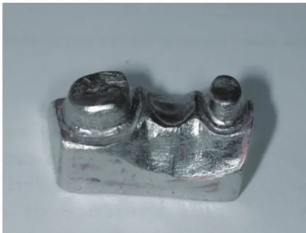
Using polyvinyl siloxane type A impression material (Elite HD, 232115, Zhermack, Ohlmuhle, Germany), an acrylic jaw (Ivoclar Vivadent, Schaan, Lichtenstein) containing a standard chamfer preparation was duplicated with plaster (Fujirock EP, 890222, GC, Chicago, IL, USA). The master base metal working model was made from the plaster dupli- cate model by conventional casting method (Fig. 1). A digi- tal impression of the master model was taken using an
intraoral scanner (Cerec Omnicam, Sirona Dental Systems, Bensheim, Germany). A 3-unit bridge framework was designed digitally with Cerec In Lab SW 15.0 with connec- tors of the dimensions 6 mm2 on both sides of the pontic.
The connectors were circular and located at the central proximal areas. A small depression was also designed on the functional cusp of the pontic for housing the testometric indenter. A power analyses were conducted for determina- tion of the number of test specimens in each group. Then, sixty zirconia frameworks were fabricated using uncolored zirconia blocks (Rainbow Zirconia Block, Genoss, Gyeonggi- do, Korea) by CAD/CAM system (Cerec InLab MCX5, Sirona Dental Systems, Bensheim, Germany) and divided into 4 groups randomly (n = 15). The first group (G2) was subjected to coloring liquid (Rainbow Coloring Liquid A2, Dentium, Gyeonggi-do, Korea) for 2 minutes, the second group (G4) for 4 minutes, and the third group (G6) for 6 minutes. The control group (CFS) was not subjected to any coloring procedures. The lot numbers of the zirconia blocks and the coloring liquid were listed in Table 1. After immer- sion in coloring liquid, test samples were fired in a zirconia sintering furnace (InFire HTC Speed, Sirona Dental Systems, Bensheim, Germany). The test samples were inspected for Fig. 1. Metal master model used in the study.
Table 1. Lot numbers of zirconia blocks and coloring liquid used in the study
Product Lot numbers
Rainbow Zirconia Block
14A17-02, 14A02-02,14B05-02, 14A16-02,14B05-02, 14A16-01, 14A17-02,14A17-02
Rainbow Shade Block 14H13-02, 14H13-01 Rainbow Coloring Liquid 14K07-06
surface defects, and the connector dimensions were assured with a digital caliper (Mitutoyo Corp, Tokyo, Japan). For the comparison of color between precolored zirconia (Rainbow Shade Block A2, Genoss, Gyeonggi-do, Korea) and zirconia colored by immersion in coloring liquid, the same digital data 3 unit bridge frameworks were fabricated with precol- ored zirconia blocks as well (n = 15). This group was the control group (CPZ) for color comparisons. The coloring and sintering procedures were given in Table 2. After zirco- nia was subjected to coloring liquids, color and spectral dis- tributions were evaluated by using Spectroshade Micro spectrophotometer (Version 2.4, MHT S.p.a, Verona, Italy).
Commission Internationale de l’Eclairage(CIE) 1976 L*a*b* co-ordinates were recorded for color evaluation. L*a*b* coordinates were measured in five different areas of each specimen including the center, which was specifically designed flat on the occlusal surface of frameworks (Fig. 2).
Average L* a* b* values of five measurements were used to calculate the color (E) of each test sample. The average E values of 15 specimens represented the color of the group.
The color (E) of each test specimen was determined by the following equation.
E = L* 2 + a*2 + b*2
Fracture test was conducted immediately after color evaluation with a universal testing device (M500-25KN;
Testometric, Rochdale, UK) at a cross-head speed of 1 mm/sec. The test samples were cemented on the metal master model with a dual cure resin cement (Panavia V5, 000011, Kuraray Medical, Tokyo, Japan) prior to fracture test. The test samples were seated on the master model with finger pressure; excessive resin material was meticulously removed; an air barrier was applied; and light polymeriza- tion (Bluephase, Ivoclar Vivadent, Schaan, Liechtenstein) was performed at 450 mW/cm2 for 20 seconds on each side. Each fracture test was conducted on the same metal master model (Fig. 3).
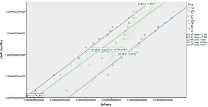
Statistical analysis for evaluating color (E) and fracture load were performed by using one way ANOVA followed by Tukey HSD test (P ≤ .05). Weibull analysis was also con- ducted and Weibull modulus was calculated for each group using SPSS 21.0 (Fig. 4).
Table 2. The coloring and sintering procedures of test specimens
Groups Coloring liguids Sintering Process
CPZ
CFS No immersion Heating rate 5 - 8 °C/min. Hold time at final temperature at 1450 - 1550 °C for 2 hours. Cooling rate 1.67 °C/min (100 °C/hr) to 1000 °C then furnace cooling.
G2 2 min immersion
3 hours of bench top drying after color liquid immersion. Heating rate 5 - 8 °C/min. Hold time at final temperature at 1450 - 1550 °C for 2 hours. Cooling rate 1.67 °C/min (100 °C/hr) to 1000 °C then furnace cooling.
G4 4 min immersion
G6 6 min immersion
Fig. 2. Test Specimens prepared for G6 test group. Fig. 3. Testing of specimens with Testometric. The indenter was located on the depression of the functional cusp.
RESULTS
The means and standard deviations of test groups for color comparisons were given in Table 3. ANOVA analysis showed no color difference between CPZ and G2 groups (P
≥ .907). G4 and G6 showed statistically significant color differences from CPZ and G2 group (P ≤ .05) but showed no difference between them (P = .678).
The mean values of fracture load according to immer- sion time in coloring liquids were given in Table 4.
According to ANOVA analysis, there was no statistically sig- nificant difference between CFS and G2 in terms of frac- ture load (P = .113). CFS group showed the highest fracture
load compared to other groups (1176.681 N). G4 and G6 groups were statistically different from CFS and G2 group (P ≤ .05). Immersion of the test samples in coloring liquids for prolonged time negatively affected the fracture load of the test groups significantly (P ≤ .05). G4 groups showed the lowest fracture load (779.340 N); however, there was no significant difference between G4 and G6 groups (P = .274). The pre-colored zirconia group (CPZ) showed statis- tically lower fracture load than CFS, showing the values 875.203 N and 1176.681 N, respectively (P = .002); howev- er, there was no significant difference between CPZ and G2 and between G4 and G6 test groups in terms of fracture load (P ≥ .05)
Fig. 4. Weibull graph.
Table 4. The mean and standard deviations of fracture load (in N)
Groups N Mean (N) SD
CFS 15 1176.681a* 301.1388
CPZ 15 875.203b 209.9250
G2 15 985.638a,b 168.0885
G4 15 779.340b 217.8654
G6 15 935.491b 129.0211
*Different superscripted lowercase letters indicate statistically different means (P
< .05).
Table 3. The mean and standard deviation of color measurements
Groups N Mean SD
CPZ 15 81.340a* .8692
G2 15 81.140a .7935
G4 15 79.340b .8846
G6 15 79.673b .6943
*Different superscripted lowercase letters indicate statistically different means (P
< .05).
Since the fracture data was not normally distributed, Weibull Analysis was conducted. The highest Weibull modu- lus was observed for G6 group (7.16) followed by G2 (5.88), CFS (3.67), G4 (3.65), and CPZ (3.52) groups as shown in Fig. 4.
DISCUSSION
The current study was performed in order to evaluate changes in color and fracture load of zirconia material when subjected to coloring liquid before sintering. This has been of great importance since esthetics and long term success of the restoration are highly dependent on this procedure.
The null hypothesis was that there would be no difference in terms of color between prefabricated shaded blocks and blocks shaded with liquids and that coloring liquids would have no effect on the fracture load of zirconia. In the study, the null hypothesis was rejected.
The results of the study revealed that the immersion of the zirconia to coloring liquids negatively affected the frac- ture load of the test specimens, emphasizing the importance of manufacturer’s instructions. Although there was a decrease in fracture load when zirconia was subjected to coloring liquids for 2 minutes, which was the manufacturer’s recommended time for coloring, the decrease was not statis- tically significant. The prolonged immersion time further decreased the fracture load of the test specimens. The results of the current study were in accordance with the study of Hjeroppe et al.,17 which reported a significant decrease in fracture strength as a result of prolonged color- ing time. The studies about the effect of coloring liquids to the mechanical properties of zirconia emphasized the rela- tionship between the inclusion of CeO2, Fe2O3, and Bi2O3 to the green zirconia and their effect on the mechanical properties.17,21 Ardlin21 investigated the effects of coloring liquid on the mechanical properties of zirconia and hypoth- esized that the inclusion of CeO2,Fe2O3, and Bi2O3 increased the flexural strength of zirconia material. On the other hand, Wen et al.22 reported a 30% decrease in flexural strength when CeO2 and Er2O3 were added to zirconia.
Sedda et al.18 and Shah et al.20 stated that by using coloring liquids at low concentration, it was possible to change the color of the zirconia to the desired shade without any sig- nificant change in mechanical properties. It seems that, by all means, using coloring liquid is very critical.
Prolonged coloring time influenced not only the fracture load but also the color of the zirconia material. In the cur- rent study, the color of the zirconia material after coloring procedures was compared to the colored zirconia at the industrial stage. The results revealed no difference between pre-colored zirconia and G2. However, when the immersion time was extended, the color of the zirconia was shifted from the desired shade. Vita shade A2 was chosen as the control color in the current study, as this color was the most common shade used in our clinic. Hjerppe et al.17 showed a reduction in fracture strength of zirconia, although not sig- nificant, when zirconia was subjected to D4 coloring liquid
compared to the other shades; Still, further studies are need- ed for the mechanisms.
Various methods can be used for investigating the frac- ture strength of dental ceramics. Ball-on-ring test, piston on 3 ball test, and ring on ring test are preferred for testing the fracture strength of zirconia ceramics.23 All these test meth- ods were well understood and preferred by researchers in their studies.24 However, the major drawback of the test methods was insufficiency in simulating clinical conditions.
Disc shape ceramic test samples were located on different numbers of ball shape bearings according to test method and loaded at the center. Thus fracture strength can be cal- culated. However, loading the test specimens on ball sup- ports induce significant friction stress as the supports can- not rotate during loading and the disc specimen loses its contact with the support. This may lead to premature fail- ures in test specimens.23 Also, 3-point flexural strength test with a bar shape can be used for testing the strength of dental ceramics. However, flaws on the specimen edge con- cerns the reliability of the test. The 3-point bending test is largely dependent on the surface finish of the edges of the specimen. To eliminate edge failures and to simulate clinical conditions better, test specimens were prepared as 3-unit fixed partial dentures in the current study. The specimens were fixed with resin cement meticulously on the metal master model before fracture test. The margins of the res- toration were checked with a brand new explorer for com- plete seating prior to curing. Although speculations may arise for the validity of the test method used in the current study, highly standard test samples were fabricated by using CAD/CAM. Cementation with resin cement stabilized the test specimen during fracture test, resulting in better simula- tion of clinical practice. Moreover, the fracture test was conducted with indenter of testometric located exactly on the same location of every test specimen by the depression prepared on the functional cusp for more standardized test assembly. Relatively higher standard deviations from frac- ture test in the study would be attributed to the brittle nature of the zirconia ceramics. The micro-cracks in zirco- nia ceramics might have caused unexpected failures, result- ing in higher standard deviations irrespective of quality of test specimens.
The connectors are the weakest portions of the fixed partial dentures. Their dimensions should be adjusted for long-term survival of the restoration. There are studies sug- gesting various connector sizes primarily depending on the number of adjoining pontics in the restoration.25 Generally the higher the pontic number, the larger the size of the con- nector.26 In the current study, 6 mm2 circular connector was designed since this was the minimum connector dimensions for three unit fixed partial dentures recommended in litera- ture.26
In several studies, it was shown that the fracture general- ly began at the gingival portion of the connector since ten- sile type stresses was occurred at this site.25 However, com- pression type stresses were occurred simultaneously on the opposite side of the connector. This might lead to failures
in calculating fracture strength of the test specimens. Thus the author decided to use maximum fracture load in com- parison. All the test samples in the current study were frac- tured from connector and fracture was initiated from gingi- val portion.
Since fracture data of brittle materials are not normally distributed, Weibull analysis is commonly used for strength data analysis of brittle materials.27 Lower Weibull modulus indicate more spread data and higher variability among mea- surements. A higher mean Weibull modulus for G6 group indicated a less spread distribution (lower variability) com- pared to the other groups with higher reliability. The Weibull modulus of the current study was in accordance with the study of Sedda et al.,18 which reported a higher Weibull value for colored specimens than for non-colored specimens and the lowest Weibull value for pre-colored zir- conia specimens.
One limitation of the current study was that the test samples were not subjected to aging cycle prior to test pro- cedures. Aging could change the results of the study.
However, this would lead to complexity in analyzing the results, thus aging with thermal cycle was avoided.
CONCLUSION
Within the limitations of the current study, it was concluded that prolonged immersion of zirconia in A2 coloring liquids not only negatively affected the fracture load of the zirconia being tested in the current study but also affected the shade of the restoration. The study showed the importance of following manufacturer’s instructions for coloring zirconia with coloring liquids. Clinical studies are needed for investi- gating the long-term color stability of the zirconia colored with coloring liquids.
ORCID
Ekren Orhun http://orcid.org/0000-0002-3237-8003 REFERENCES
1. Isgrò G, Pallav P, van der Zel JM, Feilzer AJ. The influence of the veneering porcelain and different surface treatments on the biaxial flexural strength of a heat-pressed ceramic. J Prosthet Dent 2003;90:465-73.
2. Cardelli P, Manobianco FP, Serafini N, Murmura G, Beuer F.
Full-arch, Implant-supported monolithic zirconia rehabilita- tions: Pilot clinical evaluation of wear against natural or com- posite teeth. J Prosthodont 2015 Oct 5.
3. O’Brien WJ. Dental materials and their selection. 3rd ed.
Chicago: Quintessence; 2002. p. 210-24.
4. Piconi C, Maccauro G. Zirconia as a ceramic biomaterial.
Biomaterials 1999;20:1-25.
5. Guazzato M, Albakry M, Quach L, Swain MV. Influence of surface and heat treatments on the flexural strength of a glass-infiltrated alumina/zirconia-reinforced dental ceramic.
Dent Mater 2005;21:454-63.
6. Guazzato M, Albakry M, Ringer SP, Swain MV. Strength, fracture toughness and microstructure of a selection of all- ceramic materials. Part II. Zirconia-based dental ceramics.
Dent Mater 2004;20:449-56.
7. Denry I, Kelly JR. State of the art of zirconia for dental ap- plications. Dent Mater 2008;24:299-307.
8. Kosmac T, Oblak C, Jevnikar P, Funduk N, Marion L. The ef- fect of surface grinding and sandblasting on flexural strength and reliability of Y-TZP zirconia ceramic. Dent Mater 1999;15:426-33.
9. Hannink RHJ, Kelly PM, Muddle BC. Transformation tough- ening in zirconia-containing ceramics. J Am Ceram Soc 2000;
83:461-87.
10. White SN, Miklus VG, McLaren EA, Lang LA, Caputo AA.
Flexural strength of a layered zirconia and porcelain dental all-ceramic system. J Prosthet Dent 2005;94:125-31.
11. Sailer I, Fehér A, Filser F, Lüthy H, Gauckler LJ, Schärer P, Franz Hämmerle CH. Prospective clinical study of zirconia posterior fixed partial dentures: 3-year follow-up. Quintessence Int 2006;37:685-93.
12. Sailer I, Fehér A, Filser F, Gauckler LJ, Lüthy H, Hämmerle CH. Five-year clinical results of zirconia frameworks for pos- terior fixed partial dentures. Int J Prosthodont 2007;20:383-8.
13. Roediger M, Gersdorff N, Huels A, Rinke S. Prospective evaluation of zirconia posterior fixed partial dentures: four- year clinical results. Int J Prosthodont 2010;23:141-8.
14. Lee WF, Feng SW, Lu YJ, Wu HJ, Peng PW. Effects of two surface finishes on the color of cemented and colored ana- tomic-contour zirconia crowns. J Prosthet Dent 2016;116:
264-8.
15. Tuncel İ, Özat P, Eroğlu E. Effects of coloring procedures on zirconia/veneer ceramics bond strength. J Adv Prosthodont 2014;6:451-5.
16. Kim HK, Kim SH. Effect of the number of coloring liquid applications on the optical properties of monolithic zirconia.
Dent Mater 2014;30:e229-37.
17. Hjerppe J, Närhi T, Fröberg K, Vallittu PK, Lassila LV. Effect of shading the zirconia framework on biaxial strength and surface microhardness. Acta Odontol Scand 2008;66:262-7.
18. Sedda M, Vichi A, Carrabba M, Capperucci A, Louca C, Ferrari M. Influence of coloring procedure on flexural resis- tance of zirconia blocks. J Prosthet Dent 2015;114:98-102.
19. Pittayachawan P, McDonald A, Petrie A, Knowles JC. The bi- axial flexural strength and fatigue property of Lava Y-TZP dental ceramic. Dent Mater 2007;23:1018-29.
20. Shah K, Holloway JA, Denry IL. Effect of coloring with vari- ous metal oxides on the microstructure, color, and flexural strength of 3Y-TZP. J Biomed Mater Res B Appl Biomater 2008;87:329-37.
21. Ardlin BI. Transformation-toughened zirconia for dental in- lays, crowns and bridges: chemical stability and effect of low- temperature aging on flexural strength and surface structure.
Dent Mater 2002;18:590-5.
22. Wen N, Yi Y, Zhang W, Liu H, Tian J. Properties of dental alumina ceramics infiltrated with colored glass. Rare Metal Mater Eng 2007;36:82-4.
23. Shetty DK, Rosenfield AR, McGuire P, Duckworth WH.
Biaxial flexure test for ceramics. American Ceram Soc Bulletin 1980;59:1193-7.
24. Mijoska A, Popovska M. Evaluation of different in vitro test- ing methods for mechanical properties of veneer ceramics.
Pril (Makedon Akad Nauk Umet Odd Med Nauki). 2015;36:
225-30.
25. Gargari M, Gloria F, Cappello A, Ottria L. Strength of zirco- nia fixed partial dentures: review of the literature. Oral Implantol (Rome) 2010;3:15-24.
26. Studart AR, Filser F, Kocher P, Gauckler LJ. In vitro lifetime of dental ceramics under cyclic loading in water. Biomaterials 2007;28:2695-705.
27. Quinn JB, Quinn GD. A practical and systematic review of Weibull statistics for reporting strengths of dental materials.
Dent Mater 2010;26:135-47.