J Korean Ophthalmol Soc 2013;54(8):1282-1286 pISSN: 0378-6471
eISSN: 2092-9374
http://dx.doi.org/10.3341/jkos.2013.54.8.1282
= 증례보고 =
결절경화증환자에서 발생한 성상세포 과오종과 연관된 시신경섬유 결손
이가현⋅이나은⋅이창규⋅홍사민⋅성공제⋅김찬윤 연세대학교 의과대학 안과학교실, 시기능개발연구소
목적: 시신경 유두 및 망막의 과오종과 연관되어 시신경섬유층의 결손이 발생한 결절경화증 환자를 경험하여 이를 보고하고자 한다.
증례요약: 결절경화증으로 진단받고 항경련제 Vigabatrin을 복용 중인 6세 남아가 약제에 의한 시야변화 확인을 위해 소아신경과에서 의뢰되었다. 환아는 안저검사상 우안의 시신경유두 위쪽 테두리에 연접한 1/2 시신경 유두 크기의 성상세포 과오종이 확인되었으며 다른 특이소견은 관찰되지 않았다. 초진 당시 좌안에는 특이 소견이 없었다. 초진 1년 6개월 후 우안의 시신경유두주위 성상세포 과오 종의 크기가 다소 증가하였고 좌안에는 1.5 유두직경거리의 황반하측에 새로운 망막 과오종이 발생하였다. 초진 3년 후 우안에 크기가 증가했던 시신경유두 성상세포 과오종이 과오종부위에 상이측과 중심하부방향으로 시신경섬유결손이 발생하였다.
결론: 결절경화증 환자에서 성상세포 과오종은 일반적으로 변하지 않는다고 알려졌으나 본 증례와 같이 크기가 증가하면서 시신경섬유 결손을 야기할수도 있으므로 이를 염두에 두고 면밀히 추적관찰하여야 할 것이다.
<대한안과학회지 2013;54(8):1282-1286>
■Received: 2013. 1. 11. ■ Revised: 2013. 3. 25.
■Accepted: 2013. 6. 20.
■Address reprint requests to Chan Yun Kim, MD, PhD
The Institute of Vision Research, Department of Ophthalmology, Severance Hospital, #50 Yonsei-ro, Seodaemun-gu, Seoul 120-752, Korea
Tel: 82-2-2228-3570, Fax: 82-2-312-0541 E-mail: [email protected]
* This study was presented as a poster at the 108th Annual Meeting of the Korean Ophthalmology Society 2012.
* This study was supported by a grant of the Korea Health technology R&D Project, Ministry of Health & Welfare, Republic of Korea (A101727).
결절경화증은 신경섬유종증, 스터지웨버증후군 등과 함께 눈, 중추신경, 피부와 내장을 주로 침범하는 대표적인 눈신경 피부증후군 중의 하나로, 인구 5,000-10,000명당 한 명 꼴 로 발생하며,1,2 상염색체성 우성으로 유전된다.3,4 결절경화 증은 여러 기관에 과오종을 형성하는 질환으로, 1908년 Vogt는 간질, 지능저하 및 피지선종을 세 가지 주 증상으로 제시하였으나,5현재는 뇌의 결절, 안면부의 피지선종, 조갑 섬유종, 망막의 과오종, 심장의 횡문근종 등의 다양한 임상 양상을 포함하는 포괄적 진단 기준을 사용하고 있다.6망막 의 과오종은 결절경화증 환자의 34-87%에서 나타나는 데,7,8대부분 선천성으로 발생하여 진행하거나 변화하지 않 으며 시력에 영향을 미치지 않으므로 대부분의 경우 치료를 필요로 하지 않으나,9드물게 과오종으로 인한 망막하 삼출 이나 유리체출혈 등이보고된 바 있어 주의를 요한다.10-12
국내에도 이러한 결절경화증환자에서의 망막 과오종 예가 여러 차례 보고되었다.13-21 그러나 이러한 보고에 따르면 망막 과오종은 저시력이나 사시와 연관된 경우가 있었으
나,15,19,21진행하거나 망막신경섬유층과의 관계는 언급되지
않았다.13-21이에 저자들은 3년 이상 추적 관찰한 결절경화
증 환아에서 망막 과오종의 크기가 증가하고 그 부위에 망 막신경섬유층의 결손이 발생하는 것을 관찰하였기에 이를 보고하는 바이다.
증례보고
결절경화증으로 진단받은 6세 남아가 안저검사 및 약물 부작용 여부를 판단하기 위해 소아신경과에서 의뢰되었다.
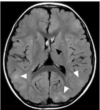
출생력상 임신 32주에 제왕절개로 분만되었으며, 신생아 황달 및 가사의 기왕력이 있었다. 뇌 자기공명영상 결과 대 뇌 반구 내 수 개의 피질 및 피질하 결절이 관찰되었으며, 양측 측뇌실 부근의 조영 증강되는 상의하세포종 소견 보 여 결절경화증에 합당하였다(Fig. 1). 환자는 생후 6개월부 터 결절경화증을 진단받고 난치성 간질발작 증상을 조절하 기 위해 Vigabatrin (Sabril Lundbeck Inc, Deerfield, IL) 500 mg을 b.i.d로 복용중이었으며, 타병원 안과에서 주기 적으로 경과 관찰한 과거력은 있었지만 구체적인 의무기록 은 없었다.
본원 안과 초진시 교정시력은 우안 0.3, 좌안 0.3이었고,
Figure 1. Axial T2-weighted images of MR show that sub-
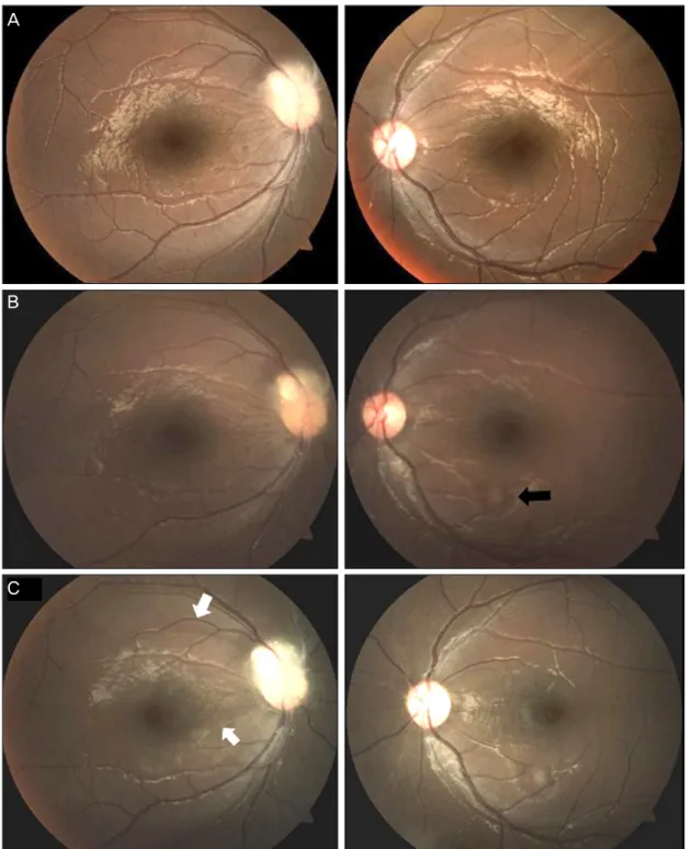
ependymal nodule (black arrow head) projecting into lumen of left lateral ventricle and multiple subtle bilateral cortical and subcortical tubers (white arrow heads).TonoPen® XL (Medtronic Solan, Jacksonville, FL, USA) 로 측정한 안압은 우안 12, 좌안 12 mmHg였다. 전안부 검 사상 특이 소견은 발견되지 않았으며, 양안 동공 반응 정상 으로 상대성 구심성 동공운동이상은 관찰되지 않았다. 조절 마비제 점안 하 시행한 타각적 굴절검사상 우안 +sph4.25 –cyl3.00 Axis180, 좌안 +sph4.50 -cyl3.00 Axis180 이 었고, 최대교정시력은 우안 0.7, 좌안 0.6이었다. 안저검사 상 우안 시신경 상이측에 연접한 1/2 유두직경거리 크기의 석회화를 동반한 뽕나무 열매 모양의 융기된 과오종이 관 찰되었으며, 좌안 망막에는 특이 이상 소견이 관찰되지 않 았다(Fig. 2A).
초진 6개월 후와 1년 후 시행한 안저 검사상 특이변화 는 관찰되지 않았다. 초진 1년 6개월 후 시행한 안저검사 결과 우안의 시신경 과오종의 크기가 다소 증가하였으며, 이전 검사상 특이 소견이 없었던 좌안에도 황반 하측에 성 상세포 과오종이 새롭게 발견되었다(Fig. 2B). 초진으로 부터 3년 경과 후 시행한 안저 검사상, 우안의 크기가 증 가한 시신경 과오종 부위에 시신경섬유층 결손이 발견되 었고 황반 아래쪽으로도 시신경섬유결손이 관찰되었다.
하지만 초진 1년 6개월 후 새로이 발생했던 좌안의 성상 세포 과오종부위에는 시신경섬유결손등의 이상소견은 없 었다(Fig. 2C).
고 찰
결절경화증 환자에서 나타나는 안과적 병변 중 가장 흔 한 것은 망막의 과오종으로 환자의 50% 이상에서 발생하 지만, 대부분 진행하지 않으며 시력에 영향을 미치지 않아 치료가 필요하지 않다고 알려졌다.7-12
본 증례의 환자의 경우, 망막의 성상세포 과오종이 추적 관찰 기간 동안 크기가 증가하였고, 이에 따라 녹내장에서 와 유사하게 시신경섬유 주행방향을 따라 나타나는 망막신 경섬유층결손이 발생하였다. 또한 대개 망막의 과오종이 선 천적으로 발생하는 것과 달리, 정상소견을 보이던 좌측 망 막에도 추적관찰 기간 동안 편평하고 투명한 결절이 새로 이 발생하였다. 아직까지 결절경화증 환자에서 시신경유두 부 과오종의 크기 증가에 따른 망막신경섬유층결손 발생은 보고된 바 없었다.
환아의 경우 결절경화증에 동반된 간질로 Vigabatrin (Sabril®)을 복용 중으로, 초진 당시 vigabatrin을 500 mg b.i.d로 복용 중이었으며, 본원에서 경과 관찰하던 기간 동 안 총 3,500 g 이상의 vigabatrin을 복용하였다. Vigabatrin 은 선택적 불가역적 GABA aminotransferase 억제제로서, 뇌에서 작용하는 억제성 신경전달물질인 GABA의 농도를 높이는 작용을 하는 약제로,22,23주로 난치성 부분간질 환자 에 효과적으로 작용한다. 그러나 이 약제를 사용한 경우, 10-50%의 환자에서 비가역적인 시야 손상을 특징으로 하 는 망막병증이 발생한다.24-26 Vigabatrin과 연관되어 발생 하는 시신경 손상은 비측의 신경섬유층 두께의 감소를 주 로 동반하는데, 이측의 신경섬유층 두께는 정상인 것이 특 징이다.27또한 2012년 Clayton et al28이 발표한 연구에 따 르면 Vigabatrin을 총 1,000 g 이상 복용한 환자들에서 그 렇지 않은 군에 비하여 시신경 주변부의 시신경섬유층 두 께 감소의 정도는 Vigabatrin의 총용량에 비례하였는데, 특 히 상비측의 시신경섬유두께가 가장 감소되어 있으며, 상대 적으로 이측, 상이측 및 하이측은 덜 침범하는 모습을 보였 다. 본 증례의 경우 환아가 복용한 Vigabatrin의 총 용량이 1,000 g을 초과하므로 신경섬유층 두께의 감소를 유발하였 을 수 있으나, 본 증례의 망막신경섬유층결손은 Vigabatrin 복용 시 일반적으로 나타나는 것과 달리 비측의 신경섬유 층 결손이 뚜렷하지 않은데 반하여, 시신경 과오종이 있는 상이측 부위에 한정되어 뚜렷하게 나타나며, 시신경 과오종 의 크기가 증가함에 따라 망막신경섬유층의 결손 또한 진 행되는 모습을 보이기 때문에 환아의 망막신경섬유층 결손 은 항경련 약물보다는 시신경 과오종의 영향을 받은 것으 로 생각한다. 본 증례의 망막신경섬유층 결손은 안저사진만 으로 판단하였는데 불행하게도 시행한 시야검사는 신뢰도
A
B
C
Figure 2. Development of nerve fiber layer defect associated with astrocytic hamartoma in a patient with tuberous
sclerosis. (A) Fundus photograph of the initial visit shows a 1/2 disc diameter-sized “mulberry-like” protruding tu- mor above the right optic disc and normal fellow eye. (B) One year and 6 months after the first visit, fundus photo- graph shows enlarged right optic disc hamartoma. Newly onset retinal astrocytic hamartoma (black arrow) is found on the fundus of the left eye. (C) Three years later, fundus photograph shows development of retinal nerve fiber layer defects (white arrows).가 낮아 판별하기가 힘들었고 광간섭단층촬영은 환아가 거 부하여 시행할 수 없었다. 하지만 사진상 시신경섬유결손이 명확하여 정성적인(quantitative) 판단을 하기에는 충분하 다고 생각한다.
결절경화증환자의 과오종이 어떻게 시신경섬유결손을 야기하는지는 기존의 보고가 없었기에 기전을 정확히 알 수 는 없다. 과오종과의 감별해야하는 질환 중에 하나인 시신 경 드루젠의 경우 53-75%의 환자에서 시야 결손을 보이는
데, 그 기전으로 제시되는 것은 드루젠으로 인한 직접적 인 압박 효과로 드루젠이 시신경의 혈관을 압박하여 시신경 의 혈액 공급에 이상을 일으키기 때문에 발생하는 것으로 알려졌다.29,31 지금까지 결절경화증 환자에서 시신경 부위 의 과오종에 의한 시신경섬유층두께의 감소는 보고된 바가 없으나, 본 증례에서 발견된 시신경섬유층의 두께 감소도 시신경 드루젠과 마찬가지로 시신경 및 시신경 혈관의 압박 으로 인한 시신경섬유층의 손상에 의해 발생하지 않았을까 추정된다. 물론 이 환자의 시신경 주위 과오종은 융기된 형 태로 드루젠과는 명확히 구별되어 드루젠에 의한 시신경섬 유결손은 아닌 것으로 생각한다. 또한 Carol et al32은 2006 년 망막의 성상세포 과오종은 망막내층 혹은 망막의 전층 을 침범하여 정상 망막 조직 구조의 혼란(disorganization) 을 발생시키는 것을 보고하였는데, 본 증례에서 발생한 신 경섬유층의 결손도 이와 마찬가지로 과오종에 의하여 발생 한 정상 망막 구조의 혼란과 연관되어 발생했을 가능성이 있다.
결론적으로 이 증례에서 경험하였듯이 일반적으로 진행 하지 않는 것으로 알려진 결절경화증 환자의 망막 과오종 이 진행하는 경우가 있으며, 특히 시신경 주위에 발생한 성 상세포 과오종은 드물게 녹내장에서 나타나는 시신경섬유 층결손과 유사하게 시신경섬유 주행방향을 따라 시신경섬 유층결손을 유발하고 진행시킬 수 있으므로, 이러한 환자는 주의 깊은 관찰이 필요할 것으로 생각한다.
REFERENCES
1) Curatolo P, Bombardieri R, Jozwiak S. Tuberous sclerosis. Lancet 2008;372:657-68.
2) Osborne JP, Fryer A, Webb D. Epidemiology of tuberous sclerosis.
Ann N Y Acad Sci 1991;615:125-7.
3) van Slegtenhorst M, de Hoogt R, Hermans C, et al. Identification of the tuberous sclerosis gene TSC1 on chromosome 9q34. Science 1997;277:805-8.
4) European Chromosome 16 Tuberous Sclerosis Consortium.
Identification and characterization of the tuberous sclerosis gene on chromosome 16. Cell 1993;75:1305-15.
5) Vogt H. Zur diagnostik der tuberosen sclerose. Z Erforsch Benhandl Fugendl Schwachsinns 1908;2:1-16.
6) Roach ES, Gomez MR, Northrup H. Tuberous sclerosis complex consensus conference: revised clinical diagnostic criteria. J Child Neurol 1998;13:624-8.
7) Rowley SA, O'Callaghan FJ, Osborne JP. Ophthalmic manifes- tations of tuberous sclerosis: a population based study. Br J Ophthalmol 2001;85:420-3.
8) Kiribuchi K, Uchida Y, Fukuyama Y, Maruyama H. High incidence of fundus hamartomas and clinical significance of a fundus score in tuberous sclerosis. Brain Dev 1986;8:509-17.
9) Töteberg-Harms M, Sturm V, Sel S, et al. Retinal astrocytomas:
long-term follow-up. Klin Monbl Augenheilkd 2011;228:337-9.
10) Gass JDM. Stereoscopic atlas of macular disease: Diagnosis and treatment, 1st ed. St. Louis: Mosby, 1997;836-43.
11) Lagos JC, Gomez MR. Tuberous sclerosis: Reappraisal of a clin- ical entity. Mayo Clin Proc 1967;42:26-49.
12) Nyboer JH, Robertson DM, Gomez MR. Retinal lesions in tuber- ous sclerosis. Arch Ophthal 1976;94:1277-80.
13) Yoon DK. A case of tuberous sclerosis. J Korean Ophthalmol Soc 1974;15:68-71.
14) Kim JH, Shin HH. Two cases of tuberous sclerosis. J Korean Ophthalmol Soc 1982;23:477-81.
15) Kwon OW, Kim YW. A case of tuberous sclerosis. J Korean Ophthalmol Soc 1983;24:645-9.
16) Yoo KH, Oum BS. Two cases of tuberous sclerosis. J Korean Ophthalmol Soc 1987;28:877-83.
17) Kim SM, Hahn YH, Kim SD. Two cases of tuberous sclerosis. J Korean Ophthalmol Soc 1988;29:1141-5.
18) Ku YJ, Kim EA, Han YB. A case of tuberous sclerosis. J Korean Ophthalmol Soc 1995;36:355-60.
19) Ji JY, Lee JH, Choi WS. A case of bilateral tuberous sclerosis. J Korean Ophthalmol Soc 1996;37:203-9.
20) Lee JH, Han KS, Han YB. One case of tuberous sclerosis. J Korean Ophthalmol Soc 1999;40:2337-42.
21) La TY, Kim CW, Lee YS, Kim MH. A case of retinal astrocytic ha- martoma causing blindness in tuberous sclerosis. J Korean Ophthalmol Soc 1999;40:1421-6.
22) Dichter MA, Brodie MJ. New antiepileptic drugs. N Engl J Med 1996;334:1583-90.
23) Ben-Menachem E. Vigabatrin. Epilepsia 1995;36 Suppl 2:S95-104.
24) Maguire MJ, Hemming K, Wild JM, et al. Prevalence of visual field loss following exposure to vigabatrin therapy: a systematic review.
Epilepsia 2010;51:2423-31.
25) Roubertie A, Bellet H, Echenne B. Vigabatrin-associated retinal cone system dysfunction. Neurology 1998;51:1779-81.
26) Miller NR, Johnson MA, Paul SR, et al. Visual dysfunction in pa- tients receiving vigabatrin: clinical and electrophysiologic findings. Neurology 1999;53:2082-7.
27) Lawthom C, Smith PE, Wild JM. Nasal retinal nerve fiber layer at- tenuation: a biomarker for vigabatrin toxicity. Ophthalmology 2009;116:565-71.
28) Clayton LM, Devile M, Punte T, et al. Patterns of peripapillary reti- nal nerve fiber layer thinning in vigabatrin-exposed individuals.
Ophthalmology 2012;119:2152-60.
29) Roh S, Noecker RJ, Schuman JS, et al. Effect of optic nerve head drusen on nerve fiber layer thickness. Ophthalmology 1998;105:
878-85.
30) Savino PJ, Glaser JS, Rosenberg MA. A clinical analysis of pseudopapilledema. II. Visual field defects. Arch Ophthalmol 1979;
97:71-5.
31) Sarkies NJ, Sanders MD. Optic disc drusen and episodic visual loss. Br J Ophthalmol 1987;71:537-9.
32) Shields CL, Benevides R, Materin MA, Shields JA. Optical coher- ence tomography of retinal astrocytic hamartoma in 15 cases.
Ophthalmology 2006;113:1553-7.
=ABSTRACT=
Retinal Nerve Fiber Layer Defect Associated with Astrocytic Hamartoma in a Patient with Tuberous Sclerosis
Ka Hyun Lee, MD, Naeun Lee, MD, Chang Kyu Lee, MD, Sa Min Hong, MD, Gong Je Seong, MD, PhD, Chan Yun Kim, MD, PhD
The Institute of Vision Research, Department of Ophthalmology, Yonsei University College of Medicine, Seoul, Korea
Purpose: To report the progression of an astrocytic hamartoma of the right optic nerve head as well as the retina, and the progression of retinal nerve fiber defect associated with astrocytic hamartoma in a patient with tuberous sclerosis.
Case summary: A 6-year-old boy with tuberous sclerosis and an astrocytic hamartoma of the right optic nerve head, which was found at the time of ophthalmologie examinations, was referred from the pediatric neurologist for evaluation of the vig- abatrin-associated visual field changes. Fundus examination revealed 1/2 disc diameter (DD)-sized astrocytic hamartoma located at the margin of the superior part of the optic nerve. The retina of the left eye was normal. Eighteen months after the first visit, enlarged optic disc hamartoma of the right eye and newly onset retinal astrocytic hamartoma located approx- imately 1.5 DD inferior to the fovea of the left eye were found. Three years later, an increase in the size of the astrocytic ha- martoma of the right optic nerve and development of retinal nerve fiber defects were observed.
Conclusions: Astrocytic hamartoma in patients with tuberous sclerosis is usually stable without progression. However, in our patient, astrocytic hamartoma showed progression, and development of retinal nerve fiber defects occurred. Regular follow-up is necessary for astrocytic hamartoma in patients with tuberous sclerosis.
J Korean Ophthalmol Soc 2013;54(8):1282-1286
Key Words: Astrocytic hamartoma, Retinal nerve fiber layer defect, Tuberous sclerosis
Address reprint requests to Chan Yun Kim, MD, PhD
The Institute of Vision Research, Department of Ophthalmology, Severance Hospital
#50 Yonsei-ro, Seodaemun-gu, Seoul 120-752, Korea
Tel: 82-2-2228-3570, Fax: 82-2-312-0541, E-mail: [email protected]