Copyright © 2014 Korean Neurological Association 171
Print ISSN 1738-6586 / On-line ISSN 2005-5013 http://dx.doi.org/10.3988/jcn.2014.10.2.171 CASE REPORT
J Clin Neurol 2014;10(2):171-173
Introduction
Spinal cord infarction is much less frequent than cerebral in- farction, accounting for only 1% of all strokes.1 The anterior spinal artery supplies the anterior two-thirds of the spinal cord via the sulcal (central) artery. This usually results in anterior spinal artery infarcts presenting with profound bilateral motor deficits, sensory disturbances, and spinothalamic sensory defi- cits.2,3 Although the mechanisms are not completely under- stood, there are some case reports of restrictive clinical syn- dromes, such as the ‘man-in-the-barrel syndrome’ and the sulcal artery syndrome.4,5 Here we describe a patient who pre- sented with bilateral hand weakness but without sensory def- icits due to left vertebral artery occlusion.
Case Report
A healthy 29-year-old man without any vascular risk factors presented with sudden neck pain and bilateral hand weakness.
After flexing his neck while tying his shoelaces, the patient experienced a sudden, severe pain (with a maximum score on a visual analog scale) that started in the posterior neck and spread rapidly across the entire head. Bilateral hand weakness that followed the pain onset prevented the patient from tying his shoelaces. The patient’s vital signs were stable upon hospi- tal admission, except for markedly elevated blood pressure (202/108 mm Hg). The initial neurological examination re- vealed weakness in both flexion and extension of the wrists and fingers bilaterally, and during left leg extension [Medical Research Council (MRC) grade 4]. However, the deep tendon reflexes were normal and the patient denied hypoesthesia in any sensory modality including pain, temperature, propriocep- tion, and vibration. A detailed examination did not reveal any cortical sensory sign such as graphesthesia. The Romberg test produced a negative result. Laboratory findings were normal,
Atypical Anterior Spinal Artery Infarction due to Left Vertebral Artery Occlusion Presenting with Bilateral Hand Weakness
Min-Ji Kim,a Mi-Hee Jang,b Mi-Song Choi,c Suk Yun Kang,c Joo Yong Kim,c Ki-Han Kwon,c Ik-Won Kang,d Soo-Jin Choc
aDepartment of Neurology, Kangnam Sacred Heart Hospital, Hallym University College of Medicine, Seoul, Korea
bDepartment of Neurology, Hallym University Sacred Heart Hospital, Anyang, Korea
cDepartments of Neurology and dRadiology, Dongtan Sacred Heart Hospital, Hallym University College of Medicine, Hwaseong, Korea
Received May 30, 2013 Revised October 24, 2013 Accepted October 25, 2013 Correspondence Soo-Jin Cho, MD, PhD Department of Neurology, Dongtan Sacred Heart Hospital, Hallym University College of Medicine, 7 Keunjaebong-gil, Hwaseong 445-907, Korea Tel +82-31-8086-2310 Fax +82-31-8086-2317 E-mail [email protected]
BackgroundzzInfarct of the anterior spinal artery is the most common subtype of spinal cord infarct, and is characterized by bilateral motor deficits with spinothalamic sensory deficits. We experienced a case with atypical anterior-spinal-artery infarct that presented with bilateral hand weakness but without sensory deficits.
Case ReportzzA 29-year-old man presented with sudden neck pain and bilateral weakness of the hands. Magnetic resonance imaging (MRI) of the brain did not reveal any lesion. His motor symptoms improved rapidly except for mild weakness in his left wrist and fingers. Magnetic resonance angiography showed proximal occlusion of the left vertebral artery; a spine MRI re- vealed left cervical cord infarction.
ConclusionszzBilateral or unilateral hand weakness can be the sole symptom of a cervical
cord infarct. J Clin Neurol 2014;10(2):171-173
Key Wordszz spinal cord infarction, vertebral artery occlusion, hands, anterior spinal artery.
Open Access
cc This is an Open Access article distributed under the terms of the Cre- ative Commons Attribution Non-Commercial License (http://creative- commons.org/licenses/by-nc/3.0) which permits unrestricted non-com- mercial use, distribution, and reproduction in any medium, provided the ori- ginal work is properly cited.
Anterior Spinal Artery Infarct with Hand Weakness
172 J Clin Neurol 2014;10(2):171-173
and chest radiography did not reveal any pathology; an elec- trocardiogram also produced no evidence of ischemia or ar- rhythmia. Furthermore, 2-D echo and transesophageal echo- cardiograms provided no evidence for an embolic stroke.
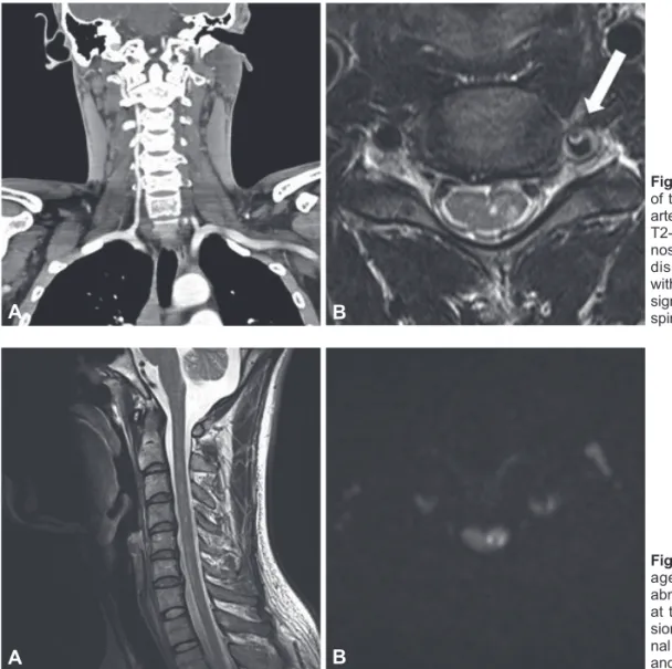
Subsequent 24-hour Holter monitoring failed to identify any arrhythmia or atrial fibrillation. His motor symptoms im- proved rapidly within 1 day except for mild weakness of his left wrist (MRC grade of flexion/extension IV/IV) and fingers (MRC grade of flexion/extension IV/IV; slightly more severe in the 5th finger). Brain computed tomography (CT) and brain magnetic resonance imaging (MRI) did not reveal any signal abnormalities or mass lesions. However, CT aortography iden- tified severe stenosis and occlusion of the left vertebral artery, and a pseudo lumen with mural thrombi was suspected on cervical MRI (Fig. 1). These clinical and radiological findings implicated arterial dissection. MRI of the cervical segment produced high signal intensities in the left gray matter of spi- nal cord at the C3, C4, and C6 levels on diffusion-weighted images (Fig. 2). The patient was discharged on an oral antico- agulant. Magnetic resonance angiography performed 3 months after symptom onset showed persistent occlusion of the left vertebral artery.
Discussion
Unique features of this spinal-cord-infarct patient were bilat- eral hand weakness, absence of sensory deficits, and rapid improvement of motor symptoms in the right hand and left leg.
Weakness of both arms has been reported previously in as- sociation with cervical cord infarcts, typically also with spi- nothalamic sensory deficit and vertebral artery occlusion.4-7 The mechanism that preserves leg strength in these cases is most likely collateral flow from the surrounding pial plexus.4 Nevertheless, weakness of both hands without sensory defi- cits is rare in infarcts of the cervical spinal cord, and its mech- anism remains to be determined.7,8
The anterior spinal artery forms at the level of the foramen magnum from the branches of the vertebral artery, and gives rise to the sulcal (central) arteries that penetrate the right and left sides of the spinal cord.3 Unlike circumferential areas of the spinal cord, its interior has no anastomosis and the sulcal arteries are essentially end arteries. Therefore, the weakness of both hands in the present patient suggests that the end-ar- teries zone or the zone bordering the sulcal artery and cir-
Fig. 1. A: Severe stenosis and occlusion of the proximal part of the left vertebral artery in a neck CT angiogram. B: Axial T2-weighted image showing severe ste- nosis of the left vertebral artery due to dissection, showing a pseudo lumen with mural thrombi (arrow), and a high signal intensity in the left gray matter of spinal cord at the C4 level.
A B
Fig. 2. A: Sagittal T2-weighted MRI im- age of the cervical spine showing an abnormal area of high signal intensities at the C3, C4, and C6 levels. B: Diffu- sion-weighted image showing high sig- nal intensities on the left side at the C3 and C4 levels.
A B
Kim MJ et al.
www.thejcn.com 173 cumferential artery corresponds to a region where hand mo-
tor neurons are located bilaterally.8 This would include the ventrolateral to lateral regions within the gray matter of the lower cervical spinal cord (Fig. 3).9,10 Alternatively, the hand motor areas may be more vulnerable than the more proximal arm motor areas. The hand motor area occupies a relatively large proportion of the cortical homunculus, so the hand mo- tor area may occupy a larger area in the spinal gray matter, or require a larger blood supply.11,12
Finally, the isolated unilateral hand weakness of this pa- tient may be related to variation in the branches of the sulcal artery. Successive sulcal arteries generally alternate in their distribution to the left or right side of the spinal cord, but not both; this would make unilateral involvement or improve- ment possible.5 Sensory deficit was absent in the present case. All subtypes of spinal cord infarcts are presumed to have sensory deficits, and therefore a pure motor presenta- tion can result in misdiagnosis.2,3 Anterolateral involvement could explain the present atypical presentation, and bilateral presentation without facial weakness may indicate a spinal cord infarct.5
Transient ischemic attacks in the spinal cord reported oc- cur primarily in the cervical cord, and recovery depends on the severity of the initial involvement and its collateral flow.1,2,6 Cervical lesions are generally less vulnerable to ischemic in- sults than are thoracic or lumbar lesions due to the intensive vascularization from the vertebral and radicular arteries.3,6,13
The rapid improvement in the present case might therefore have resulted from initial distal involvement and collateral flow from the intact right vertebral artery.
In conclusion, transient bilateral hand weakness can be the initial symptom of a spinal cord infarct, and unilateral hand weakness can be the sole symptom of a cervical cord infarct.
Conflicts of Interest
The authors have no financial conflicts of interest.
REFERENCES
1. Novy J, Carruzzo A, Maeder P, Bogousslavsky J. Spinal cord isch- emia: clinical and imaging patterns, pathogenesis, and outcomes in 27 patients. Arch Neurol 2006;63:1113-1120.
2. Kumral E, Polat F, Güllüoglu H, Uzunköprü C, Tuncel R, Alpaydin S.
Spinal ischaemic stroke: clinical and radiological findings and short- term outcome. Eur J Neurol 2011;18:232-239.
3. Satran R. Spinal cord infarction. Stroke 1988;19:529-532.
4. Berg D, Müllges W, Koltzenburg M, Bendszus M, Reiners K. Man- in-the-barrel syndrome caused by cervical spinal cord infarction.
Acta Neurol Scand 1998;97:417-419.
5. Li Y, Jenny D, Bemporad JA, Liew CJ, Castaldo J. Sulcal artery syn- drome after vertebral artery dissection. J Stroke Cerebrovasc Dis 2010;
19:333-335.
6. Machnowska M, Moien-Afshari F, Voll C, Wiebe S. Partial anterior cervical cord infarction following vertebral artery dissection. Can J Neurol Sci 2008;35:674-677.
7. Park SW, Sohn SI, Cho YW, Lee H, Lim JG, Yi SD. Spinal cord in- farction with anterior chest pain. J Korean Neurol Assoc 2005;23:
840-841.
8. Pullicino P. Bilateral distal upper limb amyotrophy and watershed in- farcts from vertebral dissection. Stroke 1994;25:1870-1872.
9. Parent A. Spinal cord: regional anatomy and internal structure. In:
Parent A, editor. Carpenter’s Human Neuroanatomy. 9th ed. Balti- more: Williams & Wilkins, 1996;325-367.
10. Alblas CL, Bouvy WH, Lycklama À Nijeholt GJ, Boiten J. Acute spinal-cord ischemia: evolution of MRI findings. J Clin Neurol 2012;
8:218-223.
11. Penfield W, Boldrey E. Somatic motor and sensory representation in the cerebral cortex of man studied by electrical stimulation. Brain 1937;60:389-443.
12. Dawson DM, Potts F. Acute nontraumatic myelopathies. Neurol Clin 1991;9:585-603.
13. Robertson CE, Brown RD Jr, Wijdicks EF, Rabinstein AA. Recovery after spinal cord infarcts: long-term outcome in 115 patients. Neurol- ogy 2012;78:114-121.
Fig. 3. Approximate locations of the motor neurons in the anterior gray horn of the cervical cord.
Substantia gelatinosa
Gamma efferent fiber Somatic efferent fiber
Trunk Shoulder
Arm Forearm
Hand