Endovascular Treatment for Ruptured Distal Anterior Inferior Cerebellar Artery Aneurysm
Jae-Sang Oh, Seok-Mann Yoon, Jai-Joon Shim, Hack-Gun Bae, Il-Gyu Yoon
Department of Neurosurgery, Soonchunhyang University Cheonan Hospital, Cheonan, Korea
A 42-year-old woman presented with Hunt and Hess grade (HHG) III subarachnoid hemorrhage (SAH) caused by a ruptured left distal anterior inferior cerebellar artery (AICA) aneurysm. Computed tomography showed a thin SAH on the cerebellopontine angle cistern, and small vermian in- tracerebral hemorrhage and intraventricular hemorrhage in the fourth ventricle. Digital subtraction angiography revealed the aneurysm on the postmeatal segment of left distal AICA, a branching point of rostrolateral and caudomedial branch of the left distal AICA. Despite thin caliber, tor- tuous running course and far distal location, the AICA aneurysm was ob- literated successfully with endovascular coils without compromising AICA flow. However, the patient developed left side sensorineural hearing loss postoperatively, in spite of definite patency of distal AICA on the final angiogram. She was discharged home without neurologic sequela except hearing loss and tinnitus.
Endovascular treatment of distal AICA aneurysm, beyond the meatal loop, is feasible while preserving the AICA flow. However, because the cochle- ar hair cell is vulnerable to ischemia, unilateral hearing loss can occur, possibly caused by the temporary occlusion of AICA flow by micro- catheter during endovascular treatment.
J Cerebrovasc Endovasc Neurosurg.
2014 March;16(1):20-25 Received : 30 December 2013 Revised : 7 February 2014 Accepted : 10 February 2014 Correspondence to Seok-Mann Yoon Department of Neurosurgery, Soonchunhyang University Cheonan Hospital, 8 Soonchunhyang 2 Gil, Dongnam-gu, Cheonan 330-721, Korea Tel : 82-41-570-3649
Fax : 82-41-572-9297 E-mail : [email protected]
ORCID : http://orcid.org/0000-0002-0048-6309
This is an Open Access article distributed under the terms of the Creative Commons Attribution Non- Commercial License (http://creativecommons.org/li- censes/by-nc/3.0) which permits unrestricted non- commercial use, distribution, and reproduction in any medium, provided the original work is properly cited.
Keywords Subarachnoid hemorrhage, Cerebellar artery, Endovascular, Hearing loss
INTRODUCTION
Distal anterior inferior cerebellar artery (AICA) aneurysms are extremely rare and account for 0.1% of all cerebral aneurysms.15) As with posterior circulation aneurysm in general, surgical treatments are often limited by the narrow angle of approach available for the cerebrovascular surgeon, the presence of brain- stem perforators, the deep location of critical neuro- vascular anatomy, and difficulty in accessing ad- equate proximal control. Evolving endovascular treat- ments add to the several available management op- tions, making a review of these lesions timely. Some
recent cases of endovascularly treated distal AICA aneurysm appear in the literature. Most require pa- rent artery occlusion to achieve adequate aneurysm obliteration. However, parent artery occlusion re- sulted in postoperative hearing loss and vertigo in most cases.3) We describe a case of AICA aneurysm distal to the meatal loop treated with endovascular coils without compromising the AICA flow.
CASE REPORT
A 42-year-old woman with no underlying diseases was referred to our department for severe headache,
Fig. 1. Computed tomography (CT) shows a subarachnoid hemorrhage (SAH) on cerebellopontine angle cistern, intraventricular hem- orrhage (IVH) in the 4th ventricle and a small vermian intracerebral hemorrhage (ICH) (Fisher grade 4).
A B
Fig. 2. Digital subtraction angiography reveals an aneurysm arising on the branch point of the caudomedial and rostrolateral branch of the left distal anterior inferior cerebellar artery (AICA), which is on distal to meatal loop. (A) On 3D rotational angiogram, the aneurysm is located on postmeatal segment of left AICA (arrow). (B) The aneurysm is 2.7×2.8 mm with a 1.5 mm neck, directed caudo-laterally. It is separated from two distal AICA branches, and the aneurysm had a fenestrated neck. The caudomedial branch of distal AICA (arrow) is slightly thicker than the rostrolateral branch.
nausea, and vomiting after coitus. She was drowsy with no neurologic deficit (Hunt and Hess grade 3).
Computed tomography (CT) and magnetic resonance image (MRI) showed a subarachnoid hemorrhage (SAH) on the cerebellopontine angle cistern, and a small vermian intracerebral hemorrhage (ICH) and in- traventricular hemorrhage (IVH) in the fourth ven- tricle (Fisher grade 4) (Fig. 1). There was no evidence of head injury. Digital subtraction angiography re- vealed an aneurysm arising on the branching point
between the caudomedial and rostrolateral branch of the left distal AICA, located distally from the meatal loop. The aneurysm was associated with fenestration (Fig. 2) and was 2.7×2.8 mm with a neck of 1.5 mm directed caudo-laterally. The diameter of AICA prox- imal to aneurysm was 0.8 mm on 3D rotational angio- gram (Fig. 2).
Under general anesthesia, a 6F Envoy guiding cathe- ter (Cordis Neurovascular, Bridgewater, NJ, United States) was placed on the right vertebral artery (VA)
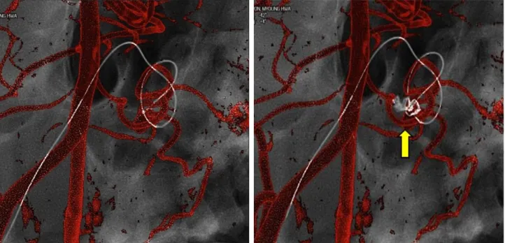
Fig. 3. Even though superselection of AICA orifice was not difficult, aneurysm selection was very difficult due to the tortuous course of AICA and relatively small diameter of AICA to accommodate microcatheter. The 2nd coil is extruded from the aneurysmal sac dur- ing coiling (arrow).
at the level of C2 vertebra. An Excelsior SL-10 micro- catheter (Boston Scientific, Natick, MA, United States) and X-pedion-14 microwire (ev3 Endovascular, Inc., Plymouth, MN, United States) were advanced to the right VA and the basilar artery. Under the roadmap guidance, aneurysm selection was tried. Even though the superselection of the AICA orifice was not diffi- cult, aneurysm selection was very difficult due to the tortuous course of AICA and relatively small diame- ter of the AICA for accommodation of the micro- catheter (Fig. 3).
After failed multiple trials of selecting the aneurysm with Excelsior SL-10 microcatheter and X-pedion-14 microwire, we changed the microwire to X-pedion-10, resulting in successful distal AICA aneurysm selection.
The aneurysmal sac was then obliterated completely without compromise of the AICA branch even though the second coil was extruded from the aneurysmal sac during coiling (Fig. 4). Postoperative CT showed slightly increased cerebellar hemorrhage, but there was no evidence of obstructive hydrocephalus. She was alert without motor weakness or brain stem sign.
However, she complained of hearing loss and tinnitus despite the patent flow to left AICA. She was dis- charged home at two weeks with remaining sensor- ineural hearing loss.
DISCUSSION
The anatomy of the AICA is highly variable.
Typically, the artery arises from the junction at the middle and the lower thirds of the basilar artery, courses along the pons and the middle cerebellar pe- duncle, to which it gives a few important perforating branches.8) The branches of the AICA that supply the inferior upper part of the olive arise 3~18 mm distal to the origin of AICA, whose occlusion could lead to a lateral inferior pontine syndrome.1)10)16)19)
Near the facial-vestibulocochlear complex, the AICA bifurcates into two major branches, called the rostro- lateral and caudomedial branch. The rostrolateral branch courses toward the internal auditory canal close to the seventh and the eighth cranial nerve com- plex and gives off the labyrinthine artery, also called
Fig. 4. The aneurysmal sac is obliterated completely without compromise of AICA branch even though 2nd coil is extruded from the aneurysmal sac during coiling (right image is the subtraction of coil). There is no occlusion or stenosis of distal AICA.
the internal auditory artery (IAA). The caudomedial branch courses medially close to the pons, to which it sends a few perforators, and terminates in cerebellar branches.11)13) The AICA also sends a few branches to the choroid plexus protruding from the foramen of Luschka.11) The hemispheric branches from the AICA frequently have anastomoses with the superior cer- ebellar artery and posterior inferior cerebellar artery.11)16) According to Zager et al.,20) if the aneur- ysm is located on the segment of the AICA that is distal to any branches coursing to the brainstem, dis- tal occlusion may be performed without neurologic sequel. However, that carries the risk of retrograde thrombosis, which can result in a devastating brain- stem infarct. Major complications can be prevented by antithrombotic therapy after the procedure.4) Smaltino et al.14) reported anatomical studies on the IAA, per- formed by Mezzogiorno and DeLula. In their study, the IAA arose from the AICA 87% of the time, and originated directly from the basilar artery (BA) in
10%. This vessel usually gives off one branch to the cerebellum before joining the internal auditory canal.
Therefore, it is called the cerebello-labyrinth artery.14) Wende et al.,18) using angiography of the IAA, found that in 45.4% of the cases, it arose from the AICA, in 24.4% from the superior cerebellar artery, and in 16%
directly from the basilar artery. In addition, it had an average lumen of only 200~300 μ. In our case, even though the IAA could not be visualized on fully mag- nified angiogram and microcatheter angiogram of the AICA, the IAA was presumed to originate from the distal AICA based on the sensorineural hearing loss occurring after embolization.
Distal AICA aneurysms are frequently dissecting in nature, thus usual treatment is direct surgical trapping.
Compared with a surgical approach of distal AICA aneurysm, endovascular treatment prevents massive bleeding and cerebellar injury during craniotomy and can spare the distal AICA without trapping.
Embolization without parent artery occlusion is diffi-
cult, and if the aneurysm originated from arterial dissection, it has a high risk of regrowth and rerupture.2)5)7) Choi et al.2) reported a distal AICA aneurysm at the meatal loop treated with parent ar- tery preservation. Even though the dome of the aneurysm was occluded successfully, that patient ex- perienced a recurrent hemorrhage from the recanal- ized aneurysm one month after the embolization. The parent artery at the proximal portion of the aneurysm was then occluded with excellent results.
However, in our case, the distal AICA aneurysm was treatable with endosaccular coil embolization, since it was a saccular aneurysm with a definite neck rather than a dissecting aneurysm. We performed suc- cessful coil embolization of the distal AICA aneurysm without compromise of the parent artery. However, our patient developed unilateral hearing loss. Ishii et al.4) reported that after coil embolization of a post- meatal AICA, their patient presented with a high grade sensorineural hearing loss, probably related to the sacrifice of the labyrinthine artery.
Perlman et al.12) showed that interruption of the blood supply to the cochlea caused immediate and complete loss of function and concluded that the hair cells were particularly vulnerable to ischemic injury.17) Takeda et al.17) used a gerbil model of cochlear ische- mia wherein animals were subjected to lethal cochlear ischemia for 15 minutes of extracranial occlusion of bilateral vertebral arteries. Hair cells on the basal turn are more vulnerable to ischemia than those at the apical turn,6) since the rate of oxygen consumption in basal turn is higher (approximately 2.5-fold) than at the ap- ical turn, and the aerobic metabolism more active.9)
In our case, the left AICA parent artery mean diam- eter was about 0.8 mm, while the proximal and distal diameter of Excelsior SL-10 microcatheter were about 0.8 and 0.56 mm, respectively. Because the AICA di- ameter could not accommodate the microcatheter, there was a high risk of vasospasm and catheter- ization failure if we remove the microcatheter during the procedure. Thus, the microcatheter was placed in
the AICA during the entire procedure time of two hours. We think that another reason for the ischemic change is the AICA traction due to its tortuous course. A possible reason for postoperative hearing loss, in spite of the patent distal two branches on postoperative angiogram, is hemodynamic ischemia to the cochlea hair cells.
CONCLUSIONS
Endovascular treatment of distal AICA aneurysm beyond the meatal loop is feasible for preserving the AICA flow. However, because the cochlear hair cell is vulnerable to ischemia, unilateral hearing loss can oc- cur, possibly from temporary reduction of the AICA flow by the microcatheter during endovascular treatment.
REFERENCES
1. Akar ZC, Dujovny M, Gomez-Tortosa E, Slavin KV, Ausman JI. Microvascular anatomy of the anterior sur- face of the medulla oblongata and olive. J Neurosurg.
1995 Jan;82(1):97-105.
2. Choi CH, Cho WH, Choi BK, Lee SW. Rerupture fol- lowing endovascular treatment for dissecting aneurysm of distal anterior inferior cerebellar artery with parent artery preservation: Retreatment by parent artery occlusion with Guglielmi detachable coils. Acta Neurochir (Wien) 2006 Mar;148(3):363-6; discussion 366.
3. Fukushima S, Hirohata M, Okamoto Y, Yamashita S, Ishida S, Shigemori M. Anterior inferior cerebellar artery dissecting aneurysm in a juvenile: Case report. Neurol Med Chir (Tokyo). 2009 Feb;49(2):81-4.
4. Ishii D, Takechi A, Shinagawa K, Sogabe T. Endovascular treatment for ruptured distal anterior inferior cerebellar artery aneurysm - Case report. Neurol Med Chir. 2010;50(5) :396-9.
5. Jinbo Y, Zheng Z, Jiaquan H, Hui Y, Jun L. Embolization of a ruptured aneurysm of the distal anterior inferior cerebellar artery with parent artery preservation. Neurol India. 2010 Sep-Oct;58(5):786-8.
6. Koga K, Hakuba N, Watanabe F, Shudou M, Nakagawa T, Gyo K. Transient cochlear ischemia causes delayed cell death in the organ of Corti: An experimental study in gerbils. J Comp Neurol. 2003 Feb;456(2):105-11.
7. Kusaka N, Maruo T, Nishiguchi M, Takayama K, Maeda Y, Ogihara K, et al. [Embolization for aneurismal dilata- tion associated with ruptured dissecting anterior inferior cerebellar artery aneurysm with preservation of the pa- rent artery: Case report]. No Shinkei Geka. 2006 Jul;34(7):729-34. Japanese.
8. Matsuyama T, Okuchi K, Norimoto K, Ueyama T.
Ruptured dissecting anterior inferior cerebellar artery aneurysm - Case report. Neurol Med Chir (Tokyo). 2002 May;42(5):214-6.
9. Mizukoshi O, Daly JF. Oxygen consumption in normal and kanamycin damaged cochleae. Acta Oto-laryngol.
1967 Jul;64(1):45-54.
10. Nishimoto A, Fujimoto S, Tsuchimoto S, Matsumoto Y, Tabuchi K, Higashi T. Anterior inferior cerebellar artery aneurysm. Report of three cases. J Neurosurg. 1983 Oct;59(4):697-702.
11. Peluso JP, van Rooij WJ, Sluzewski M, Beute GN. Distal aneurysms of cerebellar arteries: Incidence, clinical pre- sentation, and outcome of endovascular parent vessel occlusion. AJNR Am J Neuroradiol. 2007 Sep;28(8):1573-8.
12. Perlman HB, Kimura R, Fernandez C. Experiments on temporary obstruction of the internal auditory artery.
Laryngoscope. 1959 Jun;69(6):591-613.
13. Sarkar A, Link MJ. Distal anterior inferior cerebellar ar- tery aneurysm masquerading as a cerebellopontine angle tumor: Case report and review of literature. Skull Base.
2004 May;14(2):101-6; discussion 106-7.
14. Smaltino F, Bernini FP, Elefante R. Normal and patho- logical findings of the angiographic examination of the internal auditory artery. Neuroradiol. 1971 Sep;2(4):216-22.
15. Suzuki J, Hori S, Sakurai Y. Intracranial aneurysms in the neurosurgical clinics in Japan. J Neurosurg. 1971 Jul;35(1):34-9.
16. Suzuki K, Meguro K, Wada M, Fujita K, Nose T.
Embolization of a ruptured aneurysm of the distal ante- rior inferior cerebellar artery: Case report and review of the literature. Surg Neurol. 1999 May;51(5):509-12.
17. Takeda S, Hata R, Cao F, Yoshida T, Hakuba N, Hato N, et al. Ischemic tolerance in the cochlea. Neurosci Lett. 2009 Oct;462(3):263-6.
18. Wende S, Nakayama N, Schwerdtfeger P. The internal auditory artery: (embryology, anatomy, angiography, pathology). J Neurol. 1975 Aug;210(1):21-31.
19. Yokoyama S, Kadota K, Asakura T, Kawazoe K. Aneurysm of the distal anterior inferior cerebellar artery - Case report. Neurol Med Chir (Tokyo). 1995 Aug;35(8):587-90.
20. Zager EL, Shaver EG, Hurst RW, Flamm ES. Distal an- terior inferior cerebellar artery aneurysms. Report of four cases. J Neurosurg. 2002 Sep;97(3):692-6.