Received:June 27, 2018, Revised:July 11, 2018, Accepted:July 22, 2018 Corresponding to:Youn-Soo Hahn http://orcid.org/0000-0003-1332-6221
Department of Pediatrics, Chungbuk National University College of Medicine, 1 Sunhwan-ro, Seowon-gu, Cheongju 28644, Korea. E-mail:[email protected]
Copyright ⓒ 2018 by The Korean College of Rheumatology. All rights reserved.
This is a Open Access article, which permits unrestricted non-commerical use, distribution, and reproduction in any medium, provided the original work is properly cited.
Enthesitis-related Arthritis
Youn-Soo Hahn
Department of Pediatrics, Chungbuk National University College of Medicine, Cheongju, Korea
Enthesitis-related arthritis (ERA) is a disease predominantly affecting the joints and entheses of the lower extremities and has the potential to eventually affect the sacroiliac joints and spine evolving to juvenile ankylosing spondylitis. ERA is also charac- terized by rheumatoid factor seronegativity, paucity of antinuclear antibody, and a strong association with the human leukocyte antigen-B27. ERA accounts for a higher proportion of juvenile idiopathic arthritis (JIA) cases in the Asian population compared to other populations. Advances in the understanding of ERA pathogenesis continue to progress and have led to the development of new treatments targeting pro-inflammatory cytokines. In particular, tumor necrosis factor-α inhibitors have become a main- stay of therapy for patients in whom therapy with anti-inflammatory drugs and/or disease-modifying anti-rheumatic drugs are inadequate or contraindicated. Compared to other JIA subtypes, ERA is associated with a poorer quality of life, worse function, and a higher likelihood of ongoing active disease after the initial treatment. Because the current guidelines for the management of ERA is not considered separately from other categories of JIA, there is a need for treatment guidelines specific to ERA to im- prove the overall disease outcomes. (J Rheum Dis 2018;25:221-230)
Key Words. Juvenile arthritis, Enthesitis-related arthritis, Pathogenesis, Therapeutics, Prognosis
INTRODUCTION
Enthesitis-related arthritis (ERA) is a term introduced in the International League of Associations for Rheuma- tology (ILAR) classification of juvenile idiopathic arthri- tis (JIA) [1]. The definition of ERA in the ILAR classi- fication criteria for JIA is based on the inclusion and ex- clusion criteria (Table 1). ERA is characterized by the presence of predominantly lower limb arthritis and en- thesitis and can eventually affect the sacroiliac (SI) joints and spine evolving to juvenile ankylosing spondylitis (JAS). For this reason, identification of individuals with axial disease is required for early and appropriate ther- apeutic interventions. Both ERA and JAS are charac- terized by rheumatoid factor (RF) seronegativity, paucity of antinuclear antibody (ANA), and a strong association with the human leukocyte antigen-B27 (HLA-B27).
However, the fact that a radiologic evidence of bilateral SI joint inflammation is a prerequisite for the diagnosis of
JAS discriminates JAS from ERA. ERA and JAS may repre- sent the peripheral and axial forms of spondyloarthritis (SpA) based on the Assessment in Spondyloarthritis International Society (ASAS) classification system [2,3].
Epidemiological studies of ERA have been hampered by a lack of standardized criteria and case ascertainment, re- sulting in wide-ranging results [4-11]. The proportion of patients with ERA was 8.6%∼39.2% among children with JIA in previous published studies. A study with mul- ti-ethnic cohort reported that ERA was the commonest category of JIA in children of Asian descent [8], suggest- ing inherent genetic differences between Asian pop- ulation and other populations. The mean age at diagnosis of ERA is around 10∼15 years. There is a predominance of boys in ERA as reflected by high male-to-female ratios among cohorts from several countries. However, this gen- der-based difference in the prevalence of ERA may result from underestimation of the occurrence of diseases in girls.
This review will provide an update on the latest under-
Table 1. International League of Associations for Rheumatology (ILAR) classification criteria for enthesitis-related arthritis Arthritis and enthesitis or Arthritis or enthesitis with at least
two of the following:
• Sacroiliac joint tenderness and/or inflammatory spinal pain
• Presence of HLA-B27
• Onset of arthritis in a male over 6 years of age • Family history in at least one first-degree relative of
ankylosing spondylitis, Enthesitis-related arthritis, sacroiliitis with inflammatory bowel disease, reactive arthritis, or acute anterior uveitis
• Acute anterior uveitis Exclusions
• Psoriasis or a history of psoriasis in the patient or a first-degree relative
• Presence of IgM RF on at least two occasions at least 3 months apart
• Systemic JIA in the patient
HLA: human leukocyte antigen, RF: rheumatoid factor, JIA:
juvenile idiopathic arthritis.
standing regarding pathogenesis, clinical manifestations, treatments and expected outcomes in children with ERA.
MAIN SUBJECTS
Pathogenesis
The etiology of ERA is unknown. The role of enteric bac- teria in the pathogenesis of ERA has been an area of active investigation. In particular, the association between en- teric bacteria and ERA was reported by one study show- ing altered microbiota in children with ERA [12].
Elevation of a marker of intestinal inflammation such as fecal calprotectin in children with ERA indicates the link- age of ERA with enteric infectious conditions as well [13]. Interestingly, a complex interrelationship between enteric bacteria and HLA-B27 has been described in the HLA-B27 transgenic rat with a disease similar to AS [14,15] and human with AS [16,17]. Alteration in the repertoire of the gut microbiome by HLA-B27 is one pos- sible mechanism for this interaction [18].
HLA-B27 remains the major genetic factor in ERA. Of over 105 molecular subtypes of HLA-B27, HLA-B27*04 and B27*05 appear to be the most common subtypes in patients with ERA [19,20]. Although numerous theories about the pathogenic role of HLA-B27 have been pro- posed, the exact mechanism underlying the effect of HLA-B27 on disease susceptibility has still not been
determined. It has been proposed that molecular mimicry between a microbial antigen and the HLA-B27 molecule or peptides it presents brings about an HLA-B27-re- stricted cytotoxic T-cell response found only in joints and other affected tissues (arthritogenic peptide hypothesis) [21-23]. Another theory comes from misfolding of the HLA-B27 heavy chain in the endoplasmic reticulum [23-25]. As a result of this, the accumulation of misfolded protein leads to induction of the proinflammatory re- sponse and stimulates the release of proinflammatory cy- tokines through the interleukin (IL)-23/IL-17 axis. In ad- dition, an alternative concept implies that expression of HLA-B27 β2 microglobulin-free homodimers on the cell surface leads to activation of the killer cell immunoglob- ulin-like receptors (KIRs) such as KIR3DL1/2 and pro- motes the production of Th17 cytokines involved in the pathogenesis of SpA [26-28]. Besides HLA-B27, endo- plasmic reticulum aminopeptidase 1 (ERAP1) has been known to be associated with ERA, but not with other JIA subtypes [29]. However, its role in the pathogenesis in ERA has not been determined.
In contrast to other subtypes, ERA is not characterized by autoantibodies. Instead, high levels of mono- cyte-derived cytokines such as IL-1 and IL-6 in synovial fluid reflect the close relationship between the innate im- mune system and ERA [30]. Additionally, the increases of Th17 cells and γδ T cells in ERA patients suggest the con- tribution of these cells to the development of ERA [31-33].
Clinical manifestations 1) General features
ERA usually has insidious onset, but may begin abruptly [34]. Systemic symptoms including low-grade fever, fa- tigue, and rashes may develop although these symptoms are usually absent at onset. Intermittent musculoskeletal pain and stiffness of peripheral joints develop along with enthesitis mainly in knee, ankle and foot. Arthritis in the back (spondylitis) and at the SI joint (sacroiliitis) is not common at first, but can occur later [35,36].
2) Enthesitis
Enthesitis is defined as inflammation of an enthesis, which is a site where a tendon, ligament, or joint capsule attaches to bone. It is characterized by pain and swelling especially at knee, foot, or heel. The presence of enthesitis is helpful to differentiate ERA from other subtypes of JIA.
However, tenderness or pain at entheseal sites may occur
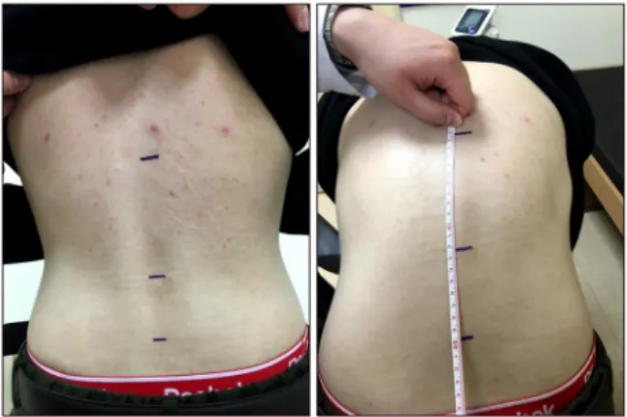
Figure 1. Modified Schober’s test. With patient standing up- right, two points are marked on the back 5 cm below and 10 cm above the horizontal line joining the dimples of Venus which is used as a mark for the lumbosacral junction. On max- imal forward flexion, keeping the knees straight, an increase of the distance between the top and bottom points reflects lum- bosacral spinal mobility and is considered normal if more than 5 cm.
in other rheumatic diseases including other JIA subtypes and even in children without diseases [37]. In addition, Osgood-Schlatter syndrome and fibromyalgia should be excluded because these conditions are also accompanied by similar symptoms [38]. Enthesitis can be documented by the careful examination of localized tenderness or swelling at the entheseal insertion sites. The most com- mon anatomic regions for enthesitis in children are the infrapatellar ligament insertion to patella, the plantar fas- cia and the Achilles tendon insertions to calcaneus, and the plantar fascia insertion to the metatarsal heads [39].
Avoidance of weight bearing on inflamed entheses can be detected during stance and gait tasks in patients with en- thesitis in the lower limb.
3) Peripheral arthritis
Peripheral arthritis in ERA is usually asymmetric and mainly involves large joints of lower extremities. Although polyarticular involvement may occur at onset or during the course of disease, oligoarticular disease is more com- mon in most instances [9,40]. Male sex, age at onset, and distribution of the affected joints may become useful tools in distinguishing ERA from other subtypes. The usually involved joints are knee, hips, ankles, and small joints of the foot and toes. Contrasting to other subtypes of JIA, hip involvement in ERA is relatively frequent [41].
Because the small joints of the hands are rarely affected, disease of these joints is less likely to be ERA [34].
Tarsitis, manifested as pain and tenderness in the mid- foot, is more common than other subtypes of JIA [42,43].
4) Axial arthritis
Involvement of spine and SI joints is relatively rare at disease onset, but may occur several years after onset [44]. Axial involvement may be asymptomatic [45-47], although it is manifested by pain particularly at the lower back and buttock areas or stiffness and limitation of mo- tion of axial skeleton. Prolonged periods of inactivity can aggravate these symptoms. Clinical examination may re- veal tenderness over SI joint, pain on flexion, abduction and external rotation of hip (Patrick or FABER test), re- stricted forward flexion (modified Schober’s test) (Figure 1), or abnormal spinal contour on maximal forward flexion.
Arthritis of the cervical spine may occur and cause severe damage [48].
5) Uveitis
The uveitis is the most common extra-articular manifes-
tations of ERA. It has been reported in around 7% of ERA patients [10,49]. Acute symptomatic anterior uveitis is characteristic of ERA and its common presentations in- clude ocular pain, redness of eye, and photophobia. It is usually unilateral and can occur repeatedly. In contrast to anterior uveitis seen in other JIA subtypes, the uveitis in ERA is more frequently symptomatic and seldom leaves any significant sequelae [34]. It may be present at diag- nosis, develop during the course of disease, or be an ini- tial manifestation usually detected in the course of rou- tine ophthalmologic examination.
Laboratory findings
There are no diagnostic laboratory tests for ERA. A com- plete blood count (CBC) with differential counts is a part of the initial evaluation of any child with a joint disease.
Mild anemia is a usual finding and suggests the presence of chronic disease. The number of white blood cells re- mains normal or increases with normal differential. The substantial elevation of inflammatory markers such as C-reactive protein or erythrocyte sedimentation rate may be noted, although these markers may be normal or only minimally increased even in patients with clinically active disease. RF are characteristically absent [38]. Compared with healthy population, the occurrence of ANAs is not more frequent in children with ERA [34]. Children with ERA were found to have higher levels of fecal calprotectin than those with other JIA subtypes [13,50]. While
Figure 2. Anteroposterior view of the pelvis shows joint space narrowing, bone erosions, and reactive sclerosis in the sacroil- iac joints.
Figure 3. Corner lesion in 15 year old male with en- thesitis-related arthritis. Plain radiograph (A) and T1-weighted magnetic resonance imaging (B) of spine show increased sig- nal at the anterior aspect of the superior endplate of L2 (arrows).
Figure 4. Achilles tendon enthesitis in a 14 year old boy. (A) Longitudinal grayscale ultrasound image demonstrates in- creased thickness of Achilles tendon (arrowheads) with hypo- echogenicity and loss of fibrillar pattern in deep layer. (B) Power Doppler images demonstrates increased signals within the tendon consistent with hyperemia.
HLA-B27 is present in more than 95% of adults with AS, a somewhat lower prevalence of HLA-B27 was reported in recent studies of children with SpA [51,52].
Radiologic findings 1) Plain radiographs
Although plain radiographs are limited by their inability to show inflammation or soft tissue changes, it can reveal subtle thickening of tendon and ligaments and a loss of the distinct margins at the tendon insertion site. In addi- tion, chronic bony changes such as enthesophyte for- mation or occasional erosions can be detected. Although radiographic evidence of sacroiliitis is often found several
years after the onset of peripheral arthritis, it may be the first radiologic finding. For radiologic evaluation of the SI joints, a standard anteroposterior view of the pelvis is usually obtained (Figure 2) and may demonstrate hazi- ness or a punched-out appearance of the cortical margins, widening of the joint space as a result of erosion of the subchondral bone, and joint sclerosis reflecting an osteo- blastic reaction. These findings are usually bilateral and symmetrical, although unilateral involvement can be seen initially. Radiological changes in the lumbosacral spine are less common and usually follow the abnormal- ities of the SI joints. The anterior corners of vertebral bod- ies may initially look bright due to periostitis with new bone formation (shiny corner sign) (Figure 3A) [38,53].
Subsequently, the anterior margin of the vertebral body loses its normal concavity and becomes flat or squared.
Formation of syndesmophyte, calcifications or hetero- topic ossifications inside a spinal ligament or of the annu- lus fibrosus, is less frequent in the pediatric population but can occur later during adulthood [54].
2) Ultrasonography with Doppler (USD)
Ultrasound with power Doppler is a highly useful and sensitive tool in the evaluation of clinical or subclinical enthesitis (Figure 4). It has the advantage of being non- invasive and easily accessible in the clinic, although it is highly operator-dependent and needs specialized training.
Figure 5. Coronal T1-weighted magnetic resonance imaging of pelvis in 15 year old male with enthesitis-related arthritis.
There is active sacroiliitis with bone marrow edema (arrows) on both iliac and sacral sides of the sacroiliac joints.
The common abnormalities of entheses detected by USD are hypoechoic (and/or thickened) tendon at its bony at- tachment and bony changes including enthesophytes, erosions or irregularity [55].
3) Magnetic resonance imaging (MRI)
Early as well as chronic inflammatory changes in the SI joints and the spine can be identified by MRI even in pa- tients with normal conventional radiographs. MRI with gadolinium enhancement is helpful to detect active syno- vitis [56,57], whereas MRI using short-tau inversion re- covery (STIR) images has an advantage of not requiring gadolinium administration in detecting bone marrow edema (BME) [58,59]. Subchondral or periarticular BME on STIR imaging or osteitis on T1 post-gadolinium im- ages of the SI joints is most indicative of active sacroiliitis (Figure 5) [58,60]. In addition, tendon or ligament thick- ening, adjacent soft-tissue swelling and edema, synovitis, enthesitis, and capsulitis around the SI joints and other sites within the pelvis can be detected by MRI. However, these findings are compatible with sacroiliitis but not suf- ficient for making a diagnosis of active sacroiliitis in the absence of bone marrow edema [61]. Because spinal in- volvement is less frequent in children and adolescents than that in adults, a spinal MRI is not usually required in these population unless there is a substantial possibility of spinal disease [58]. The typical findings in children with ERA are fatty lesions at the vertebral edge (corner le-
sions) (Figure 3B).
Treatment
1) General managements
Children with ERA should be managed by a team of health professionals who can provide patient and family education, medical treatment, physical therapy, and psy- chosocial support. The goal of therapy should be to re- lieve pain, preserve joint function, achieve disease re- mission, and prevent long-term damage caused by the disease or its treatment. An optimal management strat- egy includes differentiation or stratification of children according to the severity and risk of functional disability.
Although there is no medication that can cure the disease, prognosis of ERA has improved recently due to the prog- ress in treatment modalities. The current managements for ERA are based on evidences from studies in adults with AS or rheumatoid arthritis and children with other JIA subtypes. However, application of data from these populations to children with ERA may not be ideal, as evi- denced by recognized clinical differences between ERA and AS or other categories of JIA. In the 2011 American College of Rheumatology Recommendations for the treatment of JIA [62], the management of children with ERA is not considered separately from that for children with other categories of JIA and primarily based on the number of active joints and the presence of sacroiliitis.
For this reason, there is a critical need for a rational ther- apeutic strategy focused specifically on the ERA population.
The flow of treatment and the suggestions for the use of drugs based on the current recommendations for the treatment of patients with ERA are summarized in Figure 6.
2) Non-steroidal anti-inflammatory drugs (NSAIDs) NSAIDs are often the first line of therapy for relief of both axial and peripheral symptoms and also effective for enthesitis [63]. Although disease remission may be in- duced by continuous use of NSAIDs, use of NSAIDs as monotherapy for more than 2 months is discouraged if disease does not remit [62]. Among various NSAIDs, naproxen (15∼20 mg/day, maximum 500 mg/day) is usually selected as an initial medication because it has lower toxicity and can be administered using a convenient twice daily regimen. Toxicity monitoring includes CBC, creatinine measurement, liver function test (LFT), and urinalysis every 4∼6 weeks after initiation of treatment and then every 6∼12 months thereafter.
Figure 6. Flow of treatments for patients with enthesitis-related arthritis. NSAIDs: non-steroidal anti-inflammatory drugs, IA: in- traarticular, SSZ: sulfasalazine, MTX: methotrexate, TNF: tumor necrosis factor.
3) Glucocorticoids
Corticosteroids can be used as a short-term oral or intra- venous therapy for an acute flare and as topical treatment of acute uveitis. In addition, intraarticular administration of corticosteroids is recommended for the localized joint disease to rapidly relieve joint symptoms, to restore func- tion, and to avoid systemic use of corticosteroids [64-66].
Triamcinolone hexacetonide (for large joints, 1 mg/kg, maximum 40 mg/dose; for smaller joints, 0.5 mg/kg, maximum 20 mg/dose) is used as the drug of choice for intraarticular steroid injections. Local injections of corti- costeroids at sites of enthesitis may provide relief of symptoms but should be used cautiously due to the in- creased risk of tendon rupture [34].
4) Disease-modifying anti-rheumatic drugs (DMARDs) Although DMARDs have shown efficacy for peripheral joint disease, they have not been proven effective for axial disease or enthesopathy [42,67,68]. For this reason, DMARDs are not recommended for patients with purely axial disease [66]. However, DMARDs may be used in ex- ceptional situations of axial symptoms in which there is no other pharmacological treatment option left for a par- ticular patient for reasons of toxicity, contraindications or costs. In clinical studies of children with SpA, sulfasala- zine (30∼50 mg/kg/day, maximum 3 g/day) improved both doctor and patient assessments and was found to be effective and safe compared to placebo [69,70]. Clinical improvement is usually expected several weeks after ini- tiation of sulfasalazine. Because side effects of sulfasala-
zine include bone marrow suppression and hepatotox- icity, CBC and LFT must be regularly monitored. Despite absence of controlled studies in children with ERA, me- thotrexate or leflunomide can be considered for the man- agement of peripheral disease of ERA in case of no re- sponse to sulfasalazine.
5) Biologic agents
Biologic agents blocking tumor necrosis factor (TNF)-α (TNF-α inhibitors) have offered a powerful therapeutic option for the management of patients with ERA. They have demonstrated efficacy for peripheral disease re- fractory to NSAIDs and DMARDs as well as symptomatic treatment of axial disease [71-74]. However, the role of TNF-α inhibitors in impeding progression of structural damage in children remains to be elucidated. If a patient is in sustained remission, tapering of TNF-α inhibitors can be considered [66,75,76]. However, complete dis- continuation of TNF-α inhibitors seems to lead to a high percentage of patients that experience flares [77,78]. The choice of TNF-α inhibitors can be made according to pa- tient preference and associated extra-articular manifes- tations including uveitis. It is required to perform base- line screening for tuberculosis (tuberculin skin test or in- terferon-γ release assays) before commencing treatment with the TNF-α inhibitor. The frequently recommended TNF-α inhibitors include etanercept (0.8 mg/kg/week, subcutaneously, maximum 50 mg), adalimumab (20 mg or 40 mg subcutaneously every other week for weight less or greater than 30 kg, respectively), and infliximab (5
mg/kg initial loading every 2 weeks for 3 infusions and then every 8 weeks). So far, the evidence for the efficacy of other biologic agents such as rituximab, anakinra, abata- cept, tocilizumab, ustekinumab (anti IL-12/23 human monoclonal antibody), and secukinumab (anti-IL-17A antibody) is insufficient to support the use of these medi- cations in patients with ERA.
Prognosis
Children and adolescents with ERA shows variable course and prognosis. The disease remission within five years of diagnosis has been reported in less than 20% of patients [79], but its rate will likely increase with the ad- vent of additional biologic therapies. The prognosis of ERA appears to be worse than those of other JIA catego- ries [9,80-82]. Thus, ERA is usually associated with worse physical function, poorer quality of life, higher pain scores, and a higher possibility of ongoing active disease after initial treatment compared with other JIA subtypes.
Family history of AS, disease onset after 8 years of age, HLA-DRB1*08, HLA-B27, ankle and hip arthritis within the first 6 months of disease, and tarsitis are associated with a poorer prognosis [36,47,82]. ERA can progress to AS within 10 years of disease onset [36,44].
CONCLUSION
ERA is characterized by the presence of arthritis and en- thesitis predominantly at the lower extremities and has a potential to eventually affect the SI joints and spine evolv- ing to JAS. Its most common extra-articular manifes- tation is uveitis. Advances in the understanding of ERA pathogenesis continue to evolve and have led to the devel- opment of new treatments targeting pro-inflammatory cytokines. In particular, the TNF-α inhibitors become a mainstay of therapy for patients in whom therapy with NSAID and/or DMARDs is inadequate or contraindicated.
Because the current guidelines for management of ERA is not considered separately from other categories of JIA, there is a need for treatment guidelines specific for ERA to improve the overall course of disease.
CONFLICT OF INTEREST
No potential conflict of interest relevant to this article was reported.
REFERENCES
1. Petty RE, Southwood TR, Manners P, Baum J, Glass DN, Goldenberg J, et al. International League of Associations for Rheumatology classification of juvenile idiopathic arthritis:
second revision, Edmonton, 2001. J Rheumatol 2004;31:
390-2.
2. Rudwaleit M, van der Heijde D, Landewé R, Listing J, Akkoc N, Brandt J, et al. The development of Assessment of SpondyloArthritis international Society classification cri- teria for axial spondyloarthritis (part II): validation and final selection. Ann Rheum Dis 2009;68:777-83.
3. Rudwaleit M, van der Heijde D, Landewé R, Akkoc N, Brandt J, Chou CT, et al. The Assessment of Spondylo- Arthritis International Society classification criteria for pe- ripheral spondyloarthritis and for spondyloarthritis in general. Ann Rheum Dis 2011;70:25-31.
4. Boiu S, Marniga E, Bader-Meunier B, Mouy R, Compeyrot- Lacassagne S, Quartier P, et al. Functional status in severe juvenile idiopathic arthritis in the biologic treatment era: an assessment in a French paediatric rheumatology referral centre. Rheumatology (Oxford) 2012;51:1285-92.
5. Demirkaya E, Ozen S, Bilginer Y, Ayaz NA, Makay BB, Unsal E, et al. The distribution of juvenile idiopathic arthri- tis in the eastern Mediterranean: results from the registry of the Turkish Paediatric Rheumatology Association. Clin Exp Rheumatol 2011;29:111-6.
6. Modesto C, Antón J, Rodriguez B, Bou R, Arnal C, Ros J, et al. Incidence and prevalence of juvenile idiopathic arthritis in Catalonia (Spain). Scand J Rheumatol 2010;39:472-9.
7. Solau-Gervais E, Robin C, Gambert C, Troller S, Danner S, Gombert B, et al. Prevalence and distribution of juvenile idi- opathic arthritis in a region of Western France. Joint Bone Spine 2010;77:47-9.
8. Saurenmann RK, Rose JB, Tyrrell P, Feldman BM, Laxer RM, Schneider R, et al. Epidemiology of juvenile idiopathic arthritis in a multiethnic cohort: ethnicity as a risk factor.
Arthritis Rheum 2007;56:1974-84.
9. Weiss PF, Beukelman T, Schanberg LE, Kimura Y, Colbert RA; CARRA Registry Investigators. Enthesitis-related ar- thritis is associated with higher pain intensity and poorer health status in comparison with other categories of juve- nile idiopathic arthritis: the Childhood Arthritis and Rheumatology Research Alliance Registry. J Rheumatol 2012;39:2341-51.
10. Yu HH, Chen PC, Wang LC, Lee JH, Lin YT, Yang YH, et al.
Juvenile idiopathic arthritis-associated uveitis: a nation- wide population-based study in Taiwan. PLoS ONE 2013;
8:e70625.
11. Gmuca S, Xiao R, Brandon TG, Pagnini I, Wright TB, Beukelman T, et al. Multicenter inception cohort of en- thesitis-related arthritis: variation in disease characteristics and treatment approaches. Arthritis Res Ther 2017;19:84.
12. Stoll ML, Kumar R, Morrow CD, Lefkowitz EJ, Cui X, Genin A, et al. Altered microbiota associated with abnormal hu- moral immune responses to commensal organisms in en- thesitis-related arthritis. Arthritis Res Ther 2014;16:486.
13. Stoll ML, Patel AS, Punaro M, Dempsey-Robertson M. MR enterography to evaluate sub-clinical intestinal inflammation in children with spondyloarthritis. Pediatr Rheumatol
Online J 2012;10:6.
14. Hammer RE, Maika SD, Richardson JA, Tang JP, Taurog JD.
Spontaneous inflammatory disease in transgenic rats ex- pressing HLA-B27 and human beta 2m: an animal model of HLA-B27-associated human disorders. Cell 1990;63:1099- 112.
15. Breban M, Hammer RE, Richardson JA, Taurog JD. Transfer of the inflammatory disease of HLA-B27 transgenic rats by bone marrow engraftment. J Exp Med 1993;178:1607-16.
16. Geczy AF, Seager K, Bashir HV, de Vere-Tyndall A, Edmonds J. The role of Klebsiella in the pathogenesis of ankylosing spondylitis. II Evidence for a specific B27-associated marker on the lymphocytes of patients with ankylosing spondylitis.
J Clin Lab Immunol 1980;3:23-8.
17. Cameron FH, Russell PJ, Easter JF, Wakefield D, March L.
Failure of Klebsiella pneumoniae antibodies to cross-react with peripheral blood mononuclear cells from patients with ankylosing spondylitis. Arthritis Rheum 1987;30:300-5.
18. Rosenbaum JT, Davey MP. Time for a gut check: evidence for the hypothesis that HLA-B27 predisposes to ankylosing spondylitis by altering the microbiome. Arthritis Rheum 2011;63:3195-8.
19. Stanevicha V, Eglite J, Zavadska D, Sochnevs A, Lazareva A, Guseinova D, et al. HLA B27 allele types in homogeneous groups of juvenile idiopathic arthritis patients in Latvia.
Pediatr Rheumatol Online J 2010;8:26.
20. Srivastava R, Agnihotry S, Aggarwal R, Bajpai P, Aggarwal A. HLA-B27 subtypes in enthesitis-related arthritis cat- egory of juvenile idiopathic arthritis and ankylosing spondy- litis in northern India. Clin Exp Rheumatol 2015;33:931-5.
21. Braun J, Bollow M, Neure L, Seipelt E, Seyrekbasan F, Herbst H, et al. Use of immunohistologic and in situ hybrid- ization techniques in the examination of sacroiliac joint bi- opsy specimens from patients with ankylosing spondylitis.
Arthritis Rheum 1995;38:499-505.
22. Colbert RA. The immunobiology of HLA-B27: variations on a theme. Curr Mol Med 2004;4:21-30.
23. Bowness P. HLA-B27. Annu Rev Immunol 2015;33:29-48.
24. Chatzikyriakidou A, Voulgari PV, Drosos AA. What is the role of HLA-B27 in spondyloarthropathies? Autoimmun Rev 2011;10:464-8.
25. Colbert RA, Tran TM, Layh-Schmitt G. HLA-B27 misfolding and ankylosing spondylitis. Mol Immunol 2014;57:44-51.
26. Kollnberger S, Bird L, Sun MY, Retiere C, Braud VM, McMichael A, et al. Cell-surface expression and immune re- ceptor recognition of HLA-B27 homodimers. Arthritis Rheum 2002;46:2972-82.
27. Bowness P, Ridley A, Shaw J, Chan AT, Wong-Baeza I, Fleming M, et al. Th17 cells expressing KIR3DL2+ and re- sponsive to HLA-B27 homodimers are increased in ankylos- ing spondylitis. J Immunol 2011;186:2672-80.
28. Kollnberger S, Bowness P. The role of B27 heavy chain dim- er immune receptor interactions in spondyloarthritis. Adv Exp Med Biol 2009;649:277-85.
29. Hinks A, Martin P, Flynn E, Eyre S, Packham J. Subtype spe- cific genetic associations for juvenile idiopathic arthritis:
ERAP1 with the enthesitis related arthritis subtype and IL23R with juvenile psoriatic arthritis. Arthritis Res Ther 2011;13:R12.
30. Saxena N, Aggarwal A, Misra R. Elevated concentrations of monocyte derived cytokines in synovial fluid of children
with enthesitis related arthritis and polyarticular types of ju- venile idiopathic arthritis. J Rheumatol 2005;32:1349-53.
31. Mahendra A, Misra R, Aggarwal A. Th1 and Th17 predom- inance in the enthesitis-related arthritis form of juvenile idi- opathic arthritis. J Rheumatol 2009;36:1730-6.
32. Gaur P, Misra R, Aggarwal A. Natural killer cell and gamma delta T cell alterations in enthesitis related arthritis category of juvenile idiopathic arthritis. Clin Immunol 2015;
161:163-9.
33. Weiss PF. Update on enthesitis-related arthritis. Curr Opin Rheumatol 2016;28:530-6.
34. Petty RE, Laxer RM, Lindsey CB, Wedderburn L. Textbook of pediatric rheumatology. 7th ed. Amsterdam, Elsevier, 2016, p. 238-55.
35. Burgos-Vargas R, Vazquez-Mellado J. The early clinical rec- ognition of juvenile-onset ankylosing spondylitis and its dif- ferentiation from juvenile rheumatoid arthritis. Arthritis Rheum 1995;38:835-44.
36. Flatø B, Hoffmann-Vold AM, Reiff A, Førre Ø, Lien G, Vinje O. Long-term outcome and prognostic factors in enthesitis- related arthritis: a case-control study. Arthritis Rheum 2006;54:3573-82.
37. Sherry DD, Sapp LR. Enthesalgia in childhood: site-specific tenderness in healthy subjects and in patients with sero- negative enthesopathic arthropathy. J Rheumatol 2003;30:
1335-40.
38. Weiss PF. Diagnosis and treatment of enthesitis-related arthritis. Adolesc Health Med Ther 2012;2012:67-74.
39. Aggarwal A, Misra DP. Enthesitis-related arthritis. Clin Rheumatol 2015;34:1839-46.
40. Oen K, Duffy CM, Tse SM, Ramsey S, Ellsworth J, Chédeville G, et al. Early outcomes and improvement of pa- tients with juvenile idiopathic arthritis enrolled in a Canadian multicenter inception cohort. Arthritis Care Res (Hoboken) 2010;62:527-36.
41. Rostom S, Amine B, Bensabbah R, Abouqal R, Hajjaj- Hassouni N. Hip involvement in juvenile idiopathic arthritis. Clin Rheumatol 2008;27:791-4.
42. Ramanathan A, Srinivasulu H, Colbert RA. Update on juve- nile spondyloarthropathy. Rheum Dis Clin N Am 2013;
39:767-88.
43. Alvarez-Madrid C, Merino R, De Inocencio J, García-Consuegra J. Tarsitis as an initial manifestation of juvenile spondy- loarthropathy. Clin Exp Rheumatol 2009;27:691-4.
44. Minden K, Niewerth M, Listing J, Biedermann T, Bollow M, Schöntube M, et al. Long-term outcome in patients with ju- venile idiopathic arthritis. Arthritis Rheum 2002;46:
2392-401.
45. Chen HA, Chen CH, Liao HT, Lin YJ, Chen PC, Chen WS, et al. Clinical, functional, and radiographic differences among juvenile-onset, adult-onset, and late-onset ankylosing spondylitis. J Rheumatol 2012;39:1013-8.
46. Weiss PF, Xiao R, Biko DM, Chauvin NA. Sacroiliitis at di- agnosis of juvenile spondyloarthritis assessed by radiog- raphy, magnetic resonance imaging, and clinical examination.
Arthritis Care Res (Hoboken) 2016;68:187-94.
47. Stoll ML, Bhore R, Dempsey-Robertson M, Punaro M.
Spondyloarthritis in a pediatric population: risk factors for sacroiliitis. J Rheumatol 2010;37:2402-8.
48. El Maghraoui A, Bensabbah R, Bahiri R, Bezza A, Guedira N, Hajjaj-Hassouni N. Cervical spine involvement in ankylos-
ing spondylitis. Clin Rheumatol 2003;22:94-8.
49. Heiligenhaus A, Niewerth M, Ganser G, Heinz C, Minden K;
German Uveitis in Childhood Study Group. Prevalence and complications of uveitis in juvenile idiopathic arthritis in a population-based nation-wide study in Germany: suggested modification of the current screening guidelines. Rheum- atology (Oxford) 2007;46:1015-9.
50. Stoll ML, Punaro M, Patel AS. Fecal calprotectin in children with the enthesitis-related arthritis subtype of juvenile idio- pathic arthritis. J Rheumatol 2011;38:2274-5.
51. Weiss PF, Klink AJ, Behrens EM, Sherry DD, Finkel TH, Feudtner C, et al. Enthesitis in an inception cohort of en- thesitis-related arthritis. Arthritis Care Res (Hoboken) 2011;63:1307-12.
52. Weiss PF, Colbert RA, Xiao R, Feudtner C, Beukelman T, DeWitt EM, et al. Development and retrospective validation of the juvenile spondyloarthritis disease activity index.
Arthritis Care Res (Hoboken) 2014;66:1775-82.
53. Schueller-Weidekamm C. [Inflammatory spinal disease:
Spondyloarthritis: Importance of imaging]. Radiologe 2015;55:337-46. German.
54. Baek HJ, Shin KC, Lee YJ, Kang SW, Lee EB, Yoo CD, et al.
Juvenile onset ankylosing spondylitis (JAS) has less severe spinal disease course than adult onset ankylosing spondyli- tis (AAS): clinical comparison between JAS and AAS in Korea. J Rheumatol 2002;29:1780-5.
55. Eder L, Barzilai M, Peled N, Gladman DD, Zisman D. The use of ultrasound for the assessment of enthesitis in pa- tients with spondyloarthritis. Clin Radiol 2013;68:219-23.
56. Kim HK, Zbojniewicz AM, Merrow AC, Cheon JE, Kim IO, Emery KH. MR findings of synovial disease in children and young adults: Part 1. Pediatr Radiol 2011;41:495-511.
57. Jaganathan S, Goyal A, Gadodia A, Rastogi S, Mittal R, Gamanagatti S. Spectrum of synovial pathologies: a pictorial assay. Curr Probl Diagn Radiol 2012;41:30-42.
58. Mandl P, Navarro-Compán V, Terslev L, Aegerter P, van der Heijde D, D'Agostino MA, et al. EULAR recommendations for the use of imaging in the diagnosis and management of spondyloarthritis in clinical practice. Ann Rheum Dis 2015;
74:1327-39.
59. Sung S, Kim HS, Kwon JW. MRI assessment of sacroiliitis for the diagnosis of axial spondyloarthropathy: comparison of fat-saturated T2, STIR and contrast-enhanced sequences.
Br J Radiol 2017;90:20170090.
60. Rudwaleit M, Jurik AG, Hermann KG, Landewé R, van der Heijde D, Baraliakos X, et al. Defining active sacroiliitis on magnetic resonance imaging (MRI) for classification of axial spondyloarthritis: a consensual approach by the ASAS/
OMERACT MRI group. Ann Rheum Dis 2009;68:1520-7.
61. Sieper J, Rudwaleit M, Baraliakos X, Brandt J, Braun J, Burgos-Vargas R, et al. The Assessment of Spondylo- Arthritis international Society (ASAS) handbook: a guide to assess spondyloarthritis. Ann Rheum Dis 2009;68 Suppl 2:ii1-44.
62. Beukelman T, Patkar NM, Saag KG, Tolleson-Rinehart S, Cron RQ, DeWitt EM, et al. 2011 American College of Rheumatology recommendations for the treatment of juve- nile idiopathic arthritis: initiation and safety monitoring of therapeutic agents for the treatment of arthritis and sys- temic features. Arthritis Care Res (Hoboken) 2011;63:
465-82.
63. Gmuca S, Weiss PF. Evaluation and Treatment of Childhood Enthesitis-Related Arthritis. Curr Treatm Opt Rheumatol 2015;1:350-64.
64. Huppertz HI, Tschammler A, Horwitz AE, Schwab KO.
Intraarticular corticosteroids for chronic arthritis in chil- dren: efficacy and effects on cartilage and growth. J Pediatr 1995;127:317-21.
65. Allen RC, Gross KR, Laxer RM, Malleson PN, Beauchamp RD, Petty RE. Intraarticular triamcinolone hexacetonide in the management of chronic arthritis in children. Arthritis Rheum 1986;29:997-1001.
66. van der Heijde D, Ramiro S, Landewé R, Baraliakos X, Van den Bosch F, Sepriano A, et al. 2016 update of the ASAS-EULAR management recommendations for axial spondyloarthritis. Ann Rheum Dis 2017;76:978-91.
67. Gmuca S, Weiss PF. Juvenile spondyloarthritis. Curr Opin Rheumatol 2015;27:364-72.
68. Dougados M, Baeten D. Spondyloarthritis. Lancet 2011;
377:2127-37.
69. van Rossum MA, Fiselier TJ, Franssen MJ, Zwinderman AH, ten Cate R, van Suijlekom-Smit LW, et al. Sulfasalazine in the treatment of juvenile chronic arthritis: a randomized, double-blind, placebo-controlled, multicenter study. Dutch Juvenile Chronic Arthritis Study Group. Arthritis Rheum 1998;41:808-16.
70. Burgos-Vargas R, Vázquez-Mellado J, Pacheco-Tena C, Hernández-Garduño A, Goycochea-Robles MV. A 26 week randomised, double blind, placebo controlled exploratory study of sulfasalazine in juvenile onset spondyloarthro- pathies. Ann Rheum Dis 2002;61:941-2.
71. Henrickson M, Reiff A. Prolonged efficacy of etanercept in refractory enthesitis-related arthritis. J Rheumatol 2004;31:
2055-61.
72. Horneff G, De Bock F, Foeldvari I, Girschick HJ, Michels H, Moebius D, et al. Safety and efficacy of combination of eta- nercept and methotrexate compared to treatment with eta- nercept only in patients with juvenile idiopathic arthritis (JIA): preliminary data from the German JIA Registry. Ann Rheum Dis 2009;68:519-25.
73. Otten MH, Prince FH, Twilt M, Ten Cate R, Armbrust W, Hoppenreijs EP, et al. Tumor necrosis factor-blocking agents for children with enthesitis-related arthritis--data from the dutch arthritis and biologicals in children register, 1999-2010. J Rheumatol 2011;38:2258-63.
74. Horneff G, Fitter S, Foeldvari I, Minden K, Kuemmerle- Deschner J, Tzaribacev N, et al. Double-blind, placebo-con- trolled randomized trial with adalimumab for treatment of juvenile onset ankylosing spondylitis (JoAS): significant short term improvement. Arthritis Res Ther 2012;14:R230.
75. Yates M, Hamilton LE, Elender F, Dean L, Doll H, MacGregor AJ, et al. Is Etanercept 25 mg once weekly as ef- fective as 50 mg at maintaining response in patients with an- kylosing spondylitis? A randomized control trial. J Rheumatol 2015;42:1177-85.
76. Cantini F, Niccoli L, Cassarà E, Kaloudi O, Nannini C.
Duration of remission after halving of the etanercept dose in patients with ankylosing spondylitis: a randomized, pro- spective, long-term, follow-up study. Biologics 2013;7:1-6.
77. Song IH, Althoff CE, Haibel H, Hermann KG, Poddubnyy D, Listing J, et al. Frequency and duration of drug-free re- mission after 1 year of treatment with etanercept versus sul-
fasalazine in early axial spondyloarthritis: 2 year data of the ESTHER trial. Ann Rheum Dis 2012;71:1212-5.
78. Haibel H, Heldmann F, Braun J, Listing J, Kupper H, Sieper J. Long-term efficacy of adalimumab after drug withdrawal and retreatment in patients with active non-radio- graphically evident axial spondyloarthritis who experience a flare. Arthritis Rheum 2013;65:2211-3.
79. Flatø B, Aasland A, Vinje O, Forre O. Outcome and pre- dictive factors in juvenile rheumatoid arthritis and juvenile spondyloarthropathy. J Rheumatol 1998;25:366-75.
80. Selvaag AM, Lien G, Sørskaar D, Vinje O, Førre Ø, Flatø B.
Early disease course and predictors of disability in juvenile
rheumatoid arthritis and juvenile spondyloarthropathy: a 3 year prospective study. J Rheumatol 2005;32:1122-30.
81. Sarma PK, Misra R, Aggarwal A. Outcome in patients with enthesitis related arthritis (ERA): juvenile arthritis damage index (JADI) and functional status. Pediatr Rheumatol Online J 2008;6:18.
82. Flatø B, Smerdel A, Johnston V, Lien G, Dale K, Vinje O, et al. The influence of patient characteristics, disease variables, and HLA alleles on the development of radiographically evi- dent sacroiliitis in juvenile idiopathic arthritis. Arthritis Rheum 2002;46:986-94.