Evaluation of soft tissue asymmetry using cone-beam computed tomography after open reduction and internal fixation of
zygomaticomaxillary complex fracture
Dong Hyuck Kim1,3, Rae Hyong Kim3, Jun Lee1,2, Young Deok Chee1,2, Kyoung-Hwan Kwon1,2
1Department of Oral and Maxillofacial Surgery, College of Dentistry, Wonkwang University, 2Wonkwang Dental Research Institute,
3Department of Oral and Maxillofacial Surgery, Graduate School of Dentistry, Wonkwang University, Iksan, Korea
Abstract(J Korean Assoc Oral Maxillofac Surg 2014;40:103-110)
Objectives: In this study, we assessed soft tissue asymmetry that occurred after open reduction of unilateral zygomaticomaxillary complex (ZMC) fractures. We proposed a simple method to assess soft tissue asymmetry after reduction surgery by evaluating the symmetry between the affected and the unaffected sides. The factors affecting soft tissue contour after surgery were also analyzed.
Materials and Methods: Subjects included patients admitted to Wonkwang University Dental Hospital from 2008 to 2013. Cone-beam computed tomography (CBCT) images of asymmetric patients who underwent open reduction at least 3 months prior were compared with healthy patients.
Results: The degree of asymmetry was measured in both the open reduction and control groups. Landmarks that showed a statistically significant difference between the two groups were zygion (1.73±0.24 mm), bucclae (1.08±0.26 mm), point of cheek (2.05±0.33 mm) and frontozygomatic point (1.30±0.31 mm).
Conclusion: When compared with the normal group, asymmetry can occur in the affected side, which usually shows depression of overlying soft tis- sue and is statistically significantly different. Evaluation of soft tissue asymmetry with CBCT images after open reduction of ZMC fracture is useful.
Key words: Zygomatic fractures, Facial asymmetry
[paper submitted 2014. 4. 21 / revised 2014. 5. 18 / accepted 2014. 5. 19]
Toriumi et al.3 have described complication of the ZMC in- jury such as asymmetry of zygoma, trismus, diplopia, exoph- thalmos, enophthalmos, limitation of eye movement and hy- poesthesia due to injury of the infraorbital nerve. Reduction accuracy of fractured bone fragments were the major focus of most studies on facial asymmetry resulting from zygomatic asymmetry other than eye ball symptoms4,5. When bone frag- ments were precisely reduced, no asymmetry was reported1.
Ellis et al.6 reported that facial asymmetry which did not exceed 2 mm was difficult to perceive by experienced clini- cians and most asymmetries after reduction of ZMC were ac- ceptable. Unfortunately, the ZMC fracture pattern is diverse and soft tissue change after reduction surgery is difficult to predict. In addition, perceiving facial changes accurately is difficult due to the lack of information on patient’s face be- fore the injury. Gaziri et al.7 evaluated asymmetry of the hard tissue after ZMC fracture by comparing the affected with the unaffected side. Patients often complain of soft tissue asym- metry when comparing with the contralateral side of the face even when bone fragments were accurately reduced. There-
I. Introduction
Fracture of the zygomaticomaxillary complex (ZMC) is one of the most common facial injuries associated with man- dibular fracture1,2. Treatment of ZMC injury has improved due to various reduction methods and development of mini- plates and screws. The ZMC fracture treatment consists of the reduction and fixation of the dislocated bone fragments to their original location. Most ZMC bones are successfully repositioned with open reduction such as intraoral and trans- conjunctival approach or closed reduction such as Gillie’s approach1.
Kyoung-Hwan Kwon
Department of Oral and Maxillofacial Surgery, Wonkwang University Dental Hospital, 895 Muwang-ro, Iksan 570-711, Korea
TEL: +82-63-859-2921 FAX: +82-63-857-4002 E-mail: [email protected]
This is an open-access article distributed under the terms of the Creative Commons Attribution Non-Commercial License (http://creativecommons.org/licenses/by-nc/3.0/), which permits unrestricted non-commercial use, distribution, and reproduction in any medium, provided the original work is properly cited.
CC
Copyright Ⓒ 2014 The Korean Association of Oral and Maxillofacial Surgeons. All rights reserved.
the reduction surgery. Inclusion criteria for the patient group were lack of previous ZMC or maxilla fracture, the patient did not complain of asymmetry before the injury, diagnosis of unilateral ZMC fracture, and patient had open reduction surgery after the injury.
Inclusion criteria for the control group were absence of previous hard tissue surgery in the head and neck region and absence of facial asymmetry.
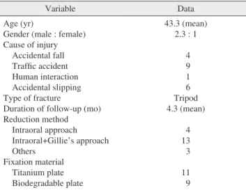
Intraoral, Gillie’s and transconjunctival approaches were used for open reduction. Bone fragments were fixated with titanium and biodegradable plates. Fixation locations were zygomaticomaxillary buttress, frontozygomatic buttress and infraorbital rim.(Table 1)
2. Materials
1) Image acquisition
In this study, we used images obtained using the CBCT (The Alphad VEGA; Asahi Roentgen, Kyoto, Japan) system at Wonkwang University Dental Hospital. Images were taken under the following settings: field of view, 200×179 mm; 80 kV; 5.00 mA; exposure time, 17 seconds, voxel size, 0.39 mm; slice thickness, 1.00 mm.
2) Construction of 3D planes
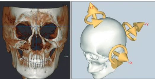
CT images were analyzed using 3D software (OnDe- mand3D; Cybermed Inc., Seoul, Korea) from DICOM format data. 3D coordinates were set up for each subject and cepha- lometric landmarks were designated. Nasofrontozygomatic (NFZ) plane was used as a reference plane of the skull base.
NFZ plane was composed of the right and left frontozygo- matic points and nasion. The coordinate origin (0, 0, 0) was set on N. Based on the origin, coordinates were constructed as x, y, z planes. X-axis (transverse axis) was a line parallel to the frontozygomatic (FZ) line. Y-axis (anteroposterior axis) was a line perpendicular to the FZ line and parallel to the right Frankfort horizontal (R FH) plane. The Z-axis was per- pendicular to both FZ line and R FH plane. Using this coordi- nate system, three planes were defined. Midsagittal plane was defined as a plane perpendicular to R FH plane and NFZ line while passing through the origin. Horizontal plane (Frankfort plane) was defined as a plane passing through right porion (R Po), right orbitale (R Or), and left orbitale (L Or). Coronal plane was defined as a plane perpendicular to horizontal and midsagittal planes and passing through the origin.(Fig. 1) fore, assessment of asymmetry should be based on the unaf-
fected side of the face after surgery.
Regarding assessment of facial asymmetry, previous stud- ies evaluated facial analysis methods using plain X-ray, 3-dimensional (3D) computed tomography (CT), cone-beam CT (CBCT) and laser scan8-18. However, most of the analyses were related with orthognathic surgery or craniofacial defor- mity. Evaluating asymmetry after reduction surgery where the distance of bone fragments was unplanned is difficult.
Moreover, use of laser scan is limited due to high cost.
In this study, we assessed soft tissue asymmetry that oc- curred after open reduction of unilateral ZMC fractures. Soft tissue asymmetry of the affected side was quantitatively com- pared with the unaffected side using 3D CBCT scans. Ten- dency of soft tissue asymmetry after reduction was analyzed and usefulness of CBCT was also investigated. This study was authorized by the Wonkwang University Dental Hospital (WKDIRB 201404-01).
II. Materials and Methods
1. Subjects
A total of 60 adults were chosen to participate in this study.
The patient group consisted of 20 subjects who had open re- duction surgery after being admitted to the emergency room of Wonkwang University Dental Hospital or medical hos- pital due to ZMC fracture. The control group comprised 40 healthy adults without any facial asymmetry. Subjects in the patient group had received a CBCT at least 3 months after
Table 1. Patient data
Variable Data
Age (yr)
Gender (male : female) Cause of injury Accidental fall Traffic accident Human interaction Accidental slipping Type of fracture
Duration of follow-up (mo) Reduction method Intraoral approach Intraoral+Gillie’s approach Others
Fixation material Titanium plate Biodegradable plate
43.3 (mean) 2.3 : 1
4 9 1 6 Tripod 4.3 (mean)
4 13 3 11 9
Dong Hyuck Kim et al: Evaluation of soft tissue asymmetry using cone-beam computed tomography after open reduction and internal fixation of zygomaticomaxillary complex
3. Methods
To compare the amount of asymmetry between healthy adults and patients who underwent ZMC reduction, distance from landmarks on the right and left soft tissues to midsagit- tal and coronal planes were measured in both groups. The degree of asymmetry after ZMC surgery was assessed by comparing measurements between the two groups. Soft and hard tissues were analyzed with landmarks used in previous studies on midfacial asymmetry10,11,15. Next, factors affecting soft tissue convexity such as reduction techniques, presence of comminuted fractures, amount of existing deviation and fixation materials were evaluated. Relationship between subjective perception and actual amount of asymmetry was statistically examined.
1) Hard tissue landmarks
Nasion (Na), Or, Po, zygion (Zy) and frontozygomatic point (FZP) were used as hard tissue landmarks.(Table 2)
Fig. 1. Constructed coordinate system used. X-axis (transverse axis) is a line parallel to the frontozygomatic (FZ) line.
Y-axis (anteroposterior axis) is a line perpendicular to FZ line while parallel to right Frankfort horizontal (R FH) plane.
Z-axis is perpendicular to both FZ line and R FH plane.
Dong Hyuck Kim et al: Evaluation of soft tissue asymmetry using cone-beam computed tomography after open reduction and internal fixation of zygomati- comaxillary complex fracture. J Korean Assoc Oral Maxillofac Surg 2014
Table 2. Landmarks
Landmark Definition
Nasion (Na) Porion (Po) Zygion (Zy)
Frontozygomatic point (FZP) Orbitale (Or)
Zygion’ (Zy’) Buccale (Bc) Point of cheek (Ch)
Frontozygomatic point’ (FZP’)
The middle point of the frontonasal suture in the frontal plane The most superior point of the right external auditory meatus The most lateral point where the zygomatic arch is the widest
The intersection of the frontozygomatic suture and the inner rim of the orbit in the frontal plane The lowest point of the infraorbital margin
The most lateral point where the zygomatic arch is the widest on the external surface
The point on the external surface of each zygomatic arch where the arch turns medially and directly starts sweeping backwards
The most anterior point of the cheek
External surface of the projection of frontozygomatic points
Dong Hyuck Kim et al: Evaluation of soft tissue asymmetry using cone-beam computed tomography after open reduction and internal fixation of zygomaticomaxillary complex fracture.
J Korean Assoc Oral Maxillofac Surg 2014
Fig. 2. Soft tissue landmarks, nasion (N), zygion’ (Zy’), buccale (Bc), point of cheek (Ch), and frontozygomatic point’ (FZP’). (R:
right, L: left)
Dong Hyuck Kim et al: Evaluation of soft tissue asymmetry using cone-beam computed tomography after open reduction and internal fixation of zygomaticomaxillary complex fracture. J Korean Assoc Oral Maxillofac Surg 2014
peated again at 2 weeks by the same investigator to prevent intra-observer error. Intra-observer error between the 2 mea- surements was verified with paired t-test and no significant difference was found (P>0.05). The first measurements were used in this study.
Independent t-test and analysis of variance (ANOVA) were performed to assess significant difference between the measurements. Statistical analysis was conducted using SPSS software (SPSS version 12.0; SPSS Inc., Chicago, IL, USA) with a 95% reliability.
III. Results
1. Amount of asymmetry in control and patient groups
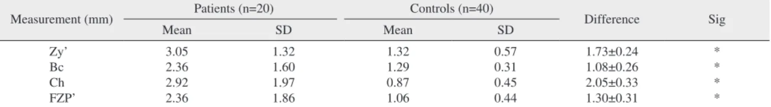
The average amount of asymmetry in the control group was less than 2 mm (Zy’: 1.31±0.57 mm, Bc: 1.28±0.30 mm), Ch:
0.87±0.45 mm, FZP: 1.06±0.44 mm). Asymmetry greater than 2 mm was observed in the patient group (Zy’: 3.05±1.32 mm, Bc: 2.36±1.60 mm, Ch: 2.92±1.97 mm, FZP’: 2.36±1.86 mm). The measured symmetry amounts in the patient group were statistically significantly different from the control group (P<0.05).(Table 3)
2) Soft tissue landmarks
Bucclae (Bc), cheek point (Ch), zygion’ (Zy’) and fronto- zygomatic point’ (FZP’) were used as soft tissue landmarks.
(Table 2, Fig. 2)
3) Soft tissue distance measuring
Zygomatic width was measured from midsagittal plane to Zy’. Zygoma and cheek projection were measured from the coronal plane to Bc, Ch and FZP’. In the control group, dif- ference between right and left measurements was used. In the patient group, measurement differences between the unaf- fected and affected areas were used.
4) Hard tissue reduction accuracy
Reduction amounts of bone fragments were measured at four buttresses where the ZMC fracture occurred (zygomati- cofrontal suture deviation, anterior wall of maxilla, zygomat- ic arch and infraorbital rim). The patient group was divided based on the amount of deviation (2 mm or more, less than 2 mm). Less than 2 mm of deviation was considered as the gold standard for hard tissue reduction.
5) Statistical analysis
Preparation and measurement of all coordinates were re-
Table 3. The mean difference between the patient and control groups
Measurement (mm) Patients (n=20) Controls (n=40)
Difference Sig
Mean SD Mean SD
Zy’
Bc Ch FZP’
3.05 2.36 2.92 2.36
1.32 1.60 1.97 1.86
1.32 1.29 0.87 1.06
0.57 0.31 0.45 0.44
1.73±0.24 1.08±0.26 2.05±0.33 1.30±0.31
*
*
*
* (SD: standard deviation, Sig: significance)
*Statistically significant difference between the groups (P<0.05).
Refer to Table 2 for the definitions of landmarks.
Dong Hyuck Kim et al: Evaluation of soft tissue asymmetry using cone-beam computed tomography after open reduction and internal fixation of zygomaticomaxillary complex fracture.
J Korean Assoc Oral Maxillofac Surg 2014
Table 4. The mean difference according to type of fracture
Measurement (mm) Linear fracture (n=10) Comminuted fracture (n=10)
Mean SD Mean SD Sig
Zy’
Bc Ch FZP’
3.23 2.20 2.86 2.25
1.30 1.49 1.80 2.07
2.49 2.85 3.10 2.71
1.38 1.98 2.66 1.14
NS NS NS NS (SD: standard deviation, Sig: significance, NS: non-significant)
*Statistically significant difference between the groups (P<0.05).
Refer to Table 2 for the definitions of landmarks.
Dong Hyuck Kim et al: Evaluation of soft tissue asymmetry using cone-beam computed tomography after open reduction and internal fixation of zygomaticomaxillary complex fracture.
3) Effect of fixation materials (titanium or biodegradable) Titanium and biodegradable plates were used in 10 patients, separately. Soft tissue depression was more pronounced at Zy’
and Ch’ in the titanium plate group (Table 6); however, no statistically significant difference was found (P<0.05).
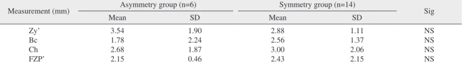
4) Effect of asymmetry perception after reduction surgery Soft tissue depression was observed irrespective of asym- metry perception after reduction surgery.(Table 7)
IV. Discussion
Various analysis methods using 3D CT scans have been 2. Comparison among ZMC reduction patients
1) Effect of comminuted fractures
When a comminuted fracture of the maxilla was present, the amount of asymmetry was increased when compared with a linear fracture.(Table 4) However, soft tissue asymmetry was not statistically significantly different regarding the pres- ence or absence of comminuted fracture (P<0.05).
2) Effect of the amount of deviation after reduction When hard tissue deviation was 2 mm or greater, soft tis- sue depression was observed on Bc (3.41±1.63 mm; Table 5) and the depression was statistically significant (P<0.05).
Table 5. The mean difference according to dislocation after surgery
Measurement (mm) <2 mm (n=9) ≥2 mm (n=11)
Mean SD Mean SD Sig
Zy’
Bc Ch FZP’
3.06 1.51 3.40 2.78
0.97 0.96 1.77 2.40
3.03 3.41 2.34 1.85
1.72 1.63 2.15 0.73
NS
* NS NS (SD: standard deviation, Sig: significance, NS: non-significant)
*Statistically significant difference between the groups (P<0.05).
Refer to Table 2 for the definitions of landmarks.
Dong Hyuck Kim et al: Evaluation of soft tissue asymmetry using cone-beam computed tomography after open reduction and internal fixation of zygomaticomaxillary complex fracture.
J Korean Assoc Oral Maxillofac Surg 2014
Table 6. The mean difference according to fixation materials
Measurement (mm) Titanium (n=12) Biodegradable (n=8)
Mean SD Mean SD Sig
Zy’
Bc Ch FZP’
3.60 2.37 3.56 2.87
1.47 1.84 2.44 2.51
2.50 2.36 2.29 1.85
0.92 1.41 1.15 0.68
NS NS NS NS (SD: standard deviation, Sig: significance, NS: non-significant)
*Statistically significant difference between the groups (P<0.05).
Refer to Table 2 for the definitions of landmarks.
Dong Hyuck Kim et al: Evaluation of soft tissue asymmetry using cone-beam computed tomography after open reduction and internal fixation of zygomaticomaxillary complex fracture.
J Korean Assoc Oral Maxillofac Surg 2014
Table 7. The mean difference according to recognition of asymmetry
Measurement (mm) Asymmetry group (n=6) Symmetry group (n=14)
Mean SD Mean SD Sig
Zy’
Bc Ch FZP’
3.54 1.78 2.68 2.15
1.90 2.24 1.87 0.46
2.88 2.56 3.00 2.43
1.11 1.37 2.06 2.15
NS NS NS NS (SD: standard deviation, Sig: significance, NS: non-significant)
*Statistically significant difference between the groups (P<0.05).
Refer to Table 2 for the definitions of landmarks.
Dong Hyuck Kim et al: Evaluation of soft tissue asymmetry using cone-beam computed tomography after open reduction and internal fixation of zygomaticomaxillary complex fracture.
J Korean Assoc Oral Maxillofac Surg 2014
bility of bony fragments after the surgery.
Presence of comminuted maxilla fracture did not affect the amount of asymmetry on each landmark in a statistically significant manner. According to a study by Klotch and Gil- liland28, facial expression muscles were detached from hard tissue and fibrosis occurred at an abnormal location if open reduction was not performed when a maxillary comminuted fracture occurred leading to inappropriate soft tissue contour.
They recommended immediate reconstruction of hard tissue and best results were obtained when open reduction surgery was performed within 48 hours. Their recommendation is in contrast to conventional treatment of closed reduction to prevent demineralization of bone tissue when mandibular comminuted fracture occurs29. In the present study, open re- duction was immediately performed when the comminuted fracture occurred. Slight soft tissue depression was observed compared with the control group. However, no significant difference was found when compared with cases where a comminuted fracture did not occur. Immediate open reduc- tion is required in the maxilla for esthetic soft tissue contour because the maxilla has ample blood supply.
When soft tissue asymmetry was compared according to the amount of deviation after the reduction surgery, soft tis- sue was more conspicuous on the landmark Bc when hard tissue deviation exceeded 2 mm after surgery (Bc: 3.41±1.63 mm). Statistically significant difference was found (P<0.05).
Soft tissue depression around zygoma would be more notice- able if reduction was not accurate. The majority of previous studies reported that hard tissue asymmetry less than 2 mm was not perceivable30,31; in the present study, patients with less than 2 mm deviation did not complain of asymmetry.
Location of the landmarks in patients with less than 2 mm of deviation was not considerably different from patients with 2 mm or greater deviation. The amount of deviation in the patient group where deviation was 2 mm or greater ranged from 2 mm to 3.78 mm indicating that approximately 3 mm of soft tissue asymmetry can be expected if reduction is not precisely performed.
With respect to plate materials, titanium and biodegradable plates were used for 10 patients, separately. Soft tissue de- pression was more noticeable on Zy’ and Ch when titanium was used in comparison to biodegradable plates (P<0.05) because titanium plates were used when the ZMC fracture dislocation amount was larger. Previous studies investigated tissue response to fixation materials. Wittwer et al.32 reported that a biodegradable plate achieved satisfactory fixation ef- introduced to evaluate soft tissue change after surgery8-18 and
are useful despite possible image errors19,20. Most of these studies focused on assessment of soft tissue change after or- thognathic surgery or surgery for correction of craniofacial deformity. Evaluation of soft tissue change after ZMC frac- ture reduction surgery employing the same principles used in those studies is difficult. Recently, laser scans are gaining popularity to evaluate facial soft tissue. However, the high cost of laser scans can be a limiting factor. In this study, soft tissue was assessed using CBCT images with OnDemand3D software and relatively accurate results were obtained.
Furst and colleagues reported no statistically significant difference between ZMC CT images and dry skull21. Gwil- liam et al.22 reported that cephalometric landmarks on the soft tissue were statistically reproducible. The present study also revealed no statistically significant difference when the measurements were repeated after 2 weeks to avoid intra- observer errors (P<0.05). 3D planes for image analysis were constructed using the same method described by Cho23.
CBCT images used in this study were taken after at least 3 months of follow-up. Modabber et al.24 investigated swell- ing after ZMC fracture reduction according to two different cooling therapy methods. They reported that swelling was the most severe 3 days after the surgery and no statistically significant difference was found between the two groups 28 days after the surgery. Generally soft tissue assessed at 6 months after surgery in orthognathic surgery is considered a relapse. This study evaluated CBCT images taken 3 months after surgery to minimize the effects of postoperative swell- ing and relapse was not considered because rigid fixation was used.
Hwang et al.25 evaluated asymmetry in adults with normal occlusion and reported a difference in distance from the mid- sagittal plane to right and left bilateral landmarks such as Bc, Ch and Zy’ (Bc: 1.43±1.06 mm, Ch: 0.85±0.69 mm); similar results were obtained in our study for normal adults (Zy’: 1.32
±0.57 mm, Bc: 1.29±0.31 mm, Ch: 0.87±0.45 mm, FZP’:
1.06±0.44 mm).(Table 3)
A variety of studies have investigated the accuracy of ZMC reduction surgery on the hard tissue and the majority reported satisfactory results26,27. However, patients perceive facial asymmetry in their soft tissue. In the present study, asymmetry was assessed on the soft tissue and the patient group showed statistically significantly more pronounced facial asymmetry than the control group (P<0.05). Soft tissue asymmetry is affected by soft tissue change after trauma or
different (P<0.05) regardless of whether patients complained of asymmetry.
Soft tissue asymmetry occurs after open reduction surgery and various factors are associated. Asymmetry of ZMC frac- ture patients after surgery was evaluated using CBCT.
Conflict of Interest
No potential conflict of interest relevant to this article was reported.
References
1. Ellis E 3rd, Kittidumkerng W. Analysis of treatment for isolated zygomaticomaxillary complex fractures. J Oral Maxillofac Surg 1996;54:386-400.
2. Chae YP, Kim SK. Clinical study on surgical treatment of zygoma fractures. Taehan Chikkwa Uisa Hyophoe Chi 1989;27:949-57.
3. Toriumi M, Nagasao T, Itamiya T, Shimizu Y, Yasudo H, Sakamoto Y, et al. 3-D analysis of dislocation in zygoma fractures. J Cranio- maxillofac Surg 2013. doi: 10.1016/j.jcms.2013.06.003.
4. Czerwinski M, Ma S, Williams HB. Zygomatic arch deforma- tion: an anatomic and clinical study. J Oral Maxillofac Surg 2008;66:2322-9.
5. Zhang QB, Dong YJ, Guan JB, Li ZB, Zhao JH, Dong FS. Epi- demiology and treatment of fractures of the zygomatic complex.
Asian J Oral Maxillofac Surg 2008;20:59-64.
6. Ellis E 3rd, el-Attar A, Moos KF. An analysis of 2,067 cases of zygomatico-orbital fracture. J Oral Maxillofac Surg 1985;43:417- 28.
7. Gaziri DA, Omizollo G, Luchi GH, de Oliveira MG, Heitz C. As- sessment for treatment of tripod fractures of the zygoma with mi- crocompressive screws. J Oral Maxillofac Surg 2012;70:e378-88.
8. Miller L, Morris DO, Berry E. Visualizing three-dimensional facial soft tissue changes following orthognathic surgery. Eur J Orthod 2007;29:14-20.
9. Solem RC, Marasco R, Guiterrez-Pulido L, Nielsen I, Kim SH, Nelson G. Three-dimensional soft-tissue and hard-tissue changes in the treatment of bimaxillary protrusion. Am J Orthod Dentofacial Orthop 2013;144:218-28.
10. Baik HS, Jeon JM, Lee HJ. Facial soft-tissue analysis of Korean adults with normal occlusion using a 3-dimensional laser scanner.
Am J Orthod Dentofacial Orthop 2007;131:759-66.
11. Li H, Yang Y, Chen Y, Wu Y, Zhang Y, Wu D, et al. Three- dimensional reconstruction of maxillae using spiral computed tomography and its application in postoperative adult patients with unilateral complete cleft lip and palate. J Oral Maxillofac Surg 2011;69:e549-57.
12. Damstra J, Oosterkamp BC, Jansma J, Ren Y. Combined 3-dimen- sional and mirror-image analysis for the diagnosis of asymmetry.
Am J Orthod Dentofacial Orthop 2011;140:886-94.
13. Cavalcanti MG, Rocha SS, Vannier MW. Craniofacial measure- ments based on 3D-CT volume rendering: implications for clinical applications. Dentomaxillofac Radiol 2004;33:170-6.
14. Park SH, Yu HS, Kim KD, Lee KJ, Baik HS. A proposal for a new analysis of craniofacial morphology by 3-dimensional computed tomography. Am J Orthod Dentofacial Orthop 2006;129:600. e23- 34.
15. You KH, Lee KJ, Lee SH, Baik HS. Three-dimensional computed tomography analysis of mandibular morphology in patients with facial asymmetry and mandibular prognathism. Am J Orthod Den- tofacial Orthop 2010;138:540.e1-8.
induced inflammatory reaction or tissue response against soft tissue. Langford and Frame33 stated no evidence was found indicating titanium induced soft tissue inflammation. Accord- ingly, asymmetry on Ch was less prominent when a biode- gradable plate was used because soft tissue response caused swelling and increased soft tissue volume when compared with titanium.
Irrespective of perception of asymmetry, soft tissue depres- sion was observed after surgery. The average location of right and left landmarks was not statistically significantly different according to asymmetry complaint (P<0.05). In the present study, we evaluated the effects of surgical techniques, pres- ence of comminuted fracture, fixation materials and accuracy of soft tissue contour reduction after ZMC fracture reduc- tion surgery. In addition to those factors, presence of exist- ing asymmetry and masseter muscle and facial expression muscles attached to the zygoma can also influence soft tissue contour34. In this study, five patients complained of asym- metry and their average deviation on Zy’ was significantly different from patients who did not complain, indicating asymmetry caused by lateral zygoma protrusion was more easily perceived than asymmetry caused by posteroanterior zygoma protrusion. A study by Choi et al.35 and Zheng et al.36 also reported that people perceived reduced lateral zygoma protrusion as more esthetic.
Asymmetry in the surgery group was generally more pro- nounced than in the control group. Postoperative soft tissue asymmetry was observed even when reduction was less than 2 mm. The amount of asymmetry was not considerably dif- ferent regardless if the patient complained. These findings can be used in clinics when informing patients that asymme- try is inevitable irrespective of reduction accuracy.
V. Conclusion
The amount of soft tissue asymmetry after open reduction of ZMC fracture was evaluated with CBCT scans taken 3-6 months postoperatively to exclude effects of postoperative swelling. OnDemand3D software was used and the following conclusions were reached;
1. Asymmetry was increased in the patient group with sta- tistical significance (P<0.05) when compared with the control group.
2. Amount of protrusion on Bc was influenced by the amount of hard tissue dislocation after surgery.
3. Soft tissue contour was not affected by fixation materials.
4. Degree of asymmetry was not statistically significantly
27. Kubota Y, Kuroki T, Akita S, Koizumi T, Hasegawa M, Rikihisa N, et al. Association between plate location and plate removal following facial fracture repair. J Plast Reconstr Aesthet Surg 2012;65:372-8.
28. Klotch DW, Gilliland R. Internal fixation vs. conventional therapy in midface fractures. J Trauma 1987;27:1136-45.
29. Alpert B, Tiwana PS, Kushner GM. Management of comminuted fractures of the mandible. Oral Maxillofac Surg Clin North Am 2009;21:185-92.
30. Ferrario VF, Sforza C, Schmitz JH, Miani A Jr, Serrao G. A three- dimensional computerized mesh diagram analysis and its applica- tion in soft tissue facial morphometry. Am J Orthod Dentofacial Orthop 1998;114:404-13.
31. af Geijerstam B, Hultman G, Bergström J, Stjärne P. Zygomatic fractures managed by closed reduction: an analysis with postop- erative computed tomography follow-up evaluating the degree of reduction and remaining dislocation. J Oral Maxillofac Surg 2008;66:2302-7.
32. Wittwer G, Adeyemo WL, Yerit K, Voracek M, Turhani D, Watz- inger F, et al. Complications after zygoma fracture fixation: is there a difference between biodegradable materials and how do they compare with titanium osteosynthesis? Oral Surg Oral Med Oral Pathol Oral Radiol Endod 2006;101:419-25.
33. Langford RJ, Frame JW. Tissue changes adjacent to titanium plates in patients. J Craniomaxillofac Surg 2002;30:103-7.
34. Dal Santo F, Ellis E 3rd, Throckmorton GS. The effects of zygo- matic complex fracture on masseteric muscle force. J Oral Maxil- lofac Surg 1992;50:791-9.
35. Choi BK, Goh RC, Moaveni Z, Lo LJ. Patient satisfaction after zygoma and mandible reduction surgery: an outcome assessment. J Plast Reconstr Aesthet Surg 2010;63:1260-4.
36. Zheng W, Xiang L, Fadare O, Kong B. A proposed model for en- dometrial serous carcinogenesis. Am J Surg Pathol 2011;35:e1-14.
16. Lee MS, Chung DH, Lee JW, Cha KS. Assessing soft-tissue char- acteristics of facial asymmetry with photographs. Am J Orthod Dentofacial Orthop 2010;138:23-31.
17. Baik HS, Kim SY. Facial soft-tissue changes in skeletal class III orthognathic surgery patients analyzed with 3-dimensional laser scanning. Am J Orthod Dentofacial Orthop 2010;138:167-78.
18. Meyer-Marcotty P, Alpers GW, Gerdes AB, Stellzig-Eisenhauer A.
Impact of facial asymmetry in visual perception: a 3-dimensional data analysis. Am J Orthod Dentofacial Orthop 2010;137:168.e1-8.
19. Peck S, Peck L, Kataja M. Skeletal asymmetry in esthetically pleas- ing faces. Angle Orthod 1991;61:43-8.
20. Ferrario VF, Sforza C, Poggio CE, Serrao G. Facial three-dimen- sional morphometry. Am J Orthod Dentofacial Orthop 1996;109:
86-93.
21. Furst IM, Austin P, Pharoah M, Mahoney J. The use of computed tomography to define zygomatic complex position. J Oral Maxil- lofac Surg 2001;59:647-54.
22. Gwilliam JR, Cunningham SJ, Hutton T. Reproducibility of soft tissue landmarks on three-dimensional facial scans. Eur J Orthod 2006;28:408-15.
23. Cho HJ. A three-dimensional cephalometric analysis. J Clin Orthod 2009;43:235-52.
24. Modabber A, Rana M, Ghassemi A, Gerressen M, Gellrich NC, Hölzle F, et al. Three-dimensional evaluation of postoperative swelling in treatment of zygomatic bone fractures using two dif- ferent cooling therapy methods: a randomized, observer-blind, prospective study. Trials 2013;14:238.
25. Hwang HS, Yuan D, Jeong KH, Uhm GS, Cho JH, Yoon SJ.
Three-dimensional soft tissue analysis for the evaluation of facial asymmetry in normal occlusion individuals. Korean J Orthod 2012;
42:56-63.
26. Zachariades N, Mezitis M, Anagnostopoulos D. Changing trends in the treatment of zygomaticomaxillary complex fractures: a 12-year evaluation of methods used. J Oral Maxillofac Surg 1998;56:1152-6.