Vol.12, No.2, April, 1999
소아 상완골 원위부 골단의 골절 및 분리
조현오・곽경덕・김병용・손수민・오필환
울산동강병원 정형외과
= Abstract =
Fracture-Separation of the Distal Humeral Epiphysis in Young Children
Hyoun-Oh Cho, M.D., Kyoung-Duck Kwak, M.D., Byung-Yong Kim, M.D., Su-Min Sohn, M.D., Pill-Whan Oh, M.D.
Department of Orthopaedic Surgery, Dong Kang General Hospital, Ulsan, Korea
We have reviewed seven cases of fracture-separation of the distal humeral epiphysis, two of which were initially misdiagnosed as a fracture of the lateral condyle and one as a fracture of the supracondyle. The four cases were treated by closed reduction and cast immobilization, and three cases by open reduction and internal fixation. The one case with conservative treatment had cubitus varus, other were good result. To distinguish the fracture-separation from a fracture of lat. condyle and from a dislocated elbow is impossible using clinical signs alone. For reducing misdiagnosis, it is important to consider age and there could be need further evaluation such as arthrogram, USG, CT and MRI.
Key Words: Distal Humeral Epiphysis, Fracture-Separation
※통신저자 : 오필환
울산시중구태화동 123-3 (681-320) 동강병원정형외과
Tel : (052) 241-1733
*본논문의요지는 1 9 9 6년대한정형외과추계학술대회에서구연되었음
서 론
소아상완골원위부골단의골절및분리는매우드 문 손상으로써, 초진시 암상소견 및 방사선 소견상 주관절부위의다른 손상들과 감별이어려워서상완 골 외과골절, 주관절 탈구 및 과상부골절 등으로 오 진하는경우가종종있다. 1818년 S t i m s o n에의해처음 으로서술되었던소아상완골원위부골단의골절및 분리는그후 1 9 8 6년 DeLee 등8 )이가장많은 1 6례, 1991 년 J a g e r와 H o f f m a n7 )이 1 2례를보고하였다. 소아상완 골 원위부 골단의 골절 및 분리는 상완골 원위부의 연골이방사선촬영시보이지않기때문에보다정확 한 진단을 위해서는 방사선촬영, 관절조영술, 초음 파, 컴퓨터 단층촬영, 자기공명영상 등을 이용할 수 있다(Fig. 1-4). 저자들은 1 9 9 2년 1월부터 1 9 9 6년 1 2월 까지 5년간 울산동강병원에서치료한7례에대해서, 정확한진단과적절한치료에다양한방사선학적검 사방법이유용하여그결과를문헌고찰과함께보고 하는바이다.
연구대상 및 방법
1 9 9 2년 1월부터 1 9 9 6년 1 2월까지 본울산동강병원 정형외과에서 치료한 7례를 대상으로 하였다. 초진 시연령은 1 3개월에서4세까지였으며평균 2 5 . 5개월
이었다. DeLee 등8 )의 분류에 의하면 Group A, B, C가 각각 0, 5, 2례였다(Table 1). 7례중 남아가 5레, 여아가 1례 였고, 수상원인은 7례에서 낙상이 3례, 추락이 4 례였으며 7례중 3례에서처음진단시상완골외과골 절( 2례) 및 상완골 과상부골절( 1례)로 오진되었다 (Table 2). 전례에서혈관및신경계의손상은없었다 . 입원당시 이학적 소견과 단순방사선촬영으로 확진 이 어려운 경우에는 초음파검사, 컴퓨터 단층촬영, 자기공명영상등이이용되었고, 때로는마취하에관 절조영술로도움을얻을수있었다(Fig. 1-4). 치료는4 366•대한골절학회지/ 제12권제2호
Table 1.Three groups based on roentgenographic appearance
Group A Newborn to nine months old No ossification center pressent in the capitellum
No metaphyseal fragment (Thurston-Holland sign)
Group B Nine months to three years old
Ossification center present in the capitellum Very small or no Thurston-Holland fragment Group C Three to seven years old
Well developed ossification center present in the capitellum
Large Thurston-Holland metaphyseal fragment
Table 2.Analysis of cases
Case Sex/Age Cause Initial Final Tx ROM§ Carrying
of truma diagnosis diagnosis angle
1 2 3 4 5 6 7
* Salter-Harris
†Closed reduction
‡Open reduction and internal fixation
§Range of motion F/22mo F/16mo M/22mo M/4yr M/4yr M/13mo M/33mo
slip down fall down fall down fall down slip down fall down slip down
at. condyle Fx.
S-H* type I S-H type II lat. condyle Fx.
S-H type I S-H type I supracondylar Fx.
S-H type II S-H type I S-H type II S-H type II S-H type I S-H type I S-H type II
CR† OR/IF‡
OR/IF CR CR CR OR/IF
5o-135o full full full full full full
20ovarus 5ovarus 3ovarus 5ovarus 5ovarus 5ovarus 3ovarus 25오필환 2003.5.17 5:41 PM 페이지366
Fig 1.Arthrogram of fracture-separation of the lower humeral epiphysis.
Fig 2.USG of fracture-separation of the lower humeral epiphysis. USG shows high echoic line (arrows) on epiphyseal area.
Fig 4.MRI of fracture-separation of the lower humeral epiphysis. T1 weighted coronal image shows epiphyseal plate and displaced
epiphysis Fig 3.CT shows slightly displaced fracture-
separation of the lower humeral epiphysis.
Fig 5.Diagrams of the elbow Fig A.normal elbow
Fig B.dislocation of the elbow Fig C.fracture of the lateral
condyle
Fig D. supracondylar fracture Fig E.fracture separation of the
lower humeral eipiphysis.
례에서도수정복후전완을회내전 상태로장상지석 고로약 3주간 고정을시행하였고 나머지 3례에서는 관혈적정복술후K -강선으로내고정하였다.
결 과
소아 상완골 원위부골단의 골절분리 7례의 평균 추시기간은 1 8개월( 1 2 - 3 6개월) 이었으며, 그중 1례에 서만 굴곡구축 5 o에서 굴곡 1 3 5o까지의 운동제한 및 건측과비교하여2 0o의내반주변형을보였고나머지 6례에서는 임상소견 및 방사선 촬영상 만족스러운 결과를보였다(Table 2).
증 례
증례1
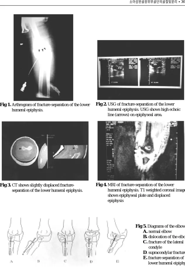
2 2개월된여아로낙상에 의한주관절부통증과종 창을 주소로내원하였다. 초진시상완골외과골절로 오진하여장상지석고고정을하였으나수상후 2주에 Salter-Harris type II 골절 및 분리로 확진되었다. 수상
후 3개월에자기공명영상으로성장판손상을추시하 였고 수상후 2년 6개월에임상소견상 굴곡구축 5o에 서굴곡 1 3 5o까지의경미한운동제한을보였고, 방사 선소견상 2 0o의내반주변형을보였다(Fig. 6-A,B,C).
증례2
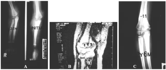
1 6개월된 여아로 추락사고후 상완골 원위부 골단 의골절분리로진단되어관혈적정복및 K -강선고정 하였고 술후 3주에 K -강선 제거하고 관절운동을 시 작하였다. 추시 1 5개월후 임상적 소견 및 방사선 소 견에서주관절의변형및운동제한을보이지않아양 호한결과를보였다(Fig. 7-A,B,C).
고 찰
소아에서 주관절부위의 손상은 대부분 과상골절, 외과골절등이며 그밖에요골두및경부골절, 내상과 골절, 척골주두골절 및 주관절탈구 등이다. 그러나 상완골원위부골단의 Salter-Harris type I, II 골절및분 리는 매우 드물뿐더러 근래에 언급되기 시작했다.
1 9 6 7년 M a c a f e e1 3 )는 3례의 유아 상완골 과상부 골절 368•대한골절학회지/ 제12권제2호
Fig 6.A 22-month-old girl with epiphyseal fracture-separation of distal humerus.
Fig A.Initial roentgenogram of the Rt. elbow.
Fig B.MRI after 3 month showing epiphyseal injury.
Fig C.Anteroposterior roentgenogram after 30 months showing cubitus varus deformity
A B C
25오필환 2003.5.17 5:41 PM 페이지368
을 보고하였는데, 후에 DeLee 등8 )에 의해 상완골 원 위부골단의 골절및분리로밝혀졌다 . 즉, 소아의상 완골 원위부 골단의 골절 및 분리는 임상소견 및 방 사선소견상주관절부위의다른 손상들과 구별이어 려워종종잘못진단되기쉽다.
Bright 등5 )에의하면신생아 및영아에서는원위부
골단이 회전 전단력(Rotatory shear force)에 약하기 때 문에 분만 손상이나 소아 학대에의한 S a l t e r - H a r r i s type I 손상이 많으며, 자궁내 위치로 인한 주관절의 굴곡구축이 남아있기 때문에 과신전 손상은 적다고 하였다. 반면, 유소아에서는주관절의과신전으로인 하여 주로 후내방의 골간단 골절편을 동반하는 Salter-Harris type II 손상이 많이 발생한다고 하였다.
저자들의경우분만손상이나소아학대에의한손상 은 없었으며 평균 연령은 2 8 . 8개월이었고, Salter- Harris type I, II가각각 3례, 4례이었다(Table 2).
소아에서는 상완골 원위부의 연골이 방사선 촬영 시 보이지 않기 때문에 진단에 많은 어려움이 있다.
그러므로 주관절의 골화중심 출현시기에 대한 지식 은 주관절부위의 손상을 진단하는데에 필수불가결 하다. 따라서 진단은 골화 중심이 언제 생기는지를 잘알아야한다. 출생시는상완골원위부의골단에는 골화중심이없으며 6주에서 8개월사이에상완골소
두가발생하고 5세에서 7세사이에내상과 골화중심 이 나타나며 8세에서 1 0세 사이에 활차의 골화중심 이 발생하고 1 1세내지 1 2세에서외상과골화중심이 나타난다.
신생아및영아에서는 Salter-Harris type I 골절및분 리가잘발생하기때문에특히주관절탈구와감별을
요하며1 3 , 1 6 ), 유소아에서는 Salter-Harris type II의 골절
및분리가많기때문에상완골외과골절과감별진단
을요한다4 , 1 1 , 1 2 , 1 5 )이에도움이 되는 진단방법으로는
단순방사선촬영, 관절조영술, 초음파 검사, 컴퓨터 단층촬영, 자기공명영상등이있다2 ).
방사선 소견상 주관절탈구는 상완소두와 요골두 사이의 정상적인 관계가변화되며, 상완골외과골절 에서는 상완골과 전박부의 관계는 유지되나 상완소 두와요골두 사이관계는변화된다. 과상부골절에서 는 상완골 과상부에 골절선을 확인할 수 있고, 반면 에상완골원위부골단의골절및분리에서는상완골 과의관계는변화되며, 골절부위에서전위정도에따 라 상완골과 전박부의 축에 변화를 초래한다(Fig. 5).
상완골원위부골단의골절및분리를관절내골절과 구별하기 위해서는 관절 조영술(Fig. 1)이 도움이 된 다고한다3 , 4 , 6 , 1 0 , 1 5 )
. 상완골원위부의골화가되기전인 신생아나유아에서상완골 원위골단의모양을 아는 Fig 7.A 16-month-old girl with epiphyseal fracture-separation of distal humerus.
Fig A.Initial roentgenogram of the Rt. elbow.
Fig B. Roentgenogram after open reduction and internal fixation.
Fig C.Roentgenogram after 15 months showing good result.
A B C
데초음파검사를사용할수있다8 )고하며건측의골 단과비교하여보면골단의분리가있는지를 결정할 수있다고한다(Fig. 2). 컴퓨터 단층촬영또는자기공 명영상 역시 골단의 골절 및 분리를 다른 손상과 감 별하는데사용될수가있다(Fig. 3,4). 저자들의경우 3 례에서 초진시 과상부골절( 1례)과 외과골절( 2례)로 오진하여 초음파 검사, 관절 조영술, 자기공명영상 등을 이용하여 Salter-Harris type II 골절 및 분리로 확 진되었다(Table 2).
상완골원위부골단의골절및분리의치료는여러 저자들이도수정복및석고고정또는견인과같은보 존적인 치료를 권유하였다6 , 7 , 8 , 1 1 , 1 2 , 1 5 , 1 6 ). DeLee, Holda
등8 , 1 1 , 1 2 , 1 5 )에 의하면 전박부의 고정위치는 외측골절
편을 안정시키기 위해 회내전위치로 고정하여 고정 기간 동안 전위를 방지 한다고 하였다. Sutherland와 W r o b e l1 7 )은 대부분 보존적 치료가 적응되지만 전위 가심할때나부분적치유가있을때는수술적 요법의 적응증이된다고하였다. 저자들의경우에도보존적 치료를 원칙으로 하였고 관혈정복하였던3례중 2례 는초진시오진하였던예였다(Table 2).
합병증으로혈관및신경계손상의보고는없었다 . Mizuno 등1 5 )은 3개월에발생한불유합 1례를보고하 였으나상완골원위부에는혈관이많고골화가잘되 기때문에불유합이아주드물다 . Holda 등1 1 )은 7례중 5례에서 1 0o- 1 5o정도의 내반주가 발생하였다고 보고 하였다. 저자들의경우 1례에서 건측에비해 1 5o이상 의차이를보이는내반주변형이 1례있었다.
요 약
평균 연령은 2 5 . 5개월 이었고, 수상원인은 추락 4 례, 낙상 3례였으며전 7례중 3례는초진시오진되었 다. 4례는 보존적 치료를 하였고 3례는 관혈적 치료 를 하여 1례를 제외하고는 모두 만족스러운 결과를 보였다(Table 2). 상완골 원위골단의 골절 및 분리는 진단에 어려움이 많고 임상증상만으로는 주관절 주 위의다른손상과감별하기힘들므로오진을줄이기 위해서는발생연령의고려및관절조영술, 초음파검 사, 컴퓨터 단층 촬영, 자기공명영상 등의 세심한 검 사가필요할것으로사료된다.
R E F E R E N C E S
1) 이석현, 장재석, 송해룡, 전재영: 소아 상완골 원 위부 골단의 Salter-Harris type I, II 골절분리에 대 한 임상적 연구. 대한정형외과학회지 , 23(1) : 248-254, 1988
2) 이동우, 김세동: 소아 상완골 원위부 골단의 골 절및분리 , 대한골절학회지, 7(1) : 72-78, 1994 3) Akbarnia B.A., Siberstein M.J., Rende R.J. :
Arthrography in the diagnosis of fractures of the distal end of the humerus in infants. J Bone Joint S u r g, 68A : 599-620, 1986.
4) Barrett W.P., Almquist I.A., Staheli L.T. : Fracture separation of the distal humeral physis in the newborn. J Pediatric Orthop 4 : 617-619, 1984.
5) Bright R.W. : Epiphyseal plate cartilage : a biomechanical and histological analysis of failure modes. J Bone Joint Surg, 56 : 688-703, 1974.
6) Chand K. : Epiphyseal separation of distal humeral epiphysis in and infant : a case report and review of literature. J Trauma, 14(6) : 521-526, 1974.
7) De Jager L.T., Hoffman E.B. : Fractures separation of the distal humeral epiphysis. J Bone Joint Surg, 73B(1) : 143-146, 1991.
8) DeLee J.C., Wilkins K.E., Rogers L.F., Rockwood C . A . : Fractureseparatio of the distal humeral epiphysis. J Bone Joint Surg, 62A(1) : 46-51, 1980.
9) Dias J.J., Lamont A. C., Jones J.M. : Ultrasonic diagnosis of neonatal separation of the distal humeral epiphysis. J Bone Joint Surg, 70B : 825-828, 1988.
10) Hansen P.E., Barnes D.A., Tullos H.S. : case report arthrographic diagnosis of an injury pattern in the distal humerus of an infant. J Pediatric Orthop, 2 : 569-572, 1982.
11) Holda M.E., Manoli A. Jr., LaMont R.L. : Epiphyseal separation of the distal end of the humerus with medial displacement. J Bone Joint S u r g, 62A(1) : 52-57, 1980.
12) Kaplan S.S., Reckling F.W. : Fracture separation of the lower humeral epiphysis with medial displacement. J Bone Joint Surg, 53A(6) : 1105- 370•대한골절학회지/ 제12권제2호
25오필환 2003.5.17 5:41 PM 페이지370
1108, 1971.
13) Macafee, A.L. : Infantile supracondylar fracture. J Bone Joint Surg, 49B : 768-770, 1967.
14) Marmor J., Bechtol C.O. : Fracture separation of the lower humeral epiphysis : report of a case. J Bone Joint Surg, 42A(2) : 323-326, 1960.
15) Mizuno K., Hirohata K., Kashiwagi D. : Fracture separation of the distal humeral epiphysis in young
child. J Bone Joint Surg, 61A(4) : 569-573, 1979.
16) Siffer R.S. : Displacement of the distal humeral epiphysis in the newborn infant. J Bone Joint Surg, 45A(1) : 165-169, 1963.
17) Sutherland D.H., Wrobel L. : Displacement of entire distal humeral epiphysis, In proceedings of the Western Orthopedic Association. J Bone Joint Surg, 56A : 206-210, 1974.