- 165 - Received October 28, 2010 Accepted November 18, 2010
∙Jong Min Yoo, M.D
Department of Orthopaedic Surgery, Seoul St. Mary’s Hospital, The Catholic University of Korea School of Medicine, 505 Banpo-dong, Seocho-gu, Seoul 137-040, Korea
Tel: +82-2-2258-2837 Fax: +82-2-535-9834 E-mail: [email protected]
대한족부족관절학회지: 제14권 제2호 2010
J Korean Foot Ankle Soc. Vol. 14. No. 2. pp.165-168, 2010
만성 족부 질환이 환측 하지의 골밀도에 미치는 영향
가톨릭대학교 의과대학 서울성모병원 정형외과, 가톨릭대학교 의과대학 여의도성모병원 정형외과*
주인탁⋅유종민⋅강민구⋅정진화*
Effect of Chronic Foot Disease to Bone Mineral Density of the Affected Lower Limb
In-Tak Chu, M.D., Jong Min Yoo, M.D., Min Gu Kang, M.D., Jin Wha Chung, M.D.*
Department of Orthopaedic Surgery, Seoul St. Mary’s Hospital, The Catholic University of Korea School of Medicine, Seoul, Korea Department of Orthopaedic Surgery, Yeouido St. Mary’s Hospital, The Catholic University of Korea School of Medicine, Seoul, Korea*
=Abstract=
Purpose: Pain or discomfort caused by foot diseases may lead to abnormal gait, resulting in decreased bone mineral density (BMD) of the affected lower limb. We analyzed the effect of foot affection to BMD and its clinical significance.
Materials and Methods: Bilateral hip BMD was evaluated in 93 patients with unilateral chronic foot disease. To minimize statistical errors, we excluded patients with medical histories that had influence on BMD. Analysis was based on the results of BMD tests at the first visit. All patients denied past medical intervention for osteoporosis. The difference in density between bilateral limbs was determined by comparing BMDs of the neck, upper neck, trochanter and total area of hip.
Results: Test results revealed the decrease of BMD in the lower limb with the affected foot, compared to the unaffected side. This decrease was significant in the area of the trochanter (p <0.05). There was no marked difference of BMD in relation with duration of affection, underlying disease or age. Pertaining the location of foot affection, the hindfoot group showed significant decrease in BMD compared to the forefoot group. The group with affection in bone and joint also showed a marked decrease in BMD compared to the soft tissue group (p <0.05).
Conclusion: Pain and discomfort caused by chronic foot diseases can lead to a decrease in the BMD of the affected lower limb. This may increase the risk of complications such as osteoporotic fracture and muscular atrophy.
Key Words: Disuse osteoporosis, Bone mineral density, Chronic foot disease, Osteopenia
서 론
골밀도 유지를 위해서는 유전적 인자 및 환경적 인자가
작용하며 환경적 인자에는 칼슘과 비타민등의 영양인자, 비 만도, 호르몬 인자, 흡연 등의 기호식품이나 운동 등이 작용 할 수 있으며, 최근 골다공증 예방을 위한 운동의 중요성이 특히 강조되고 있다.1) 지속적인 운동을 통한 하지의 골격근 계의 체중부하를 통해 골밀도 감소를 예방할 수 있으며 이 를 위해서는 정상적인 보행 및 하지의 운동기능이 중요하 다. 고관절이나 슬관절과 같은 대관절에 이환된 질환의 경 우 통증이나 불편감으로 인한 보행이상이나 체중부하의 감 소는 환측 하지의 전체적인 골밀도 감소나 근육량의 감소 를 초래할 수 있으나2,3) 족부 질환과 같이 소관절에 이환된
주인탁⋅유종민⋅강민구⋅정진화
- 166 - Table 1. Difference between Affected Side and Contralateral Side
Difference of T-score in detailed area* mean (n=93) Standard deviation p value neck
upper neck wards trochanter total†
-0.0170 -0.0042 -0.0031 -0.1377 -0.0420
0.3551 0.4119 0.3254 0.4898 0.3774
0.6433 0.9204 0.9245 0.0076‡
0.2832
*T-score of affected side minus T-score of contralateral side; †Total T-score in BMD analysis is not the sum of the other areas but the independent measurement; ‡We use the term of “statistically significant” in this paper when the p-value was lower than 0.05.
Figure 1. Distribution of T-score of patients matched with age. The light gray zone (n=31) is the area of osteopenia (T-score≤-1.0) and the dark gray zone (n=48) is the area of osteoporosis (T-score≤
-2.5).
경우에선 골밀도에 미치는 영향에 대해 보고된 바는 거의 없다. 이에 저자들은 양측 하지의 골밀도 비교를 통해 만성 족부 질환이 하지의 골밀도에 미치는 영향을 보고자 하였다.
대상 및 방법
2008년 3월부터 2010년 9월까지 본원에 내원한 편측의 만성 족부 질환을 가진 93명의 환자를 대상으로 후향적으 로 분석하였다. 무작위로 환자를 선택하여 고관절 양측을 포함한 골밀도 검사를 시행하였으며 족부 질환 이외의 다 른 골밀도 수치에 영향을 줄 수 있는 과거력을 조사하였다.
통계적 오차를 유발할 수 있는 간섭요인을 최소화하기 위 해 인위적으로 환부의 체중부하를 제한할 수 있는 석고고 정이나 수술, 골절을 포함한 외상, 슬관절 및 고관절의 질환 이 있는 경우, 측만증 등의 척추 질환 및 하지 부동으로 인 한 파행 보행을 가진 경우는 배제하였다. 분석한 골밀도 검 사의 시기는 기존의 골다공증 치료가 개입되지 않은 초진 이후 첫 검사시기를 대상으로 양측 고관절의 세부부위에 따른 환측과 건측 하지의 T값 측정치 차이의 통계적 유의 성을 짝지은 t-검정법(paired t-test)을 이용하여 검토하였다.
또한, 나이 및 족부 질환의 이환 기간에 따른 연관성을 각 각 피어슨 상관계수(Pearson correlation coefficient) 및 스 피어만 상관계수(Spearman correlation coefficient)를 이용 한 상관분석을 하였고 내과적 기저질환의 유무에 따른 골 밀도의 차이는 t-검정을 이용하여 분석하였다. 또한, 전체 대상 환자를 전족부 및 후족부의 이환 부위에 따른 질환군 및 골관절과 연부조직 질환에 따른 그룹으로 나누어 t-검정 을 이용하여 비교 분석하였다.
결 과
대상 환자는 93명으로 평균나이는 56.8세였으며 성별은 남성이 6, 여성이 87명이었다. 총 93명의 환자 중 골밀도 검사상 T값이 -2.5 이하인 골다공증 진단 기준에 속하는 환
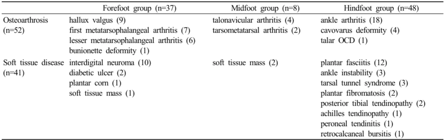
자는 48명, -1.0 이하인 골감소증 진단 기준에 속하는 환자 는 31명이었다(Fig. 1). 만성 족부 질환을 가지고 있는 환측 은 건측과 비교해 전체적으로 골밀도의 감소를 보였으며 짝지은 t-검정분석(paired t-test) 결과 건측에 대한 환측 전 자부의 골밀도 수치의 감소 정도가 평균 0.1377로 유의한 감소를 보였다(p<0.05, Table 1). 질병의 이환 기간은 평균 32.7개월로 상관계수의 검정결과 골밀도 감소와의 유의할 만한 상관관계는 보이지 않았으며, 또한 나이 및 기저질환 에 따른 유의할 만한 환측 하지의 골밀도 감소도 보이지 않 았다. 질환의 부위에 따라 후족부 질환군은 48명으로 족근 관절염과 족저 근막염이 많았고 전족부 질환군은 37명으로 지간 신경종 및 무지 외반증, 중족족지 관절염이 많았으며 골관절 조직에 이환된 질환군은 52명, 연부조직 질환군은 41명이었다(Table 2). 후족부 질환군과 전족부 질환군을 서 로 t-검정을 이용하여 비교한 결과 후족부 질환군의 골밀도 의 차이가 전족부 질환군에 비해 크게 나타났으며 이 중 대 퇴근위부에서 유의하게 환측의 골밀도 감소를 보였고 특히 후족부 질환군에서는 하위집단 분석에서도 전자부 및 대퇴 근위부에서 독립적으로 유의한 감소를 나타내었다(p<0.05,
만성 족부 질환이 환측 하지의 골밀도에 미치는 영향
- 167 - Table 2. Disease Distribution According to Affected Location
Forefoot group (n=37) Midfoot group (n=8) Hindfoot group (n=48) Osteoarthrosis
(n=52)
hallux valgus (9)
first metatarsophalangeal arthritis (7) lesser metatarsophalangeal arthritis (6) bunionette deformity (1)
talonavicular arthritis (4) tarsometatarsal arthritis (2)
ankle arthritis (18) cavovarus deformity (4) talar OCD (1)
Soft tissue disease (n=41)
interdigital neuroma (10) diabetic ulcer (2) plantar corn (1) soft tissue mass (1)
soft tissue mass (2) plantar fasciitis (12) ankle instability (3) tarsal tunnel syndrome (3) plantar fibromatosis (2) posterior tibial tendinopathy (2) achilles tendinopathy (1) peroneal tendinitis (1) retrocalcaneal bursitis (1)
Table 3. Analysis of the Subgroup According to the Affected Location
Subgroup analysis Difference of T-score
in detailed area* Comparing group A with B§ group A† (n=37) group B‡ (n=48)
t value p value Mean p value Mean p value
neck upper neck wards trochanter total
-0.27 -0.89 -0.38 1.76 2.17
0.7887 0.3769 0.7041 0.0827 0.0327
-0.0368 -0.0500 -0.0052 -0.0289 0.0605
0.5230 0.4230 0.9245 0.6669 0.2335
-0.0163 0.0286 0.0204 -0.2112 -0.1112
0.7481 0.6440 0.6221 0.0073 0.0606
*T-score of affected side minus T-score of contralateral side; †The group of patients affected at forefoot; ‡The group of patients affected at hindfoot; §The difference between group A and B. Statisically analyzed by the independent t-test.
Table 4. Analysis of the Subgroup According to the Characteristic of Disease
Subgroup analysis Difference of T-score
in detailed area* Comparing group A with B§ group A† (n=51) group B‡ (n=42)
t value p value Mean p value Mean p value
neck upper neck wards trochanter total
-1.78 -1.61 -1.63 -2.85 -2.94
0.0781 0.1098 0.1066 0.0054 0.0042
-0.0750 -0.0653 -0.0519 -0.2586 -0.1375
0.1622 0.2545 0.2906 0.0010 0.0205
0.05476 0.0714 0.0571 0.0119 0.0761
0.2580 0.2625 0.1992 0.8412 0.0947
*T-score of affected side minus T-score of contralateral side; †The group of patients with skeletal disease; ‡The group of patients with soft tissue disease; §The difference between group A and B. Statisically analyzed by the t-test.
Table 3). 골관절 조직에 이환된 질환군은 골관절 조직에 이환된 질환군에서 연부조직 질환에 비해 현저한 환측 골 밀도의 감소를 보였으며 역시 전자부 및 대퇴근위부의 전 체적인 골밀도에서 많은 감소를 보였다(p<0.05, Table 4).
고 찰
족부의 질환으로 인한 불편감이나 통증은 보행 주기중 입각기에 영향을 미칠 수 있다. 후족부의 질환은 입각기의
첫 단계인 발뒤축 닿음(heel strike)에 있어서, 전족부의 질 환은 입각기의 마지막 단계인 발가락 들림(toe off)에 영향 을 미칠 수 있다. 결국, 족부 질환으로 인한 통증이나 불편 감은 양측 하지의 유각기 및 입각기의 정상적인 균형을 와 해하여 환측의 입각기의 감소 및 유각기의 증가로 비정상 적인 보행을 야기할 수 있고4-6) 건측에 비해 환측 하지의 사 용량이 현저히 줄어들 수 있다. 이는 환측 하지 골격에 대 한 생리적인 자극이 감소되어 전체적인 골질량의 감소를 초래할 수 있으며7,8) 저자들의 연구에서도 환측은 건측에
주인탁⋅유종민⋅강민구⋅정진화
- 168 - 비해 골밀도의 감소를 보였다.
이 논문에서는 족부 질환이 후족부에 위치한 경우 전족 부에 비해 골밀도 감소에 미치는 영향이 큰 것으로 나타났 다. 이 결과는 후족부의 질환이 전족부에 비해 보행이상 및 환측 하지 사용량의 감소에 큰 영향을 미치는 것이라고 추 측해 볼 수 있다.3,9) 또한, 앞의 결과에서 보듯이 관절염이 나 무지 외반증과 같은 골관절조직에 이환된 족부 질환의 경우 연부조직에 발생한 경우보다 환측 하지의 사용량이 줄어들 수 있음을 간접적으로 알 수 있었다.
골다공증은 현대사회가 고령화되어 가면서 치료적 중요 성이 대두되고 있다. 골다공증은 골의 강도가 감소함으로 골절 및 근육 약화의 위험도가 높아지게 되는 골격계의 질
환이다.10,11) 골의 강도는 골량 및 골질을 종합적으로 평가
해야 하며 골다공증으로 인한 골절이 위험도는 골의 강도 뿐만 아니라 환경적 요인, 환자의 전신상태를 평가해야 임 상적 의의가 있으므로12,13) 이 논문에서 검토한 골밀도 검사 수치가 골절의 위험도를 전반적으로 평가하기에는 부족한 면이 있다. 아울러 골절환자의 경우 골다공증에 의한 위험 도보다는 환자의 활동량에 미치는 여러가지 요인 및 환경 적 요인이 더 크게 작용할 수 있어14-16) 골밀도 검사의 결과 는 골절에 대한 위험도의 부분적 지표로서 가치가 있을 것 이다. 예를 들어 이 연구에서 환측 하지에서 유의한 감소를 보인 전자부의 골밀도의 감소가 전자간 골절의 높은 위험 도를 반영하기 어려운 것이다. 하지만 족부 질환에 있어서 의 환측 하지의 골밀도 감소는 질환으로 인한 활동량의 제 한이 초래될 수 있다는 가설에 뒷받침하여 그 임상적 의의 가 크다고 볼 수 있다.
이 논문의 제한점은 93명 중 6명을 제외한 대다수의 환 자가 여성으로 폐경으로 인한 여성호르몬 변화가 골밀도 차이에 어느 정도 영향을 미치는지 고려하지 못했으며 환 자의 활동량에 영향을 미칠 수 있는 체질량 지수를 이용한 비만도, 시각적 통증 척도(VAS) 등을 이용한 통증 정도, 각 족부 질환별 중증도를 이용한 통계적 사정을 시행하지 못 했다는 점이다.
결 론
족부의 질환으로 야기된 환자의 불편감 및 보행이상은 환측 하지의 체중부하 감소를 야기할 수 있으며 결국 이환 된 하지의 골밀도의 감소를 유발하여 골절, 근육 약화 등의 이차적 합병증의 발병률을 높일 수 있다. 만성 족부질환 환
자에 있어 질환 자체의 치료와 더불어 골감소증으로 인한 부작용을 예방하기 위한 적극적인 치료적 접근이 요구된다.
REFERENCES
1. Kai MC, Anderson M, Lau EM. Exercise interventions:
defusing the world’s osteoporosis time bomb. Bull World Health Organ. 2003;81:827-30.
2. Lingard EA, Mitchell SY, Francis RM, et al. The prevalence of osteoporosis in patients with severe hip and knee osteoarthritis awaiting joint arthroplasty. Age Ageing. 2010;39:234-9.
3. Favier FB, Benoit H, Freyssenet D. Cellular and molecular events controlling skeletal muscle mass in response to altered use. Pflugers Arch. 2008;456:587-600.
4. Damiano J. Diagnostic procedures for painful foot. Rev Prat.
2010;60:327-33.
5. Voronov ML, Pinzur MS, Hoffman HH, et al. Static measure of foot loading. Foot Ankle Spec. 2009;2:267-70.
6. Keegan TH, Kelsey JL, Sidney S, Quesenberry CP Jr. Foot problems as risk factors of fractures. Am J Epidemiol. 2002;
155:926-31.
7. Sievänen H. Immobilization and bone structure in humans.
Arch Biochem Biophys. 2010;503:146-52.
8. Akhter MP, Alvarez GK, Cullen DM, Recker RR. Disuse- related decline in trabecular bone structure. Biomech Model Mechanobiol. 2010;Aug 4. [Epub ahead of print]
9. Horisberger M, Hintermann B, Valderrabano V. Alterations of plantar pressure distribution in posttraumatic end-stage ankle osteoarthritis. Clin Biomech (Bristol, Avon). 2009;24:303-7.
10. Newton MT, Archer JA, Scruggs M, et al. Low bone mass and fractures on foot radiographs: missed opportunities to evaluate for osteophorosis: a pilot study. J Am Podiatr Med Assoc.
2009;99:1-7.
11. Ferretti, JL, Cointry GR, Capozza RF, Frost HM. Bone mass, bone strength, muscle-bone interactions, osteopenias and osteoporoses. Mech Ageing Dev. 2003;124:269-79.
12. Cohen MM Jr. The new bone biology: pathologic, molecular, and clinical correlates. Am J Med Genet A. 2006;140:2646- 706.
13. Ralston SH. Genetics of osteoporosis. Proc Nutr Soc. 2007;
66:158-65.
14. van den Bergh JP, van Geel TA, Lems WF, Geusens PP.
Assessment of individual fracture risk: FRAX and beyond.
Curr Osteoporos Rep. 2010;8:131-7.
15. van Helden S, van Geel AC, Geusens PP, et al. Bone and fall-related fracture risks in women and men with a recent clinical fracture. J Bone Joint Surg Am. 2008;90:241-8.
16. Nguyen T, Sambrook P, Kelly P, et al. Prediction of osteoporotic fractures by postural instability and bone density.
Br Med J 1993;307:1111-5.